Review
. 2011 Jan 24;2(1):18-32.
doi: 10.1111/j.2040-1124.2010.00070.x.

Mechanism of diabetic neuropathy: Where are we now and where to go?

Affiliations
Review

Mechanism of diabetic neuropathy: Where are we now and where to go?

Soroku Yagihashi et al. J Diabetes Investig. .

Abstract

Neuropathy is the most common complication of diabetes. As a consequence of longstanding hyperglycemia, a downstream metabolic cascade leads to peripheral nerve injury through an increased flux of the polyol pathway, enhanced advanced glycation end-products formation, excessive release of cytokines, activation of protein kinase C and exaggerated oxidative stress, as well as other confounding factors. Although these metabolic aberrations are deemed as the main stream for the pathogenesis of diabetic microvascular complications, organ-specific histological and biochemical characteristics constitute distinct mechanistic processes of neuropathy different from retinopathy or nephropathy. Extremely long axons originating in the small neuronal body are vulnerable on the most distal side as a result of malnutritional axonal support or environmental insults. Sparse vascular supply with impaired autoregulation is likely to cause hypoxic damage in the nerve. Such dual influences exerted by long-term hyperglycemia are critical for peripheral nerve damage, resulting in distal-predominant nerve fiber degeneration. More recently, cellular factors derived from the bone marrow also appear to have a strong impact on the development of peripheral nerve pathology. As evident from such complicated processes, inhibition of single metabolic factors might not be sufficient for the treatment of neuropathy, but a combination of several inhibitors might be a promising approach to overcome this serious disorder. (J Diabetes Invest, doi: 10.1111/j.2040-1124.2010.00070.x, 2010).

Keywords: Diabetic neuropathy; Novel treatment; Pathogenesis.


Figures

Figure 1
Figure 1
Vascular supply of the peripheral nervous system is sparse and transperineurial arteriole penetrates into endoneurium. Autonomic nerve endings contact with the wall of arterioles, but vascular autoregulation is lacking in peripheral nerves as a result of sparse innervations. In diabetes, autonomic nerve endings to the arteriole are likely to be lost and therefore vasoregulation is further impaired (modified from Pathology of Diabetes Mellitus for Clinicians by Soroku Yagihashi, Shindan‐to‐Chiryo Co., Tokyo, 2004, page 110).
Figure 2
Figure 2
Epidermal innervation in diabetic patients as shown by immunostaining with PGP9.5. (a) In a normal subject (a 32‐year‐old man), small branching fibers (arrows) penetrating to basal lamina (arrowhead) derived from dermis distribute diffusely and end in the surface of the epidermis of the skin. (b) In contrast, in a type 2 diabetic subject with symptomatic neuropathy (a 52‐year‐old woman with 15 years duration of diabetes), fibers in the epidermis are completely lost. Only a few fibers are sparsely left in the dermis. Vascular systems also develop in the upper dermis (red color of tortuous structure). Bar, 100 mm.
Figure 3
Figure 3
Tissue‐specific regulation of polyol pathway and its metabolic cascade to diabetic neuropathy. Major regulating enzymes of the polyol pathway are differentially expressed in the epineurial artery and endoneurial tissues. Aldose reductase (AR) is strongly expressed in both the endoneurium and the wall of the epineurial artery, whereas expression of sorbitol dehydrogenase (SDH) is equivocal in the endoneurium, but clearly positive for the wall of the epineurial artery (see reference 120, with kind permission from Springer Science + Business Media: Virchows Arch, Vol. 439, 2001, page 48. Enhanced in situ expression of aldose reductase in peripheral nerve and renal glomeruli in diabetic patients; Kasajima H, Yamagishi SI, Sugai S, Yagihashi N, Yagihashi S, Figure 2). Hence, hyperglycemia in nerve tissues exerts conversion from glucose to sorbitol by AR, thereby causing the depletion of reduced glutathione (GSH) and nitric oxide (NO) consequent from the overconsumption of nicotinamide adenine di‐nucleotide phosphate (NADPH). Concurrently, intracellular myo‐inositol is depleted to cause phosphatidylinositol (PI) depletion, which further suppresses diacylglycerol (DAG) production and finally protein kinase C (PKC) activity. As a consequence, Na,K‐ATPase activity will be reduced to result in functional and structural changes of neuropathy. In contrast, the second portion of the polyol pathway regulated by SDH is activated in the vascular wall in the hyperglycemic condition. As a result of redox changes of NAD/NADH, conversion from glyceraldehyde‐3‐phosphate (Glycer‐3P) to phosphatidic acid will be promoted. Then enhanced synthesis of DAG results in increased PKC activity. In our studies, major isoforms that underwent changes in the diabetic condition are PKCα in the nerve and PKCβ in the epineurial artery (reference 122).
Figure 4
Figure 4
Implication of aldose reductase in ischemia/reperfusion injury. Recently, a new role of aldose reductase in ischemia/reperfusion and inflammatory injury was proposed. When a cell becomes ischemic, glucose uptake is enhanced to compensate energy depletion (formula image). However, because mitochondria are impaired to produce ATP as a result of oxygen depletion, surplus glucose enters the collateral pathway to sorbitol and phosphatidic acid. From the former, aldose reductase is activated to cause glutathione deficiency and redox deviation, as in the hyperglycemic condition (formula image). As a result, free radical injury and protein kinase C (PKC) activation ensue to aggravate ischemic injury (formula image). Once reperfusion starts, oxygen radicals accumulate aldehydes, which are also substrates of aldose reductase, and enhance radical injury (formula image) (adapted from reference 69 and modified by the author).
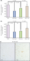
Figure 5
Figure 5
Advanced glycation end‐products (AGE) and receptor for AGE (RAGE) reactions in the pathogenesis of diabetic neuropathy. Nerve tissues, such as Schwann cells, nerve fibers and endothelial cells of vasa nervosum all express RAGE. When AGE bind with RAGE, the reaction generates oxidative stress mainly through the activation of NADPH oxidase. Complexes of IκBα‐nuclear factor‐(NF)‐κB will be separated into each fraction of IκBα and NFκB, the latter of which translocates into the nucleus as a transcription factor to activate genes related to cell death or survival. As a result, both microangiopathic processes and neural dysfunction ensue, resulting in the manifestation of pain or nerve conduction delay.
Figure 6
Figure 6
Neuropathy in normal rats given exogenous advanced glycation end‐products (AGE). When AGE were given exogenously, normal rats showed neuropathic changes, similar to those found in experimental diabetic animals. Rats given AGE showed (a) a significant delay of motor nerve conduction velocity and (b) suppression of nerve Na,K‐ATPase activity, whereas no effects were detected in bovine serum albumin (BSA)‐treated rats. Such suppression was corrected by co‐treatment with aminoguanidine, an inhibitor of glycation and nitric oxide. (c) On the sections, AGE‐treated rats showed strong expression of nuclear factor‐κB on the nuclei of endothelial cells of microvessels and Schwann cells (quoted from reference 94).
Figure 7
Figure 7
Pro‐inflammatory reactions and experimental diabetic neuropathy. In the sciatic nerve of STZ‐induced diabetic rats, there were many macrophages stained positive for ED1 (upper center). Migration of macrophages was inhibited when diabetic rats were treated with pioglitazone (upper right). Pioglitazone treatment also corrected the delay of motor nerve conduction velocity (MNCV) and sensory nerve conduction velocity (SNCV), and activation of extracellular signal‐regulated kinase (ERK), one of mitogen activated protein kinases (MAPK) (adapted from reference 134).
Figure 8
Figure 8
Summary of pathogenetic mechanisms of diabetic neuropathy. Long‐term hyperglycemia causes downstream metabolic cascades of polyol pathway hyperactivity, advanced glycation end‐products (AGE)/receptor for AGE (RAGE) reactions and increased reactive oxygen species (ROS). They compromise both endoneurial microvessels and neural tissues themselves through activation of poly‐ADP‐ribose polymerase (PARP), alterations of protein kinase C (PKC) and an increase in mitogen‐activated protein kinase (MAPK), as well as activation of nuclear factor‐(NF)‐κB, resulting in functional and structural changes of peripheral neuropathy. Metabolic aberrations in the nerve elicit pro‐inflammatory reactions, inducing release of cytokines, suppression of neurotrophins and migration of macrophages, and promote the development of neuropathy. Recently, cellular factors derived from the bone marrow were found to produce chimeric cells in peripheral nerves of diabetic animals to elicit nerve injury. There is also the possibility that other cellular components from the bone marrow have an influence on the nerve pathology in diabetes. In addition, ischemia/reperfusion might also accelerate nerve injury, in part mediated by inflammatory reactions. Risk factors represented by hypertension, hyperlipidemia, smoking and insulin resistance are also important contributors to the development of neuropathy.

Similar articles

Cited by

References

    1. Boulton AJ, Vinik AI, Arezzo JC, et al. Diabetic neuropathies: a statement by the American Diabetes Association. Diabetes Care 2005; 28: 956–962 - PubMed
    1. Strotmeyer ES, de Rekeneire N, Schwartz AV, et al. Sensory and motor peripheral nerve function and lower‐extremity quadriceps strength: the health, aging and body composition study. J Am Geriatr Soc 2009; 57: 2004–2010 - PMC - PubMed
    1. Pirart J. Diabetes mellitus and its degenerative complications: a prospective study of 4400 patients observed. Diabetes Care 1978; 1: 168–188
    1. Vinik AI, Liuzze FJ, Holland MT, et al. Diabetic neuropathies. Diabetes Care 1992; 15: 1926–1975 - PubMed
    1. Ewing DJ, Campbell IW, Clarke BF. Mortality in diabetic autonomic neuropathy. Lancet 1976; 1: 601–603 - PubMed