. 2014 Feb 12;5(1):99-107.
doi: 10.1111/jdi.12131. Epub 2013 Sep 6.

Number of circulating pro-angiogenic cells, growth factor and anti-oxidative gene profiles might be altered in type 2 diabetes with and without diabetic foot syndrome

Affiliations

Number of circulating pro-angiogenic cells, growth factor and anti-oxidative gene profiles might be altered in type 2 diabetes with and without diabetic foot syndrome

Witold N Nowak et al. J Diabetes Investig. .

Abstract

Aims/introduction: Type 2 diabetes is often complicated by diabetic foot syndrome (DFS). We analyzed the circulating stem cells, growth factor and anti-oxidant gene expression profiles in type 2 diabetes patients without or with different forms of DFS.

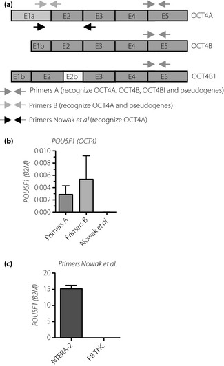
Materials and methods: Healthy volunteers (n = 13) and type 2 diabetes patients: (i) without DFS (n = 10); or with (ii) Charcot osteoneuropathy (n = 10); (iii) non-infected (n = 17); (iv) infected (n = 11); and (v) healed ulceration were examined (n = 12). Peripheral blood endothelial progenitor cells (EPC), mesenchymal stem cells (MSC), hematopoietic stem cells (HSC) and very small embryonic-like (VSEL) cells were phenotyped using flow cytometry. Plasma cytokine concentrations and gene expressions in blood cells were measured by Luminex and quantitative real-time polymerase chain reaction assays, respectively.

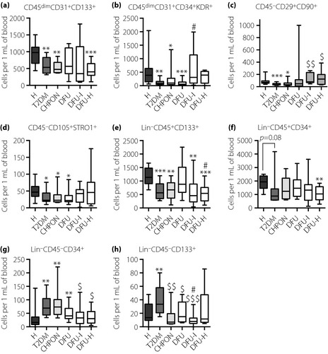
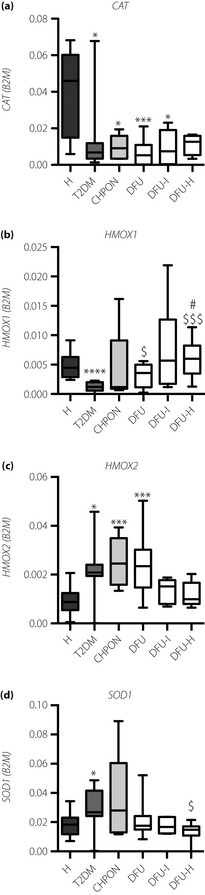
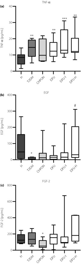
Results: Patients with non-complicated type 2 diabetes showed reduced HMOX1 expression, accompanied by HMOX2 upregulation, and had less circulating EPC, MSC or HSC than healthy subjects. In contrast, VSEL cells were elevated in the type 2 diabetes group. However, subjects with DFS, even with healed ulceration, had fewer VSEL cells, more CD45-CD29(+)CD90(+)MSC, and upregulated HMOX1 when compared with the type 2 diabetes group. Patients with Charcot osteopathy had lowered plasma fibroblast growth factor-2. Elevated plasma tumor necrosis factor-α and decreased catalase expression was found in all diabetic patients.

Conclusions: Patients with type 2 diabetes and different forms of DFS have an altered number of circulating stem cells. Type 2 diabetes might also be associated with a changed plasma growth factor and anti-oxidant gene expression profile. Altogether, these factors could contribute to the pathogenesis of different forms of DFS.

Keywords: Anti‐oxidant genes; Diabetic foot syndrome; Stem cells.


Figures

Figure 1
Figure 1
Numbers of circulating stem and progenitor cells in the peripheral blood of patients. (a) CD45dimCD31+CD133+ endothelial progenitor cells (EPC); (b) CD45dimCD31+CD34+KDR+ EPC; (c) CD45CD29+CD90+ mesenchymal stem cells (MSC); (d) CD45CD105+STRO‐1 MSC; (e) LinCD45+CD133+ hematopoietic stem cells (HSC); (f) LinCD45+CD34+ HSC; (g) LinCD45CD34+ very small embryonic‐like (VSEL) cells; (h) LinCD45CD133+ VSEL cells. Flow cytometry phenotyping. *< 0.05, **< 0.01, ***< 0.001 vs healthy volunteers (H); $< 0.05, $$< 0.01, $$$< 0.001 vs type 2 diabetes group (T2DM); #< 0.05 vs type 2 diabetes patients with non‐infected ulceration (DFU). Results are shown as box and whisker charts displaying minimum, maximum, median, upper and lower quartiles. CHPON, Charcot peripheral osteoneuropathy; DFU‐H, type 2 diabetes patients with healed ulceration; DFU‐I, type 2 diabetes patients with infected ulceration.
Figure 2
Figure 2
Schematic localization of recognized sequences in the gene encoding OCT4. (a) E1a‐E5 exons. (b) Analysis of POU5F1 expression in peripheral blood total nucleated cells (PB TNC) of patients (mean ± standard deviation, n = 8, randomly chosen patients). Primers A and B provide false‐positive signals. (c) Primers by Nowak et al. produce no signal. Comparison of signal from NTERA‐2 human embryonal carcinoma cell line (positive control) and PB TNC using primers by Nowak et al.
Figure 3
Figure 3
Expression of anti‐oxidant genes in circulating total nucleated cells and concentration of plasma cytokines in peripheral blood of patients. (a) Catalase. (b) HMOX1. (c) HMOX2. (d) SOD1. Expression of messenger ribonucleic acid was determined by quantitative real‐time polymerase chain reaction. B2M served as a constitutive control. *< 0.05, **< 0.01, ***< 0.001 vs healthy volunteers (H); $< 0.05 vs type 2 diabetes group (T2DM); #< 0.05 vs type 2 diabetes patients with non‐infected ulceration (DFU). Results are shown as box and whisker charts displaying minimum, maximum, median, upper and lower quartiles. CHPON, Charcot peripheral osteoneuropathy; DFU‐H, type 2 diabetes patients with healed ulceration; DFU‐I, type 2 diabetes patients with infected ulceration.
Figure 4
Figure 4
Plasma levels of (a) tumor necrosis factor‐α (TNF‐α); (b) epidermal growth factor (EGF); and (c) fibroblast growth fator‐2 (FGF‐2). The concentration of cytokines in plasma was measured using the Milliplex FlexMap 3D method. *< 0.05, **< 0.01, ***< 0.001, vs healthy volunteers (H); $< 0.05 vs type 2 diabetes group (T2DM); #< 0.05 vs type 2 diabetes patients with non‐infected ulceration (DFU). Results are shown as box and whisker charts displaying minimum, maximum, median, upper and lower quartiles. CHPON, Charcot peripheral osteoneuropathy; DFU‐H, type 2 diabetes patients with healed ulceration; DFU‐I, type 2 diabetes patients with infected ulceration.

References

    1. Brownlee M. The pathobiology of diabetic complications: a unifying mechanism. Diabetes 2005; 54: 1615–1625 - PubMed
    1. Fadini GP, Miorin M, Facco M, et al Circulating endothelial progenitor cells are reduced in peripheral vascular complications of type 2 diabetes mellitus. J Am Coll Cardiol 2005; 45: 1449–1457 - PubMed
    1. Nandy D, Mukhopadhyay D, Basu A. Both vascular endothelial growth factor and soluble Flt‐1 are increased in type 2 diabetes but not in impaired fasting glucose. J Investig Med 2010; 58: 804–806 - PMC - PubMed
    1. Yeboah J, Sane DC, Crouse JR, et al Low plasma levels of FGF‐2 and PDGF‐BB are associated with cardiovascular events in type II diabetes mellitus (diabetes heart study). Dis Markers 2007; 23: 173–178 - PMC - PubMed
    1. Lodovici M, Giovannelli L, Pitozzi V, et al Oxidative DNA damage and plasma antioxidant capacity in type 2 diabetic patients with good and poor glycaemic control. Mutat Res 2008; 638: 98–102 - PubMed