

Implementation of large-scale routine diagnostics using whole slide imaging in Sweden: Digital pathology experiences 2006-2013
- PMID: 24843825
- PMCID: PMC4023034
- DOI: 10.4103/2153-3539.129452
Implementation of large-scale routine diagnostics using whole slide imaging in Sweden: Digital pathology experiences 2006-2013
Abstract
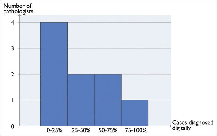
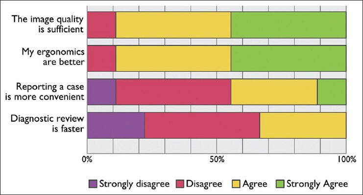
Recent technological advances have improved the whole slide imaging (WSI) scanner quality and reduced the cost of storage, thereby enabling the deployment of digital pathology for routine diagnostics. In this paper we present the experiences from two Swedish sites having deployed routine large-scale WSI for primary review. At Kalmar County Hospital, the digitization process started in 2006 to reduce the time spent at the microscope in order to improve the ergonomics. Since 2008, more than 500,000 glass slides have been scanned in the routine operations of Kalmar and the neighboring Linköping University Hospital. All glass slides are digitally scanned yet they are also physically delivered to the consulting pathologist who can choose to review the slides on screen, in the microscope, or both. The digital operations include regular remote case reporting by a few hospital pathologists, as well as around 150 cases per week where primary review is outsourced to a private clinic. To investigate how the pathologists choose to use the digital slides, a web-based questionnaire was designed and sent out to the pathologists in Kalmar and Linköping. The responses showed that almost all pathologists think that ergonomics have improved and that image quality was sufficient for most histopathologic diagnostic work. 38 ± 28% of the cases were diagnosed digitally, but the survey also revealed that the pathologists commonly switch back and forth between digital and conventional microscopy within the same case. The fact that two full-scale digital systems have been implemented and that a large portion of the primary reporting is voluntarily performed digitally shows that large-scale digitization is possible today.
Keywords: Clinical routine; digital pathology; digital pathology workflow; remote work; whole slide imaging.
Figures
References
-
- Evans AJ, Chetty R, Clarke BA, Croul S, Ghazarian DM, Kiehl TR, et al. Primary frozen section diagnosis by robotic microscopy and virtual slide telepathology: The University health network experience. Hum Pathol. 2009;40:1070–81. - PubMed
-
- Al-Janabi S, Huisman A, Nap M, Clarijs R, van Diest PJ. Whole slide images as a platform for initial diagnostics in histopathology in a medium-sized routine laboratory. J Clin Pathol. 2012;65:1107–11. - PubMed
-
- Huisman A, Looijen A, van den Brink SM, van Diest PJ. Creation of a fully digital pathology slide archive by high-volume tissue slide scanning. Hum Pathol. 2010;41:751–7. - PubMed
LinkOut - more resources
Full Text Sources
Other Literature Sources
