

Regulation of photoreceptor gap junction phosphorylation by adenosine in zebrafish retina
- PMID: 24844306
- PMCID: PMC4109651
- DOI: 10.1017/S095252381300062X
Regulation of photoreceptor gap junction phosphorylation by adenosine in zebrafish retina
Abstract
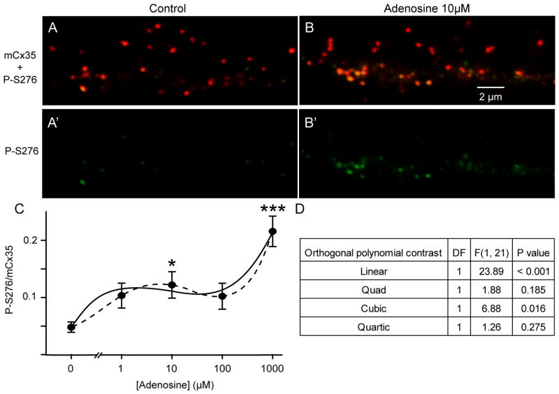
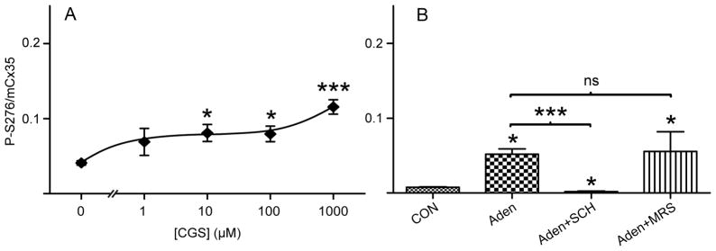
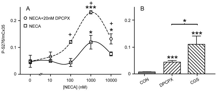
Electrical coupling of photoreceptors through gap junctions suppresses voltage noise, routes rod signals into cone pathways, expands the dynamic range of rod photoreceptors in high scotopic and mesopic illumination, and improves detection of contrast and small stimuli. In essentially all vertebrates, connexin 35/36 (gene homologs Cx36 in mammals, Cx35 in other vertebrates) is the major gap junction protein observed in photoreceptors, mediating rod-cone, cone-cone, and possibly rod-rod communication. Photoreceptor coupling is dynamically controlled by the day/night cycle and light/dark adaptation, and is directly correlated with phosphorylation of Cx35/36 at two sites, serine110 and serine 276/293 (homologous sites in teleost fish and mammals, respectively). Activity of protein kinase A (PKA) plays a key role during this process. Previous studies have shown that activation of dopamine D4 receptors on photoreceptors inhibits adenylyl cyclase, down-regulates cAMP and PKA activity, and leads to photoreceptor uncoupling, imposing the daytime/light condition. In this study, we explored the role of adenosine, a nighttime signal with a high extracellular concentration at night and a low concentration in the day, in regulating photoreceptor coupling by examining photoreceptor Cx35 phosphorylation in zebrafish retina. Adenosine enhanced photoreceptor Cx35 phosphorylation in daytime, but with a complex dose-response curve. Selective pharmacological manipulations revealed that adenosine A2a receptors provide a potent positive drive to phosphorylate photoreceptor Cx35 under the influence of endogenous adenosine at night. A2a receptors can be activated in the daytime as well by micromolar exogenous adenosine. However, the higher affinity adenosine A1 receptors are also present and have an antagonistic though less potent effect. Thus, the nighttime/darkness signal adenosine provides a net positive drive on Cx35 phosphorylation at night, working in opposition to dopamine to regulate photoreceptor coupling via a push-pull mechanism. However, the lower concentration of adenosine present in the daytime actually reinforces the dopamine signal through action on the A1 receptor.
Figures

Similar articles
-
Photoreceptor coupling is controlled by connexin 35 phosphorylation in zebrafish retina.J Neurosci. 2009 Dec 2;29(48):15178-86. doi: 10.1523/JNEUROSCI.3517-09.2009. J Neurosci. 2009. PMID: 19955370 Free PMC article.
-
Adenosine and dopamine receptors coregulate photoreceptor coupling via gap junction phosphorylation in mouse retina.J Neurosci. 2013 Feb 13;33(7):3135-50. doi: 10.1523/JNEUROSCI.2807-12.2013. J Neurosci. 2013. PMID: 23407968 Free PMC article.
-
Connexin 35/36 is phosphorylated at regulatory sites in the retina.Vis Neurosci. 2007 May-Jun;24(3):363-75. doi: 10.1017/S095252380707037X. Epub 2007 Jul 20. Vis Neurosci. 2007. PMID: 17640446 Free PMC article.
-
Electrical synapses between AII amacrine cells in the retina: Function and modulation.Brain Res. 2012 Dec 3;1487:160-72. doi: 10.1016/j.brainres.2012.05.060. Epub 2012 Jul 7. Brain Res. 2012. PMID: 22776293 Review.
-
Into the twilight zone: the complexities of mesopic vision and luminous efficiency.Ophthalmic Physiol Opt. 2006 May;26(3):225-39. doi: 10.1111/j.1475-1313.2006.00325.x. Ophthalmic Physiol Opt. 2006. PMID: 16684149 Review.
Cited by
-
Circadian clock control of connexin36 phosphorylation in retinal photoreceptors of the CBA/CaJ mouse strain.Vis Neurosci. 2015 Jan;32:E009. doi: 10.1017/S0952523815000061. Vis Neurosci. 2015. PMID: 26241696 Free PMC article.
-
Cyclic AMP mediates ovine cumulus-oocyte gap junctional function via balancing connexin 43 expression and phosphorylation.Endocr Connect. 2023 Oct 12;12(11):e230337. doi: 10.1530/EC-23-0337. Print 2023 Nov 1. Endocr Connect. 2023. PMID: 37855365 Free PMC article.
-
Molecular pathways associated with the nutritional programming of plant-based diet acceptance in rainbow trout following an early feeding exposure.BMC Genomics. 2016 Jun 13;17:449. doi: 10.1186/s12864-016-2804-1. BMC Genomics. 2016. PMID: 27296167 Free PMC article.
-
Adenosine A2A Receptor: A New Neuroprotective Target in Light-Induced Retinal Degeneration.Front Pharmacol. 2022 Mar 21;13:840134. doi: 10.3389/fphar.2022.840134. eCollection 2022. Front Pharmacol. 2022. PMID: 35387355 Free PMC article.
-
The ever-changing electrical synapse.Curr Opin Neurobiol. 2014 Dec;29:64-72. doi: 10.1016/j.conb.2014.05.011. Epub 2014 Jun 21. Curr Opin Neurobiol. 2014. PMID: 24955544 Free PMC article. Review.
References
-
- Attwell D, Borges S, Wu SM, Wilson M. Signal clipping by the rod output synapse. Nature. 1987;328:522–524. - PubMed
-
- Blazynski C. Discrete distributions of adenosine receptors in mammalian retina. J Neurochem. 1990;54:648–655. - PubMed
-
- Cohen AI, Blazynski C. Dopamine and its agonists reduce a light-sensitive pool of cyclic AMP in mouse photoreceptors. Vis Neurosci. 1990;4:43–52. - PubMed
-
- Dearry A, Burnside B. Dopaminergic regulation of cone retinomotor movement in isolated teleost retinas: I. Induction of cone contraction is mediated by D2 receptors. J Neurochem. 1986;46:1006–1021. - PubMed
Publication types
MeSH terms
Substances
Grants and funding
LinkOut - more resources
Full Text Sources
Other Literature Sources
Molecular Biology Databases
Miscellaneous
