. 2014 Jun 12;57(11):4805-18.
doi: 10.1021/jm500311e. Epub 2014 May 29.

Designing allosteric inhibitors of factor XIa. Lessons from the interactions of sulfated pentagalloylglucopyranosides

Affiliations

Designing allosteric inhibitors of factor XIa. Lessons from the interactions of sulfated pentagalloylglucopyranosides

Rami A Al-Horani et al. J Med Chem. .

Abstract

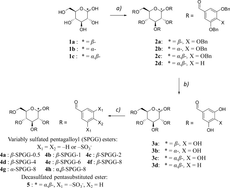
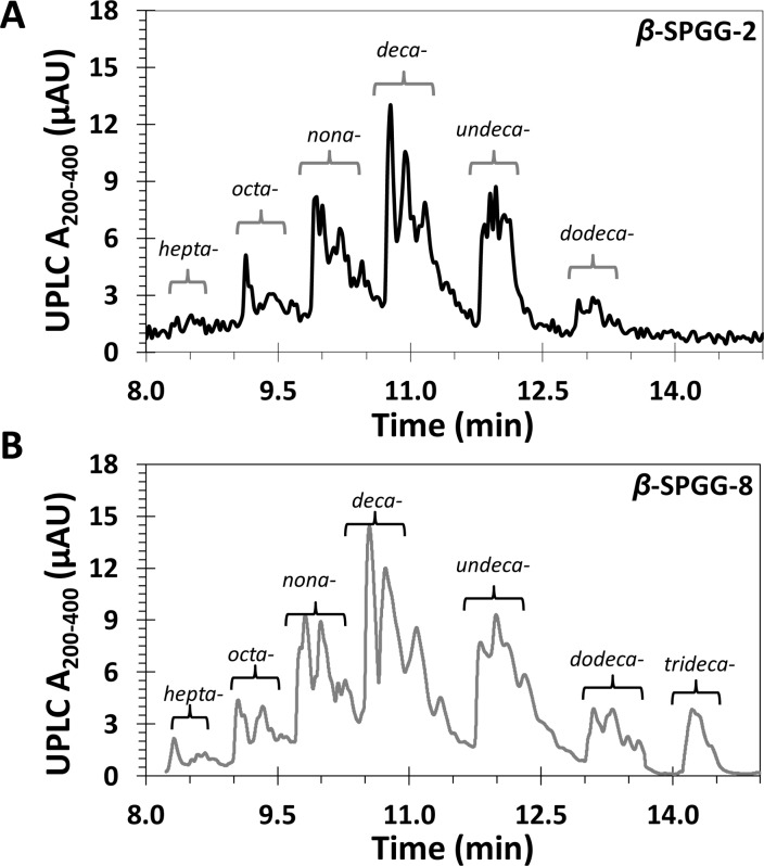
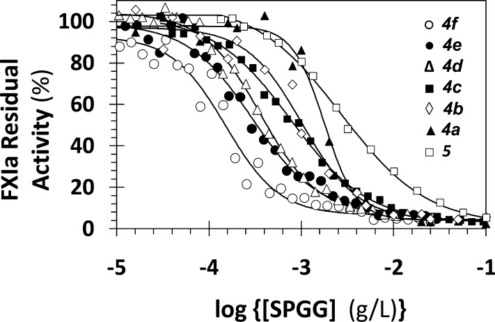
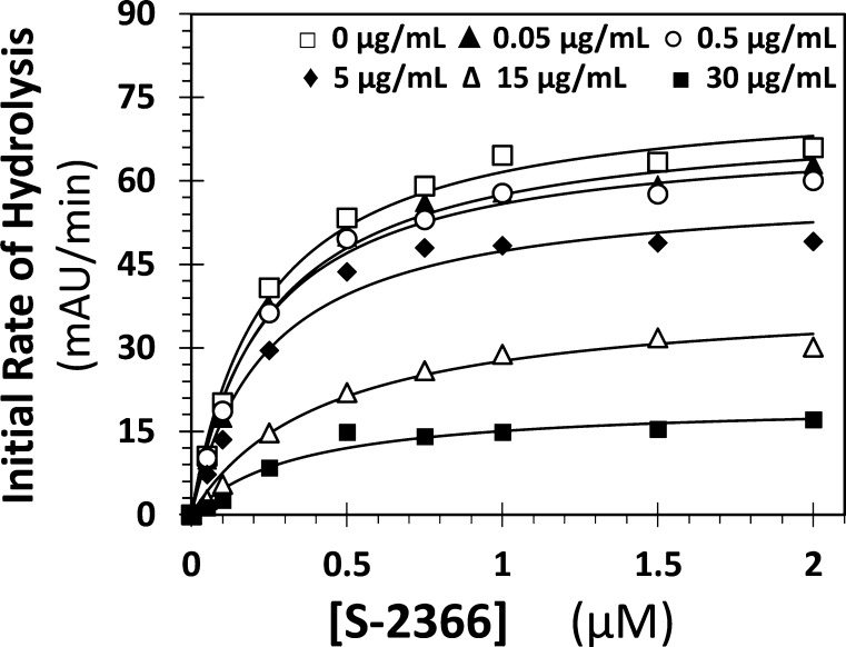
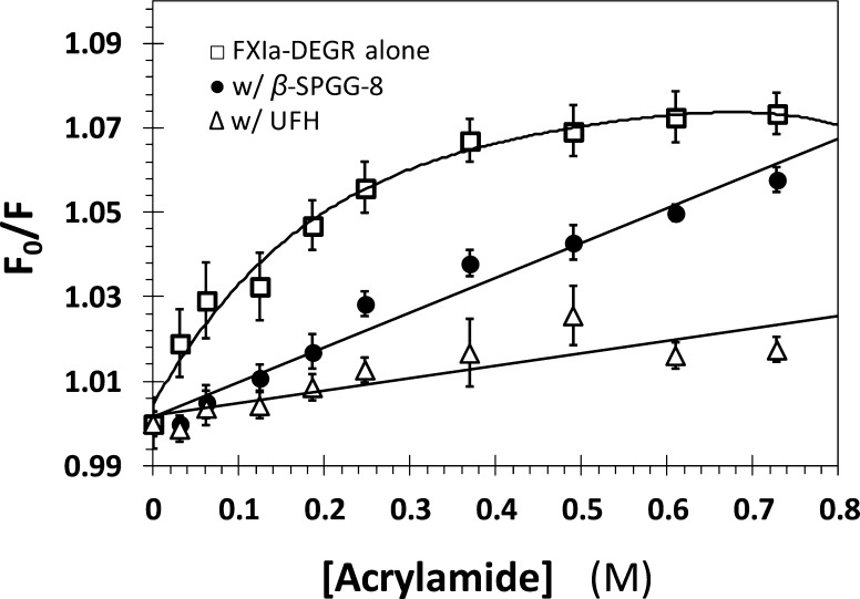
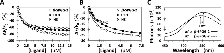
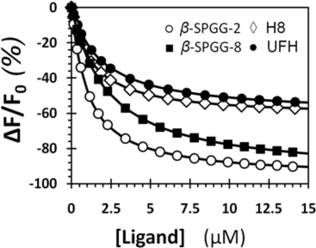
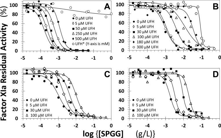
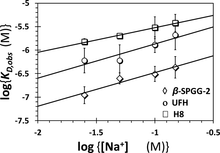
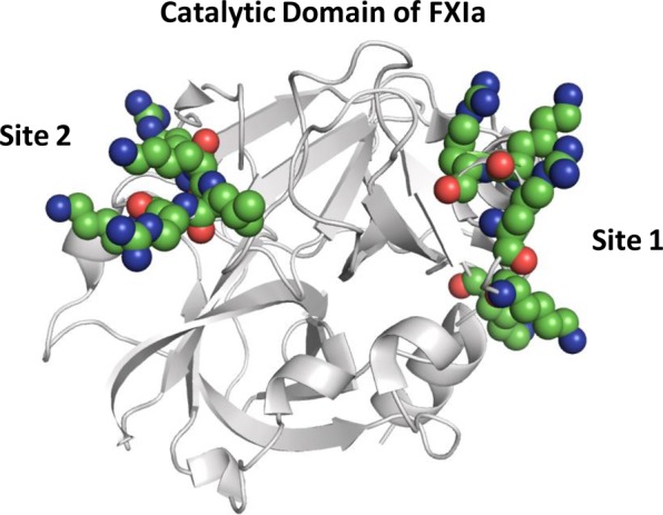
We recently introduced sulfated pentagalloylglucopyranoside (SPGG) as an allosteric inhibitor of factor XIa (FXIa) (Al-Horani et al., J. Med Chem. 2013, 56, 867-878). To better understand the SPGG-FXIa interaction, we utilized eight SPGG variants and a range of biochemical techniques. The results reveal that SPGG's sulfation level moderately affected FXIa inhibition potency and selectivity over thrombin and factor Xa. Variation in the anomeric configuration did not affect potency. Interestingly, zymogen factor XI bound SPGG with high affinity, suggesting its possible use as an antidote. Acrylamide quenching experiments suggested that SPGG induced significant conformational changes in the active site of FXIa. Inhibition studies in the presence of heparin showed marginal competition with highly sulfated SPGG variants but robust competition with less sulfated variants. Resolution of energetic contributions revealed that nonionic forces contribute nearly 87% of binding energy suggesting a strong possibility of specific interaction. Overall, the results indicate that SPGG may recognize more than one anion-binding, allosteric site on FXIa. An SPGG molecule containing approximately 10 sulfate groups on positions 2 through 6 of the pentagalloylglucopyranosyl scaffold may be the optimal FXIa inhibitor for further preclinical studies.


Figures

Scheme 1
Scheme 1. Synthesis of SPGG Derivatives (4a4h) and the Decasulfated Species (5)
(a) 3,4,5-Tribenzyloxybenzoicacid or 3,5-dibenzyloxybenzoic acid (5 equiv), DCC (5 equiv), DMAP (5 equiv), CH2Cl2, reflux, 24 h, 85–90%; (b) H2 (g) (50 psi), Pd(OH)2/C (20%), CH3OH/THF, rt, 10 h, >92%; (c) N(CH3)3-SO3 (5 equiv/OH), CH3CN (2 mL), MW, 90 °C, 0.5–8 h, 66–72%.
Figure 1
Figure 1
Reversed phase-ion pairing UPLC–MS analysis of β-SPGG-2 (4c) (A) and β-SPGG-8 (4f) (B). Both 4c and 4f (and likewise other SPGG variants 4a4h) could be resolved into peaks corresponding to components with varying levels of sulfation from hepta- to trideca-sulfated PGG scaffold (see also Supporting Information Figures S1 and S2). The proportion of higher sulfated species increases from 4a through 4h.
Figure 2
Figure 2
Direct inhibition of full-length factor XIa by variably sulfated SPGG variants as well as the synthesized decasulfated species. The inhibition of factor XIa by 4f (○), 4e (●), 4d (Δ), 4c (■), 4b (◇), 4a (▲), and 5 (□) was studied at pH 7.4 and 37 °C, as described in Experimental Procedures. Solid lines represent sigmoidal dose–response fits using eq 1 to the data to calculate the IC50, ΔY, and HS values.
Figure 3
Figure 3
Michaelis–Menten kinetics of S-2366 hydrolysis by full-length factor XIa in the presence of β-SPGG-8. The initial rate of hydrolysis at various substrate concentrations was measured in pH 7.4 buffer as described in Experimental Procedures using the wild-type full-length factor XIa. β-SPGG-8 concentrations are 0 (□), 0.05 (▲), 0.5 (○), 5 (◆), 15 (Δ), and 30 μg/mL (■). Solid lines represent nonlinear regressional fits to the data using the standard Michaelis–Menten equation to calculate the VMAX and KM.
Figure 4
Figure 4
Quenching of dansyl fluorescence of DEGR-factor XIa by acrylamide in the absence (□) and presence of 20 μM β-SPGG-8 (●) and 20 μM UFH (Δ). Fluorescence intensity at 547 (λEX = 345 nm) was recorded following sequential addition of acrylamide. Solid lines represents fits to the data using either eq 2 (●, Δ) or 3 (□).
Figure 5
Figure 5
Spectrofluorimetric measurement of the affinity of full-length factor XIa (A) and factor XIa–DEGR (B) for β-SPGG-2, UFH, and H8 at pH 7.4 and 37 °C using intrinsic tryptophan (A, λEM = 348 nm, λEX = 280 nm) or dansyl (B, λEM = 547 nm, λEX = 345 nm) fluorescence. Solid lines represent nonlinear regressional fits using quadratic eq 4. (C) Change in the fluorescence emission spectrum of DEGR-factor XIa (λEX = 345 nm) induced by the interaction with β-SPGG-2 at pH 7.4 and 37 °C.
Figure 6
Figure 6
Spectrofluorimetric measurement of the affinity of full-length factor XI for β-SPGG-2 (○), β-SPGG-8 (■), UFH (●), and H8 (◇) at pH 7.4 and 37 °C using intrinsic tryptophan fluorescence (λEM = 348 nm, λEX = 280 nm). Solid lines represent nonlinear regressional fits using quadratic eq 4
Figure 7
Figure 7
Competitive direct inhibition of factor XIa by β-SPGG-8 (4f) (A), β-SPGG-2 (4c) (B), β-SPGG-1 (4b) (C), and β-SPGG-0.5 (4a) (D) in the presence of UFH. The inhibition was determined spectrophotometrically at pH 7.4 and 37 °C. Solid lines represent fits by the dose–response eq 1 to obtain the IC50,predicted, as described in Experimental Procedures. The concentrations of UFH selected for the study are provided.
Figure 8
Figure 8
Dependence of the equilibrium dissociation constant of β-SPGG-2–DEGR-factor XIa complex on the concentration of sodium ion in the medium at pH 7.4 and 37 °C. The KD,obs of β-SPGG-2 (◇), UFH (○), and H8 (□) binding to DEGR-factor XIa was measured through spectrophotometric titrations. Solid lines represent linear regression fits using eq 5. Error bars in symbols represent standard deviation of the mean from at least two experiments. Symbols without apparent error bars indicate that the standard error was smaller than the size of the symbol.
Figure 9
Figure 9
Structure of factor XIa catalytic domain. The crystal structure of factor XIa (PDB ID: 2FDA) shows the presence of two highly electropositive sites that are hypothesized to engage SPGG variants. Site 1 is the traditional heparin-binding site and contains residues K529, R530, R532, K536, and K540, while site 2 is another site containing residues R504, K505, R507, and K509. FXIa is shown in cartoon representation (gray), where the residues in the catalytic domain I and II are shown as spheres colored by atom type.

References

    1. Ruppert A.; Lees M.; Steinle T. Clinical burden of venous thromboembolism. Curr. Med. Res. Opin. 2010, 26, 2465–2473. - PubMed
    1. Nutescu E. A.; Dager W. E.; Kalus J. S.; Lewin J. J. III; Cipolle M. D. Management of bleeding and reversal strategies for oral anticoagulants: clinical practice considerations. Am. J. Health Syst. Pharm. 2013, 70, 1914–1929. - PubMed
    1. Cove C. L.; Hylek E. M. An updated review of target-specific oral anticoagulants used in stroke prevention in atrial fibrillation, venous thromboembolic disease, and acute coronary syndromes. J. Am. Heart Assoc. 2013, 2, e000136. - PMC - PubMed
    1. Yates S.; Sarode R. Novel thrombin and factor Xa inhibitors: challenges to reversal of their anticoagulation effects. Curr. Opin. Hematol. 2013, 20, 552–557. - PubMed
    1. van Montfoort M. L.; Meijers J. C. M. Anticoagulation beyond direct thrombin and factor Xa inhibitors: indications for targeting the intrinsic pathway?. Thromb. Haemost. 2013, 110, 223–232. - PubMed

Publication types

MeSH terms