. 2014 Oct;16(10):1582-601.
doi: 10.1111/cmi.12315. Epub 2014 Jun 17.

Uptake of Shiga-toxigenic Escherichia coli SubAB by HeLa cells requires an actin- and lipid raft-dependent pathway

Affiliations

Uptake of Shiga-toxigenic Escherichia coli SubAB by HeLa cells requires an actin- and lipid raft-dependent pathway

Sayaka Nagasawa et al. Cell Microbiol. 2014 Oct.

Abstract

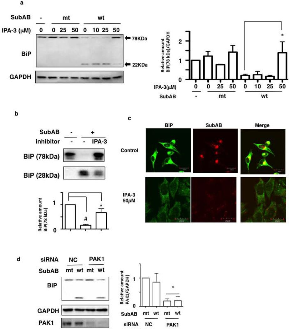
The novel cytotoxic factor subtilase cytotoxin (SubAB) is produced mainly by non-O157 Shiga-toxigenic Escherichia coli (STEC). SubAB cleaves the molecular chaperone BiP/GRP78 in the endoplasmic reticulum (ER), leading to activation of RNA-dependent protein kinase (PKR)-like ER kinase (PERK), followed by caspase-dependent cell death. However, the SubAB uptake mechanism in HeLa cells is unknown. In this study, a variety of inhibitors and siRNAs were employed to characterize the SubAB uptake process. SubAB-induced BiP cleavage was inhibited by high concentrations of Dynasore, and methyl-β-cyclodextrin (mβCD) and Filipin III, but not suppressed in clathrin-, dynamin I/II-, caveolin1- and caveolin2-knockdown cells. We observed that SubAB treatment led to dramatic actin rearrangements, e.g. formation of plasma membrane blebs, with a significant increase in fluid uptake. Confocal microscopy analysis showed that SubAB uptake required actin cytoskeleton remodelling and lipid raft cholesterol. Furthermore, internalized SubAB in cells was found in the detergent-resistant domain (DRM) structure. Interestingly, IPA-3, an inhibitor of serine/threonine kinase p21-activated kinase (PAK1), an important protein of macropinocytosis, directly inhibited SubAB-mediated BiP cleavage and SubAB internalization. Thus, our findings suggest that SubAB uses lipid raft- and actin-dependent, but not clathrin-, caveolin- and dynamin-dependent pathways as its major endocytic translocation route.


Figures

Figure 1
Figure 1. Role of dynamin and clathrin during SubAB entry
(a) HeLa cells were pretreated with the indicated concentrations of Dynasore, CPZ or MDC, and then incubated with mSubAB (mt) or SubAB (wt) for 1 h. SubAB-induced BiP cleavage was determined by immunoblots. GAPDH served as a loading control. Experiments were repeated three times with similar results. (b) Quantification of uncleaved BiP (78-kDa) by mt or wt SubAB in the presence of the indicated inhibitors was performed by densitometry. Data are the means ± SD value from three independent experiments. *P<0.05. (c) HeLa cells were pretreated with or without the indicated concentrations of inhibitors for 30min, and then treated with Alexa555-labeled SubAB for 1h. Cells were fixed and reacted with anti-BiP antibodies (green). A merged picture shows co-localization in HeLa cells. Bars represent 20 µm. Experiments were repeated two times with similar results. (d) HeLa cells were transfected with control (NC), clathrin siRNA or received no treatment, and cell lysates were subjected to immunoblotting with the indicated antibodies. GAPDH served as a loading control. Quantification by densitometry of the amounts of clathrin (192-kDa), with knockdown by the indicated siRNA. Clathrin siRNA-transfected cells were incubated with incubated with mSubAB (mt) or SubAB (wt) for the indicated times. SubAB-induced BiP cleavage was determined by immunoblots. GAPDH served as a loading control. Experiments were repeated three times with similar results. Data are the means ± SD from three separate experiments. *P<0.05. (e) The indicated siRNA-transfected HeLa cells were incubated with Alexa555-labeled SubAB and Alexa488-labeled transferrin (Tf) for 1h at 37°C. Bars represent 20 µm. Experiments were repeated two times with similar results. (f) Dynamin I/II siRNA-transfected cells were incubated with mSubAB (mt) or SubAB (wt) for 1 h. SubAB-induced BiP cleavage was determined by immunoblots with the indicated antibodies. Experiments were repeated three times with similar results. (g) The indicated siRNA-transfected HeLa cells were incubated with Alexa555-labeled SubAB or Alexa488-labeled transferrin (Tf) for 0.5 h at 37°C. Bars represent 20 µm. Experiments were repeated two times with similar results.
Figure 2
Figure 2. SubAB uptake is lipid raft-dependent, not caveolin-dependent
(a) HeLa cells were pretreated with or without the indicated concentrations of caveolin inhibitors (i.e., Filipin III, MβCD) for 30 min and then incubated with mt or wt SubAB for 1 h. SubAB-mediated BiP cleavage was determined by immunoblots with the indicated antibodies. Experiments were repeated three times with similar results. (b) HeLa cells were preincubated with Filipin III (5 µg/ml), MβCD (10 mM) or DMSO as a control for 30 min at 37°C, and then cells were incubated with Alexa555-labeled CtxB and SubAB were incubated for 1h at 37°C. Cells were fixed and observed by confocal microscopy. Bars represent 20 µm. Experiments were repeated two times with similar results. (c) The recombinant hamster BiP (0.1 µg/10 µl) was incubated with SubAB (20 ng/10 µl) in the presence or absence of Filipin III (5 µg/ml), MβCD (10 mM) or DMSO as a control for 1 h at 37°C, and then cleaved BiP was detected with anti-BiP antibody by Western blotting as described in Materials and Methods. Experiments were repeated two times with similar results. (d) Control (NC), caveolin1 (Cav1) and caveolin2 (Cav2) siRNA-transfected cells were incubated with SubAB for the indicated times at 37 °C. The expression levels of Cav1 and Cav2 and SubAB-mediated BiP cleavage were determined by immunoblots with the indicated antibodies. Experiments were repeated three times with similar results.
Figure 3
Figure 3. SubAB-induced the extension of filopodia
(a) HeLa cells were incubated with or without Alexa555-labeled SubAB for 10 or 30 min at 37 °C, fixed, and stained with F-actin by anti-stain™ 488 fluorescent phalloidin (PHDG1) antibody (green). A merged picture shows co-localization in HeLa cells. Bars represent 20 µm. Experiments were repeated three times with similar results. (b–d) Cells, treated as in (a), were subjected to quantitative measurements, including the number of filopodia per cell (b), filopodia length (c), and 2D cell size (d). Data are the means ± SD from three separate experiments, which involved quantifying two cells chosen at random. *P<0.05. (e) HeLa cells were incubated with mt or wt SubAB for 20min at 37 °C, fixed, and stained for F-actin by anti-stain™ 488 fluorescent phalloidin (PHDG1) antibody (green). Picture shows that both mt and wt SubAB induced filopodia. Experiments were repeated three times with similar results.
Figure 4
Figure 4. SubAB uptake is dependent on Na+/H+ membrane exchanger in HeLa cells
(a) Cells were pretreated with or without the indicated concentrations of EIPA for 30 min and then incubated with mt or wt SubAB for 1 h. SubAB-induced BiP cleavage was determined by Western blot analysis. GAPDH served as a loading control. Experiments were repeated three times with similar results (Top panel). HeLa cells were preincubated with 100 µM EIPA or DMSO as a control for 30 min at 37°C, and Alexa555-labeled SubAB were incubated for 1h at 37°C. Cells were fixed and observed by confocal microscopy. Bars represent 20 µm. Experiments were repeated two times with similar results (bottom panel). (b) Cells were preincubated with Na+-rich (Na+), K+-rich (K+) or NMG+-rich (NMG+) buffer for 30 min and then incubated with mt or wt SubAB for 1 h. SubAB-induced BiP cleavage was determined by Western blot analysis. GAPDH served as a loading control. Experiments were repeated two times with similar results (Top, left panel). Quantification of uncleaved BiP (78-kDa) following incubation with mt or wt SubAB in the presence of Baf A1 was performed by densitometry. Data are the means ± SD from three independent experiments (Top, right panel). *P<0.05. HeLa cells were preincubated with EMEM as a control and the indicated buffer for 30 min at 37°C, and Alexa555-labeled SubAB was added for 1h at 37 °C. Cells were fixed and analyzed by confocal microscopy. Bars represent 20 µm. Experiments were repeated two times with similar results (bottom panel). (c) HeLa cells were pretreated with the indicated concentration of Baf A1 for 30 min and then incubated with mt or wt SubAB for 1 h. SubAB-induced BiP cleavage was determined by Western blot analysis as described above (left panel). Quantification of uncleaved BiP (78-kDa) following incubation with mt or wt SubAB in the presence of Baf A1 was performed by densitometry. Data are the means ± SD from three independent experiments (right panel). *P<0.05. HeLa cells were preincubated with 0.5 µM Baf A1 or control as shown in Figure 1c for 30 min at 37°C, and then incubated with Alexa555-labeled SubAB for 1h at 37°C. Cells were fixed and observed by confocal microscopy. Bars represent 20 µm. Experiments were repeated two times with similar results (bottom panel). (d) SubAB stimulates fluid-phase macropinocytosis in HeLa cells. SubAB was bound on ice to cells, which were then incubated at 37°C for 1 h, with exposure to FITC-dextran (fDx) during the last 10 min; results were analyzed by confocal microscopy. A merged picture demonstrated that SubAB treatment enhanced fDx uptake. Transiently both SubAB and fDx were colocalized. A merged picture shows co-localization. Bars represent 20 µm. Experiments were repeated three times with similar results. (e) Quantification of fDx(dot) in cells was determined by confocal microscopy (n=10). Data are the means ± SD from three independent experiments. *P<0.05.
Figure 5
Figure 5. Effect of PI3K inhibitors and Ca2+ on SubAB uptake by HeLa cells
(a) SubAB entry was determined by BiP cleavage using Western blot in cells pretreated with the indicated concentrations of LY294002 or wortmannin. (b) Cells were pretreated with the indicated concentrations of BAPTA-AM, EGTA or ionomycin, and then incubated with mt or wt SubAB for 45 min at 37 °C. Analysis was performed by Western blot using anti-BiP and anti-GAPDH antibodies. Experiments were repeated three times with similar results. Quantification of uncleaved BiP (78-kDa) following incubation with mt or wt SubAB in the presence of the indicated inhibitors was performed by densitometry. Data are the means ± SD from three independent experiments. *P<0.05. (c) HeLa cells were pretreated with or without the indicated concentrations of inhibitors for 30min and then treated with Alexa555-labeled SubAB for 1h. Cells were fixed and reacted with anti-BiP antibodies (green). A merged picture shows co-localization in HeLa cells. Bars represent 20 µm. Experiments were repeated two times with similar results.
Figure 6
Figure 6. Effects of IPA-3 on SubAB uptake by HeLa cells
(a) HeLa cells were pretreated with the indicated concentrations of IPA-3 for 30 min, and then cells were incubated with mt or wt SubAB for 1 h and effects determined by Western blot as described above (left panel). Quantification of uncleaved BiP (78-kDa) following incubation with mt or wt SubAB in the presence of the indicated inhibitors was performed by densitometry (right panel). Data are the means ± SD from three separate experiments. *P<0.05. (b) Recombinant hamster BiP was incubated with SubAB with or without IPA-3 (50 µM) for 1h at 37°C as described in Materials and Methods. Data are the means ± SD from five separate experiments. #P<0.05, *P<0.03. (c) Cells were pretreated with 50 µM IPA-3 for 30 min and then incubated with Alexa555-labeled SubAB for 1 h at 37 °C. Cells were imaged by confocal microscopy for colocalization of SubAB (red) with BiP (green) as described in Materials and Methods. Bars represent 20 µm. Experiments were repeated two times with similar results. (d) The indicated siRNA-transfected cells were incubated with mt or wt SubAB for 1 h and effects determined by Western blot as described above (left panel). Quantification by densitometry of the amounts of PAK1 with knockdown by siRNA (right panel). Data are the means ± SD from three separate experiments. *P<0.05.
Figure 7
Figure 7. SubAB entry involves the actin cytoskeleton
(a) HeLa cells were pretreated with the indicated concentrations of cytochalasin D (Cyt D) for 30 min, and then cells were incubated with mt or wt SubAB for 1 h and effects determined by Western blot as described above (left panel). Quantification of uncleaved BiP (78-kDa) following incubation with mt or wt SubAB in the presence of the indicated inhibitors was performed by densitometry (right panel). Data are the means ± SD from three separate experiments. *P<0.05. (b) Cells were pretreated with 10 µM Cyt D or control DMSO for 30 min and then incubated with Alexa555-labeled SubAB for 1 h at 37 °C. Cells were imaged by confocal microscopy for colocalization of SubAB (red) with BiP (green) as described in Materials and Methods. Bars represent 20 or 30 µm. Experiments were repeated three times with similar results.
Figure 8
Figure 8. Internalized SubAB associated with DRMs
HeLa cells were incubated with FITC-labeled Tf (green) and Alexa555-labeled SubAB (red) for 30 min were subjected to cold Triton X-100 extraction and imaged by confocal microscopy of SubAB (red), Tf (green) and DAPI (blue) as described in Materials and Methods. Bars represent 20 µm. Experiments were repeated three times with similar results.

Similar articles

Cited by

References

    1. Boquet P, Ricci V. Intoxication strategy of Helicobacter pylori VacA toxin. Trends Microbiol. 2012;20:165–174. - PubMed
    1. Byres E, Paton AW, Paton JC, Lofling JC, Smith DF, Wilce MC, et al. Incorporation of a non-human glycan mediates human susceptibility to a bacterial toxin. Nature. 2008;456:648–652. - PMC - PubMed
    1. Chong DC, Paton JC, Thorpe CM, Paton AW. Clathrin-dependent trafficking of subtilase cytotoxin, a novel AB5 toxin that targets the endoplasmic reticulum chaperone BiP. Cell Microbiol. 2008;10:795–806. - PubMed
    1. de Vries E, Tscherne DM, Wienholts MJ, Cobos-Jimenez V, Scholte F, Garcia-Sastre A, et al. Dissection of the influenza A virus endocytic routes reveals macropinocytosis as an alternative entry pathway. PLoS Pathog. 2011;7:e1001329. - PMC - PubMed
    1. Dharmawardhane S, Schurmann A, Sells MA, Chernoff J, Schmid SL, Bokoch GM. Regulation of macropinocytosis by p21-activated kinase-1. Mol Biol Cell. 2000;11:3341–3352. - PMC - PubMed

Publication types

MeSH terms

Substances