. 2015 Jan;16(1):14-26.
doi: 10.1111/mpp.12156. Epub 2014 Jul 7.

Fungal cellulase is an elicitor but its enzymatic activity is not required for its elicitor activity

Affiliations

Fungal cellulase is an elicitor but its enzymatic activity is not required for its elicitor activity

Yanan Ma et al. Mol Plant Pathol. 2015 Jan.

Abstract

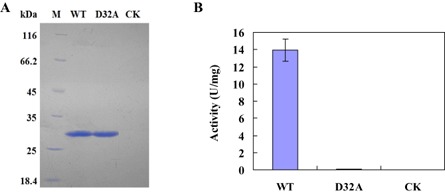
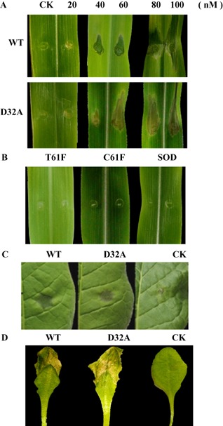
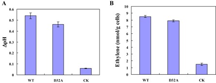
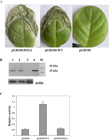
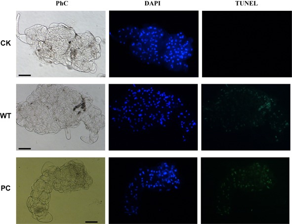
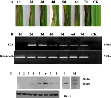
Plant-pathogenic fungi produce cellulases. However, little information is available on cellulase as an elicitor in plant-pathogen interactions. Here, an endocellulase (EG1) was isolated from Rhizoctonia solani. It contains a putative protein of 227 amino acids with a signal peptide and a family-45 glycosyl hydrolase domain. Its aspartic acid (Asp) residue at position 32 was changed to alanine (Ala), resulting in full loss of its catalytic activity. Wild-type and mutated forms of the endoglucanase were expressed in yeast and purified to homogeneity. The purified wild-type and mutant forms induced cell death in maize, tobacco and Arabidopsis leaves, and the transcription of three defence marker genes in maize and tobacco and 10 genes related to defence responses in maize. Moreover, they also induced the accumulation of reactive oxygen species (ROS), medium alkalinization, Ca(2+) accumulation and ethylene biosynthesis of suspension-cultured tobacco cells. Similarly, production of the EG1 wild-type and mutated forms in tobacco induced cell death using the Potato virus X (PVX) expression system. In vivo, expression of EG1 was also related to cell death during infection of maize by R. solani. These results provide direct evidence that the endoglucanase is an elicitor, but its enzymatic activity is not required for its elicitor activity.

Keywords: Rhizoctonia solani; elicitor; fungal cellulase.


Figures

Figure 1
Figure 1
(A) Sodium dodecylsulphate‐polyacrylamide gel electrophoresis (SDS‐PAGE) of the purified enzymes of EG1 from Rhizoctonia solani. WT, the wild‐type form of EG1; D32A, the mutated form (D32A) of EG1; M, molecular weight marker; CK, check sample. (B) Cellulase activity (U) of the wild‐type (WT) form of EG1, the mutant form (D32A) of EG1 and the check sample (CK). One unit (U) of cellulase activity was defined as the amount of cellulase that catalysed the liberation of reducing sugar equivalent to 1.0 μg glucose/min under assay conditions. Three independent biological replicates were used for each protein.
Figure 2
Figure 2
Induction of death in plant leaves by EG1 from Rhizoctonia solani and unrelated enzymes from other fungi. (A) Maize leaves 3 days after inoculation with the wild‐type (WT) form of EG1, the mutant (D32A) form of EG1 and the check sample (CK) at concentrations of 0–100 nm for WT and D32A. (B) Maize leaves 3 days after inoculation with three unrelated proteins at a concentration of 40 nm: a cellulase (C61F, KC441879) of Chaetomium thermophilum, a manganese superoxide dismutase (SOD, EF569978) of Chaetomium thermophilum and a cellulase (T61F, KF170230) of Thermoascus aurantiacus. (C) Tobacco leaves 3 days after inoculation with the wild‐type (WT) form of EG1, the mutant (D32A) form of EG1 and the check sample (CK) at a concentration of 40 nm. (D) Arabidopsis leaves 2 days after inoculation with the wild‐type (WT) form of EG1, the mutant (D32A) form of EG1 and the check sample (CK) at a concentration of 40 nm. Three independent biological replicates were used for each plant leaf.
Figure 3
Figure 3
Induction of defence response genes by EG1 from Rhizoctonia solani. (A) Detection of transcripts of three defence response genes [phenylalanine ammonia‐lyase (PAL), peroxidase (POD) and pathogen‐related 1a (PR1a)] and elongation factor 1 (EF1‐a) using reverse transcription‐polymerase chain reaction (RT‐PCR) in maize and tobacco leaves 3 days after inoculation with the wild‐type (WT) form of EG1, mutant (D32A) form of EG1 and check sample (CK) at a concentration of 40 nm. Three independent biological replicates were used for each plant leaf. (B) Patterns of gene expression of 10 selected genes (1–10) in WT and D32A compared with CK in maize leaves 3 days after inoculation with the wild‐type (WT) form of EG1, mutant (D32A) form of EG1 and check sample (CK) at a concentration of 40 nm. The fold changes of these genes were calculated as the expression ratios (Relative Fold Expression) of WT and D32A compared with CK. Real‐time quantitative RT‐PCR (RTqPCR) data from three biological replicates for each maize leaf: WT, blue columns; D32A, red columns. 1, Thaumatin (GRMZM2G006853); 2, thaumatin (GRMZM2G042631); 3, histamine receptor activity (GRMZM2G041493); 4, 1,3‐β‐glucanase (GRMZM2G065585); 5, pathogenesis‐related protein (GRMZM2G112524); 6, alternative oxidase (GRMZM2G125669); 7, zinc finger homeodomain protein (GRMZM2G353076); 8, chitinase (GRMZM2G453805); 9, pathogenesis‐related protein (GRMZM2G465226); 10, glutathione transferase (GRMZM2G475059).
Figure 4
Figure 4
Production of reactive oxygen species (ROS) and cytosolic Ca2+ accumulation induced by EG1 from Rhizoctonia solani. (A) Production of ROS induced by EG1. Suspension‐cultured tobacco cells were treated with the wild‐type (WT) form of EG1, the mutant (D32A) form of EG1 and the check sample (CK) for 30 min at 25 °C at a concentration of 40 nm. Afterwards, suspension‐cultured tobacco cells were incubated with 2′,7′‐dichlorodihydrofluorescein diacetate (H 2 DCFDA) and ROS production was visualized under a fluorescence microscope with fluorescence (top) and phase contrast (bottom). Scale bar, 50 μm. (B) Induction of cytosolic Ca2+ accumulation in suspension‐cultured tobacco cells by EG1. Cytosolic Ca2+ accumulation of suspension‐cultured tobacco cells was investigated for the wild‐type (WT) form of EG1, the mutant (D32A) form of EG1 and the check sample (CK) at a concentration of 40 nm. Afterwards suspension‐cultured tobacco cells were incubated with Fura 2‐AM and cytosolic Ca2+ accumulation was visualized under a fluorescence microscope with fluorescence (top) and phase contrast (bottom). Scale bar, 100 μm. Three independent biological replicates were used for suspension‐cultured tobacco cells.
Figure 5
Figure 5
Alkalinization response and induction of ethylene biosynthesis in suspension‐cultured tobacco cells by EG1. (A) Alkalinization response in suspension‐cultured tobacco cells to EG1. Extracellular pH was measured with a pH electrode after tobacco cells had been treated with the wild‐type (WT) form of EG1, the mutant (D32A) form of EG1 and the check sample (CK) at 25 °C for 15 min at a concentration of 40 nm. ΔpH was the difference between the final pH reading of the cells after addition of the different proteins and the pH reading of the cells before addition of the different proteins. (B) Induction of ethylene biosynthesis in suspension‐cultured tobacco cells by EG1. Ethylene accumulation in the free‐air phase of the cultures was measured after treatment with the wild‐type (WT) form of EG1, the mutant (D32A) form of EG1 and the check sample (CK) for 4 h at 25 °C at a concentration of 40 nm. Three independent biological replicates were used for suspension‐cultured tobacco cells.
Figure 6
Figure 6
Elicitor activity of EG1 produced in planta using the Potato virus X (PVX)‐based expression system. (A) Symptoms of tobacco leaves injected with Agrobacterium tumefaciens strains carrying the pGR106, pGR106/WT or pGR106/D32A vector. Photographs were taken at 5 days after inoculation. (B) Protein blot analysis of EG1 production in plants. Tobacco leaf samples were collected at 5 days after inoculation, and total protein was extracted and subjected to protein gel blot analysis using an anti‐EG1 polyclonal antiserum. Lane 1, tobacco leaves injected with the A. tumefaciens strain carrying the pGR106/D32A vector; lane 2, tobacco leaves injected with the A. tumefaciens strain carrying the pGR106/WT vector; lane 3, tobacco leaves injected with the A. tumefaciens strain carrying the pGR106 vector; lane 4, EG1; lane M, molecular weight marker. Anti‐plant actin mouse monoclonal antibody (Abbkine) was used as a loading control to detect actin expression. (C) Cellulase activity assay of tobacco leaves. Leaf samples were collected at 5 days after inoculation, and total protein was extracted and subjected to cellulase activity assay. Relative activity was the difference between the activity (A 540) of tobacco leaves inoculated with A. tumefaciens strains carrying pGR106, pGR106/WT or pGR106/D32A and the activity (A 540) of tobacco leaves not inoculated with A. tumefaciens. Three independent biological replicates were used for each tobacco leaf.
Figure 7
Figure 7
Dose–response curves of lesion size of leaf death in maize at 3 days (A), tobacco at 3 days (B) and Arabidopsis at 2 days (C). Leaves were injected with the wild‐type form (WT) of EG1 and the mutant form (D32A) of EG1. The lesion size of leaf death was estimated using the lesion length (mm) for maize leaves, the lesion diameter (mm) for tobacco leaves and the lesion length (mm) for Arabidopsis leaves. Three independent biological replicates were used for each plant leaf.
Figure 8
Figure 8
EG1‐induced DNA fragmentation in suspension‐cultured tobacco cells at 12 h. The cells were counterstained in situ with 4′,6‐diamidino‐2‐phenylindole (DAPI) (blue colour represents nuclei) followed by terminal deoxynucleotidyl transferase‐mediated dUTP nick‐end labelling (TUNEL) reagents (green colour represents DNA fragmentation). Corresponding phase contrast images (PhC) of suspension‐cultured tobacco cells are also shown. Bars, 50 μm. WT, suspension‐cultured tobacco cells treated with the wild‐type of EG1 (40 nm). CK, suspension‐cultured tobacco cells treated with the check sample. PC, positive control. Three independent biological replicates were used for suspension‐cultured tobacco cells.
Figure 9
Figure 9
Maize leaves injected by Rhizoctonia solani. The fungus was injected onto potato dextrose agar (PDA) plates. One disc (diameter, 3 mm) of PDA from the margin of an actively growing colony of the fungus on PDA was placed on maize leaves at 100% relative humidity for 12 h. (A) The symptoms of maize leaves injected with R. solani. (B) Expression of EG1 of R. solani during inoculation of maize. (C) Protein blot analysis of EG1 in maize. Maize leaf samples were collected at 1–7 days (lanes 2–8) after inoculation, and total protein was extracted and subjected to protein gel blot analysis using an anti‐EG1 polyclonal antiserum. Anti‐plant actin mouse monoclonal antibody (Abbkine) was used as a loading control to detect actin expression. M, molecular weight marker; lane 1, check sample (CK); lane 9, EG1.

Similar articles

Cited by

References

    1. Ahuja, I. , Kissen, R. and Bones, A.M. (2012) Phytoalexins in defense against pathogens. Trends Plant Sci. 17, 73–90. - PubMed
    1. Angelova, Z. , Georgiev, S. and Roos, W. (2006) Elicitation of plants. Biotechnol. Biotechnol. Equip. 20, 72–83.
    1. Annis, S.L. and Goodwin, P.H. (1997) Recent advances in the molecular genetics of plant cell wall‐degrading enzymes produced by plant pathogenic fungi. Eur. J. Plant Pathol. 103, 1–14.
    1. Ausubel, F.M. (2005) Are innate immune signaling pathways in plants and animals conserved? Nat. Immunol. 6, 973–979. - PubMed
    1. Bailey, B.A. , Dean, J.F.D. and Anderson, J.D. (1990) An ethylene biosynthesis‐inducing endoxylanase elicits electrolyte leakage and necrosis in Nicotiana tabacum cv. xanthi leaves. Plant Physiol. 94, 1849–1854. - PMC - PubMed

Publication types

MeSH terms

LinkOut - more resources