

Vertical and horizontal corneal epithelial thickness profile using ultra-high resolution and long scan depth optical coherence tomography
- PMID: 24844566
- PMCID: PMC4028229
- DOI: 10.1371/journal.pone.0097962
Vertical and horizontal corneal epithelial thickness profile using ultra-high resolution and long scan depth optical coherence tomography
Abstract
Purpose: To determine the vertical and horizontal thickness profiles of the corneal epithelium in vivo using ultra-long scan depth and ultra-high resolution spectral domain optical coherence tomography (SD-OCT).
Methods: A SD-OCT was developed with an axial resolution of ∼ 3.3 µm in tissue and an extended scan depth. Forty-two eyes of 21 subjects were imaged twice. The entire horizontal and vertical corneal epithelial thickness profiles were evaluated. The coefficient of repeatability (CoR) and intraclass correlation (ICC) of the tests and interobserver variability were analyzed.
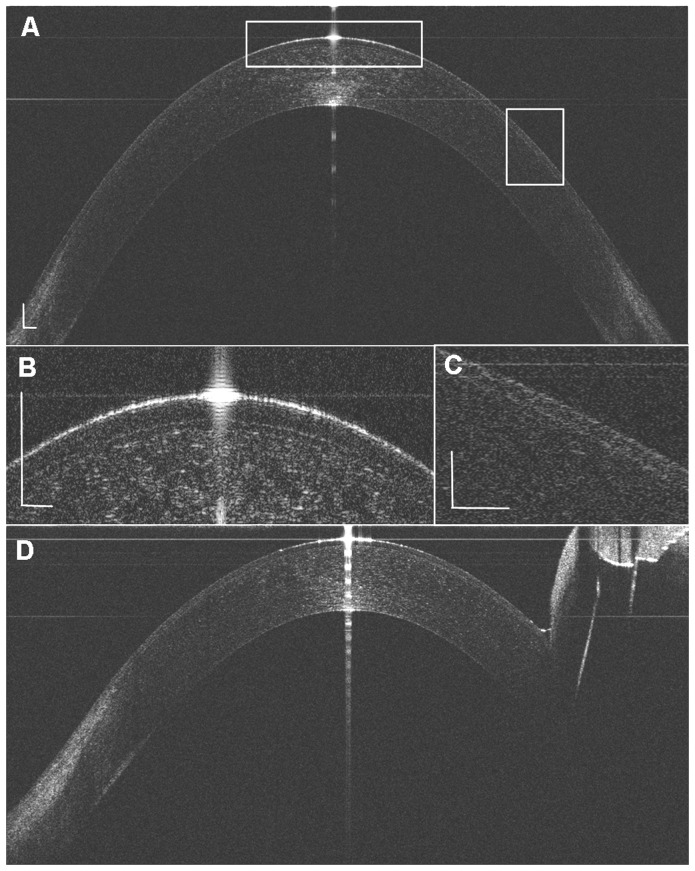
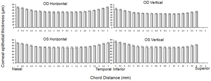
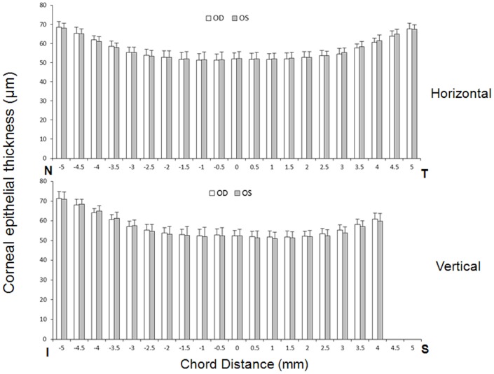
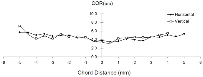
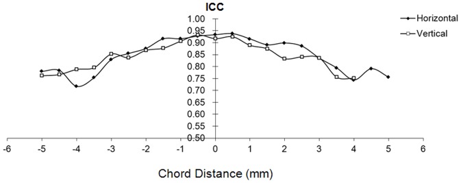
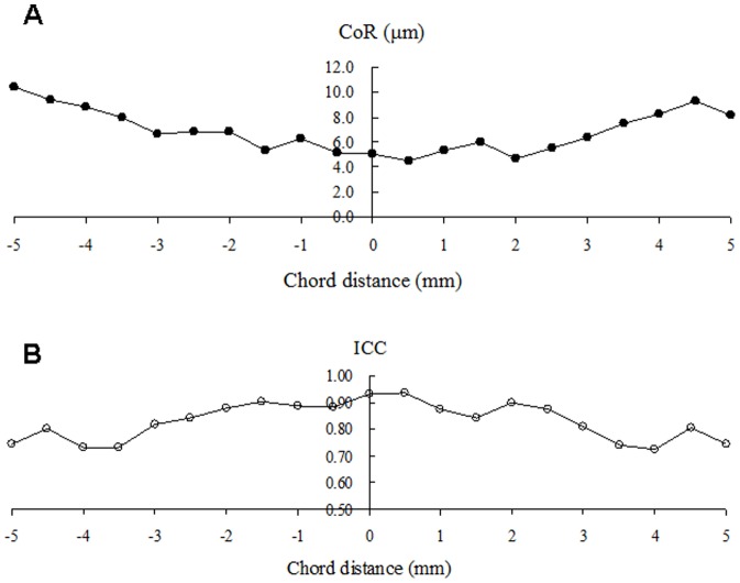
Results: The full width of the horizontal epithelium was detected, whereas part of the superior epithelium was not shown for the covered super eyelid. The mean central epithelial corneal thickness was 52.0 ± 3.2 µm for the first measurement and 52.3 ± 3.4 µm for the second measurement (P>.05). In the central zone (0-3.0 mm), the paracentral zones (3.0-6.0 mm) and the peripheral zones (6.0-10.0 mm), the mean epithelial thickness ranged from 51 to 53 µm, 52 to 57 µm, and 58 to 72 µm, respectively. There was no difference between the two tests at both meridians and in the right and left eyes (P>.05). The ICCs of the two tests ranged from 0.70 to 0.97 and the CoRs ranged from 2.5 µm to 7.8 µm from the center to the periphery, corresponding to 5.6% to 10.6% (CoR%). The ICCs of the two observers ranged from 0.72 to 0.93 and the CoRs ranged from 4.5 µm to 10.4 µm from the center to the periphery, corresponding to 8.7% to 15.2% (CoR%).
Conclusions: This study demonstrated good repeatability of ultra-high resolution and long scan depth SD-OCT to evaluate the entire thickness profiles of the corneal epithelium. The epithelial thickness increases from the center toward the limbus.
Conflict of interest statement
Figures
References
-
- Lohmann CP, Reischl U, Marshall J (1999) Regression and epithelial hyperplasia after myopic photorefractive keratectomy in a human cornea. J Cataract Refract Surg 25: 712–715. - PubMed
-
- Kanellopoulos AJ, Asimellis G (2014) In vivo 3-dimensional corneal epithelial thickness mapping as an indicator of dry eye: preliminary clinical assessment. Am J Ophthalmol 157: : 63–68 e62. - PubMed
-
- Maldonado MJ, Ruiz-Oblitas L, Munuera JM, Aliseda D, Garcia-Layana A, et al. (2000) Optical coherence tomography evaluation of the corneal cap and stromal bed features after laser in situ keratomileusis for high myopia and astigmatism. Ophthalmology 107: 81–87 discussion 88. - PubMed
-
- Haque S, Jones L, Simpson T (2008) Thickness mapping of the cornea and epithelium using optical coherence tomography. Optom Vis Sci 85: E963–976. - PubMed
Publication types
MeSH terms
Grants and funding
LinkOut - more resources
Full Text Sources
Other Literature Sources
