. 2014 May 20;12(5):e1001866.
doi: 10.1371/journal.pbio.1001866. eCollection 2014 May.

Structural and functional insights into the mode of action of a universally conserved Obg GTPase

Affiliations

Structural and functional insights into the mode of action of a universally conserved Obg GTPase

Boya Feng et al. PLoS Biol. .

Abstract

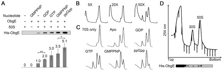
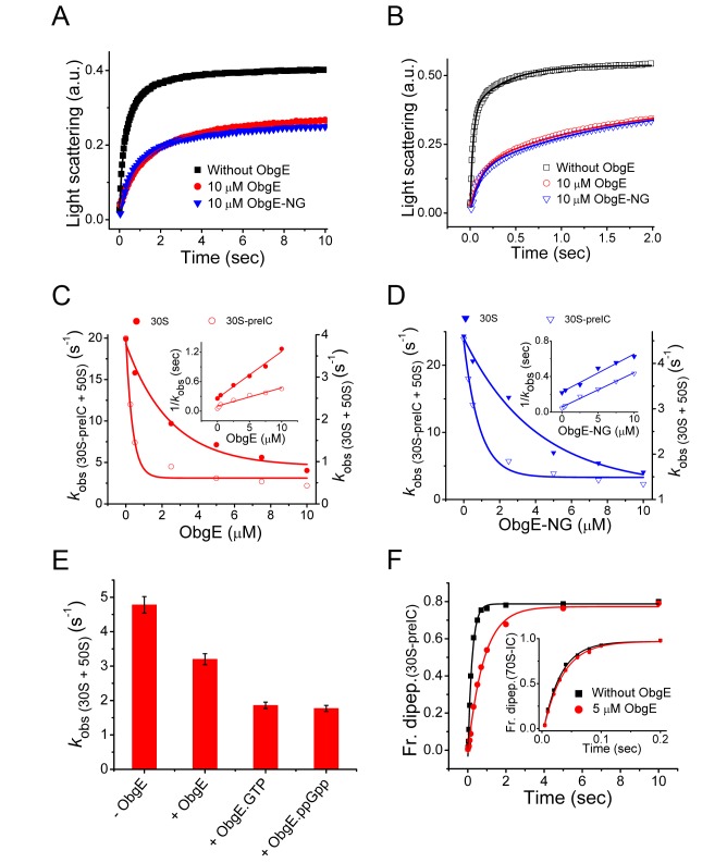
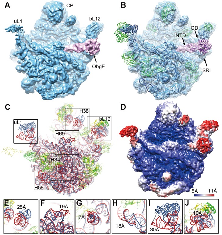
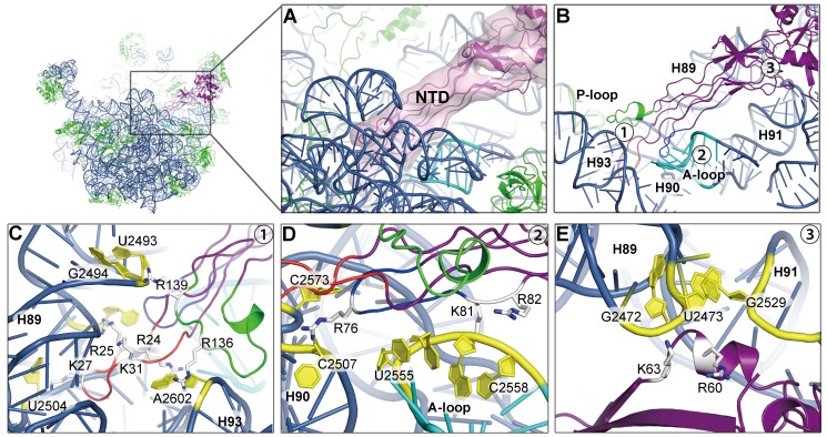
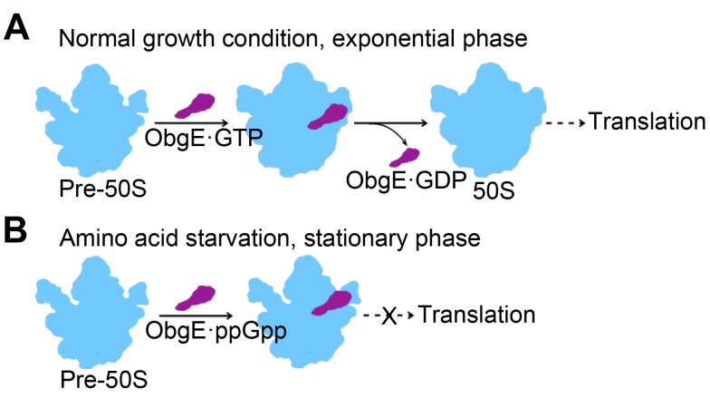
Obg proteins are a family of P-loop GTPases, conserved from bacteria to human. The Obg protein in Escherichia coli (ObgE) has been implicated in many diverse cellular functions, with proposed molecular roles in two global processes, ribosome assembly and stringent response. Here, using pre-steady state fast kinetics we demonstrate that ObgE is an anti-association factor, which prevents ribosomal subunit association and downstream steps in translation by binding to the 50S subunit. ObgE is a ribosome dependent GTPase; however, upon binding to guanosine tetraphosphate (ppGpp), the global regulator of stringent response, ObgE exhibits an enhanced interaction with the 50S subunit, resulting in increased equilibrium dissociation of the 70S ribosome into subunits. Furthermore, our cryo-electron microscopy (cryo-EM) structure of the 50S·ObgE·GMPPNP complex indicates that the evolutionarily conserved N-terminal domain (NTD) of ObgE is a tRNA structural mimic, with specific interactions with peptidyl-transferase center, displaying a marked resemblance to Class I release factors. These structural data might define ObgE as a specialized translation factor related to stress responses, and provide a framework towards future elucidation of functional interplay between ObgE and ribosome-associated (p)ppGpp regulators. Together with published data, our results suggest that ObgE might act as a checkpoint in final stages of the 50S subunit assembly under normal growth conditions. And more importantly, ObgE, as a (p)ppGpp effector, might also have a regulatory role in the production of the 50S subunit and its participation in translation under certain stressed conditions. Thus, our findings might have uncovered an under-recognized mechanism of translation control by environmental cues.


Conflict of interest statement

The authors have declared that no competing interests exist.

Figures

Figure 1
Figure 1. ppGpp stimulates the 50S subunit binding and 70S ribosome dissociation activities of ObgE.
(A) Co-sedimentation assay on the binding of ObgE (1 µM) to the 50S subunit (1 µM), in the absence or presence of GDP, GTP, GMPPNP, or ppGpp (0.5 mM). Pellets were resolved by SDS-PAGE and examined by Western blot analysis (anti-His antibody). Quantification was performed by adjusting the value of the apo state to 1.0. Error bars (standard deviation) were calculated from three independent experiments. (B) Dissociation of 70S ribosomes (1 µM) by varying amount of ObgE (from 5- to 50-fold excess), in the presence of GDP (2 mM). (C) Dissociation of 70S ribosomes (1 µM) by 30-fold excess of ObgE, in the absence or presence of GDP, GTP, GMPPNP, or ppGpp (2 mM). (D) Complete dissociation of 70S ribosomes (1 µM) by 50-fold excess of ObgE in the presence of GMPPNP (2 mM). Fractions were resolved by SDS-PAGE and examined by Western blot analysis.
Figure 2
Figure 2. ObgE is a 50S based anti-association factor.
(A) Formation of 70S ribosome by association of naked 30S subunit (0.25 µM) with 50S subunit alone (0.25 µM) (black square), or the 50S subunit preincubated with ObgE (10 µM) (red circle) or ObgE-NG (10 µM) (blue triangle), followed by Rayleigh light scattering with time in a stopped flow apparatus. The solid lines are drawn by fitting the data according to the subunit association model described in . (B) Same as (A), except that the 30S-preIC (0.25 µM) was used. (C) The observed rates of subunit association (k obs) with naked 30S (filled circle, scale on the right vertical axis) and the 30S-preIC (open circle, scale on the left vertical axis), with increasing concentration of ObgE. Inset, the reciprocal of k obs plotted as a function of ObgE concentration, fitted with the straight line equation. (D) Same as (C), except that ObgE-NG was used. (E) The rates of association of the naked subunits in the presence of ObgE with various guanine nucleotides. (F) The formation of f[3H]Met–Leu dipeptide without (black square) or with (red circle) ObgE (5 µM) starting from the 30S-preIC. The solid lines are drawn by fitting the data with single exponential function. The inset shows the f[3H]Met–Leu dipeptide formed from the 70S-IC without (black square) or with (red circle) ObgE (5 µM).
Figure 3
Figure 3. Cryo-EM structure of the 50S·ObgE·GMPPNP complex.
(A) The cryo-EM map is displayed in surface representation, with the 50S subunit and ObgE colored blue and pink, respectively. (B) The atom model of the 50S·ObgE·GMPPNP complex is displayed in cartoon representation, and superimposed with the density map. Ribosomal RNA, ribosomal proteins, and ObgE are colored blue, green, and purple, respectively. CP, central protuberance; uL1, uL1 stalk; bL12, bL12 stalk. (C) Crystal structure of the 50S subunit (rRNA in red and r-proteins in yellow) is superimposed with the atomic model of the 50S·ObgE·GMPPNP complex (rRNA in blue and r-proteins in green). (D) Local resolution map of the 50S·ObgE·GMPPNP structure. (E–J) Close-up views of regions with large scale conformational changes as boxed in (C). Distances between selected residue pairs are represented in dashed lines.
Figure 4
Figure 4. Interaction of the ObgE-NTD with the peptidyl transferase center.
(A) Binding position of the ObgE-NTD on the 50S subunit, with the ObgE density map displayed in transparent surface representation. (B) Overview of the interactions of loop 1 (red), loop 2 (dark blue), and loop 3 (green) of the ObgE-NTD with H89, H90, H91, H93, A-loop (cyan), and P-loop (lime) of the 23S rRNA. (C–E) Close-up views of numbered locations (1–3) in (B), with conserved basic residues of the ObgE-NTD highlighted in stick model. For illustration, orientations of (C–E) are set differently from that of (B). (C) Interactions of Loop 1 and Loop 3 with H89 and H93. (D) Interaction of Loop 2 with H90 and the A-loop. (E) Interaction of the ObgE-NTD β-barrel base with H89 and H91. Residues of ObgE are shown in stick representation and colored white, and residues of the 23S rRNA are shown in cartoon representation and colored yellow.
Figure 5
Figure 5. Conserved basic residues in the ObgE-NTD are required for the 50S binding.
(A) Positions of conserved arginine or lysine residues that are subjected to site-directed mutagenesis. (B) Co-sedimentation assay on the binding of various ObgE mutants to the 50S subunit. Wild type (WT) and mutant ObgE (M1–M4) were incubated with equal amount of 50S subunits in the absence or presence of saturating GMPPNP and subjected to co-sedimentation assay. M1, M2, M3, and M4 refer to ObgE mutants, K27EK31E, R76GR82G, R136GR139G, and K27EK31E R76GR82G R136GR139G, respectively.
Figure 6
Figure 6. Comparison of ObgE with the A-site tRNA and release factor 2 on the 50S subunit.
(A) Superimposition of ObgE (purple) with the A-site tRNA (red) and the P-site tRNA (yellow) [PDB 2WDK and 2WDL, [75]]. (B) Superimposition of ObgE with the P-site tRNA and RF2 (dark blue) from the crystal structure of a release complex [PDB 2X9S and 2X9R, [28]]. (C–E) Close-up views of the CCA-end of the P-site tRNA, with the A-site tRNA (C), RF2 (D), and ObgE (E) superimposed. (F–H) Close-up views of the CCA-end of the A-site tRNA (F), GGQ-motif of RF2 (G), and the NTD protrusion of ObgE (H), showing local specific interactions with the ribosomal A-loop. The A-loop, P-loop, and H92 (A2451) of the 23S rRNA are colored cyan, lime, and blue, respectively. Critical residues of RF2 and ObgE are shown in stick representation and labeled accordingly.
Figure 7
Figure 7. ObgE is a distinctive 50S-dependent GTPase.
(A and B) Two different views to illustrate the interaction of the ObgE-GD (purple) with the 50S subunit. Interactions of the GDs of IF2 (C) [PDB 1ZO3, [25]], EF-G (D) [PDB 2WRI and 2WRJ, [26]], EF-Tu (E) [PDB 2WRN and 2WRO, [27]], and RF3 (F) [PDB 3SFS and 3SGF, [29]] with the 50S subunit, shown in the same view as (B). The 23S rRNA, r-proteins, ObgE, and translational GTPases are colored blue, green, purple, and yellow, respectively. Switch I of ObgE is disordered and is shown by dashed lines. Switch II of ObgE, Switch II of other factors, SRL, and A2662 are colored, grey, orange, cyan, and red, respectively. Guanine nucleotides are shown in stick models. (G) Low intrinsic GTPase activity of ObgE, monitored by inorganic phosphate production. (H) Time-course GTP hydrolysis by ObgE, in the presence of indicated amount of purified 50S subunits (0.05-, 0.2-, 0.69-, and 1.0-fold).
Figure 8
Figure 8. Proposed model for the function of ObgE in different growth phases.
(A) In exponential phase of growth, ObgE functions primarily as a surveillance factor for the late-stage assembly the 50S subunit. In addition, it prevents the pre-50S subunit from premature association with the 30S subunit. (B) In stationary phase, ObgE acts as a ppGpp effector to delay the 50S maturation and sequesters large numbers of 50S subunits from being engaged in subunit association and thus downregulates global translation.

Comment in

Similar articles

Cited by

References

    1. Wittinghofer A, Vetter IR (2011) Structure-function relationships of the G domain, a canonical switch motif. Annu Rev Biochem 80: 943–971. - PubMed
    1. Verstraeten N, Fauvart M, Versees W, Michiels J (2011) The universally conserved prokaryotic GTPases. Microbiol Mol Biol Rev 75: : 507–542, second and third pages of table of contents. - PMC - PubMed
    1. Kint C, Verstraeten N, Hofkens J, Fauvart M, Michiels J (2013) Bacterial Obg proteins: GTPases at the nexus of protein and DNA synthesis. Crit Rev Microbiol. - PubMed
    1. Persky NS, Ferullo DJ, Cooper DL, Moore HR, Lovett ST (2009) The ObgE/CgtA GTPase influences the stringent response to amino acid starvation in Escherichia coli. Mol Microbiol 73: 253–266. - PMC - PubMed
    1. Jiang M, Sullivan SM, Wout PK, Maddock JR (2007) G-protein control of the ribosome-associated stress response protein SpoT. J Bacteriol 189: 6140–6147. - PMC - PubMed

Publication types

MeSH terms

Substances

LinkOut - more resources