. 2014 Dec;472(12):3823-34.
doi: 10.1007/s11999-014-3683-x.

Fracture reduction and primary ankle arthrodesis: a reliable approach for severely comminuted tibial pilon fracture

Affiliations

Fracture reduction and primary ankle arthrodesis: a reliable approach for severely comminuted tibial pilon fracture

Douglas N Beaman et al. Clin Orthop Relat Res. 2014 Dec.

Abstract

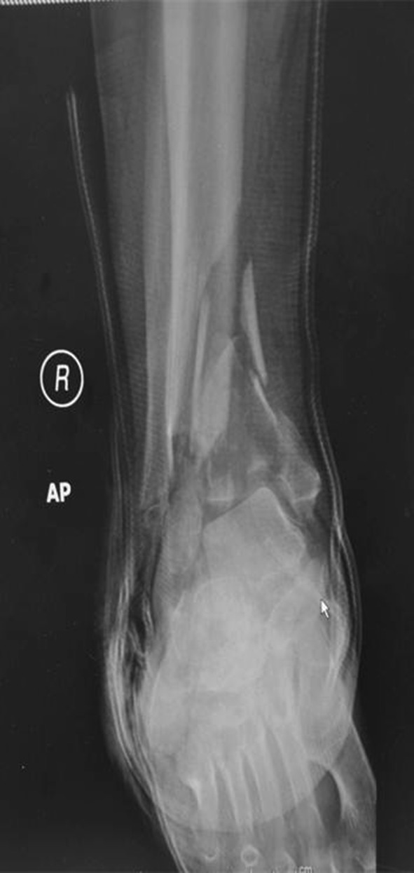
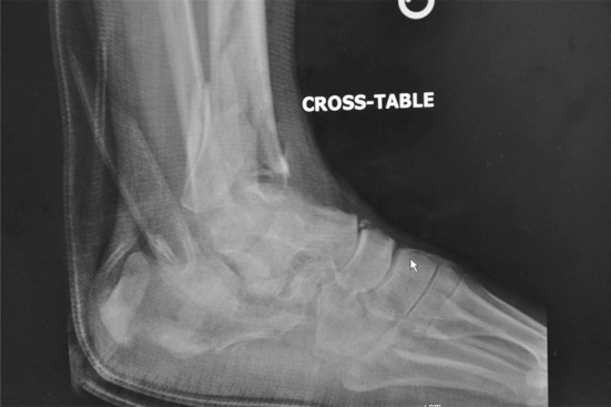
Background: Posttraumatic arthritis and prolonged recovery are typical after a severely comminuted tibial pilon fracture, and ankle arthrodesis is a common salvage procedure. However, few reports discuss the option of immediate arthrodesis, which may be a potentially viable approach to accelerate overall recovery in patients with severe fracture patterns.

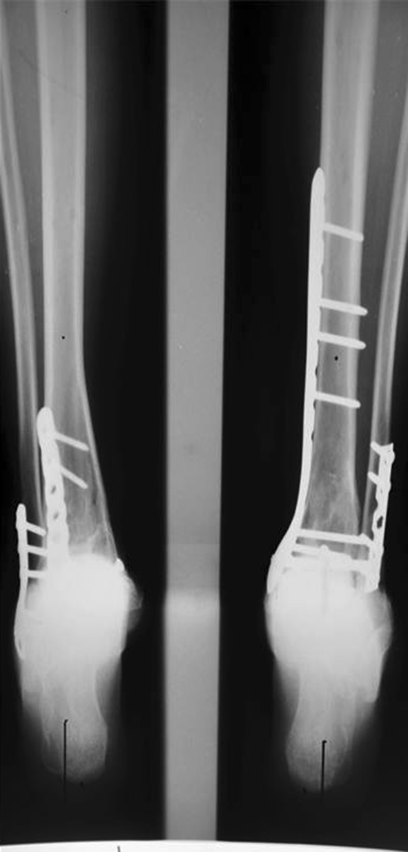
Questions/purposes: (1) How long does it take the fracture to heal and the arthrodesis to fuse when primary ankle arthrodesis is a component of initial fracture management? (2) How do these patients fare clinically in terms of modified American Orthopaedic Foot and Ankle Society (AOFAS) scores and activity levels after this treatment? (3) Does primary ankle arthrodesis heal in an acceptable position when anterior ankle arthrodesis plates are used?

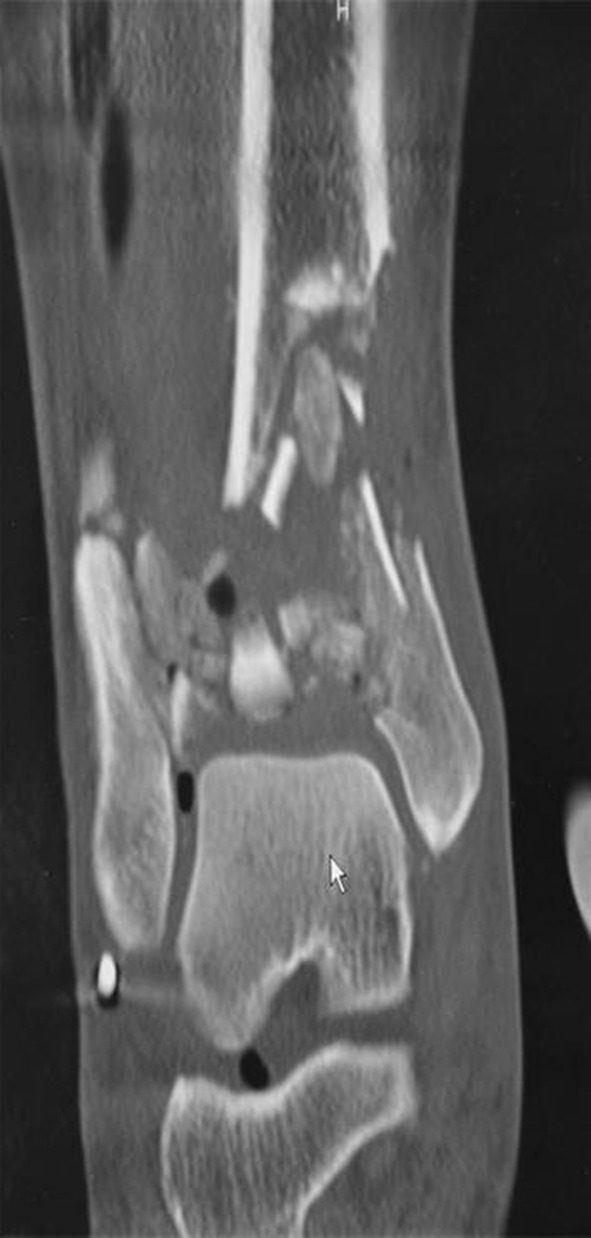
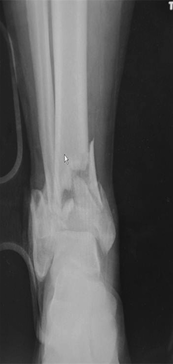
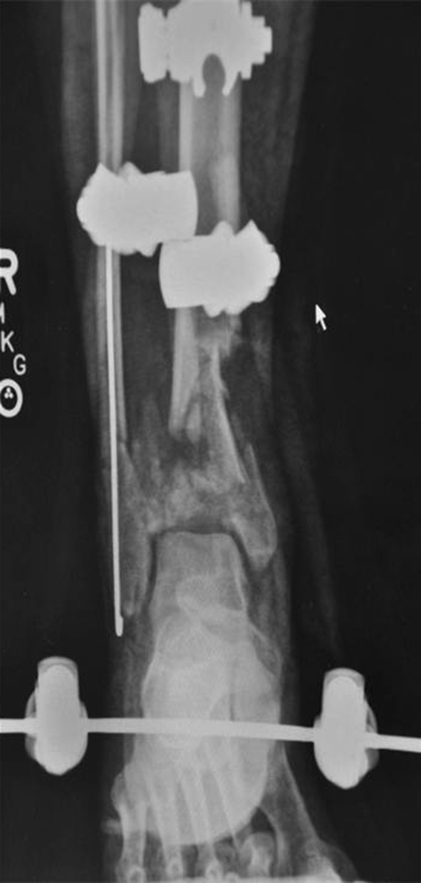
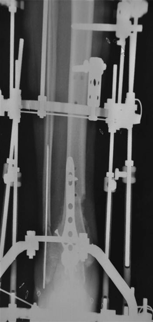
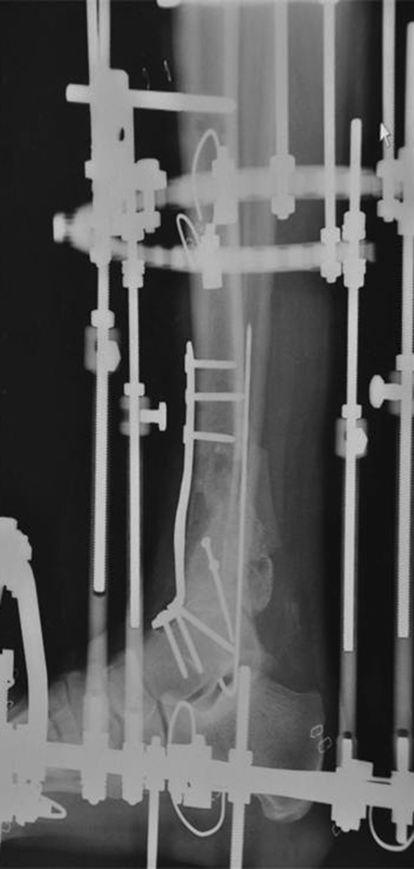
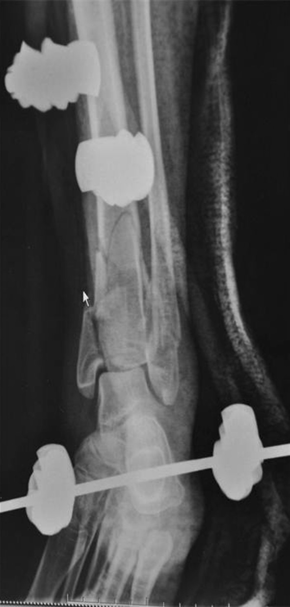
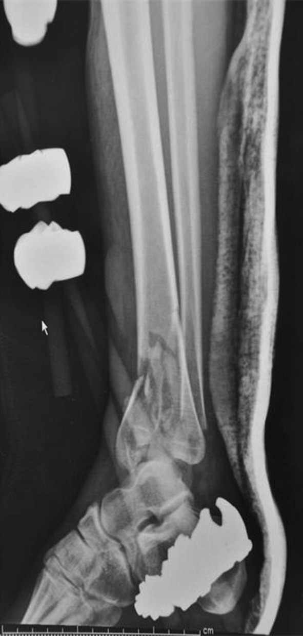
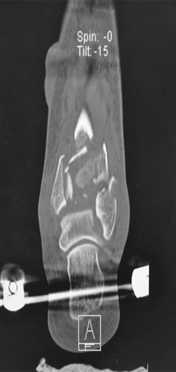
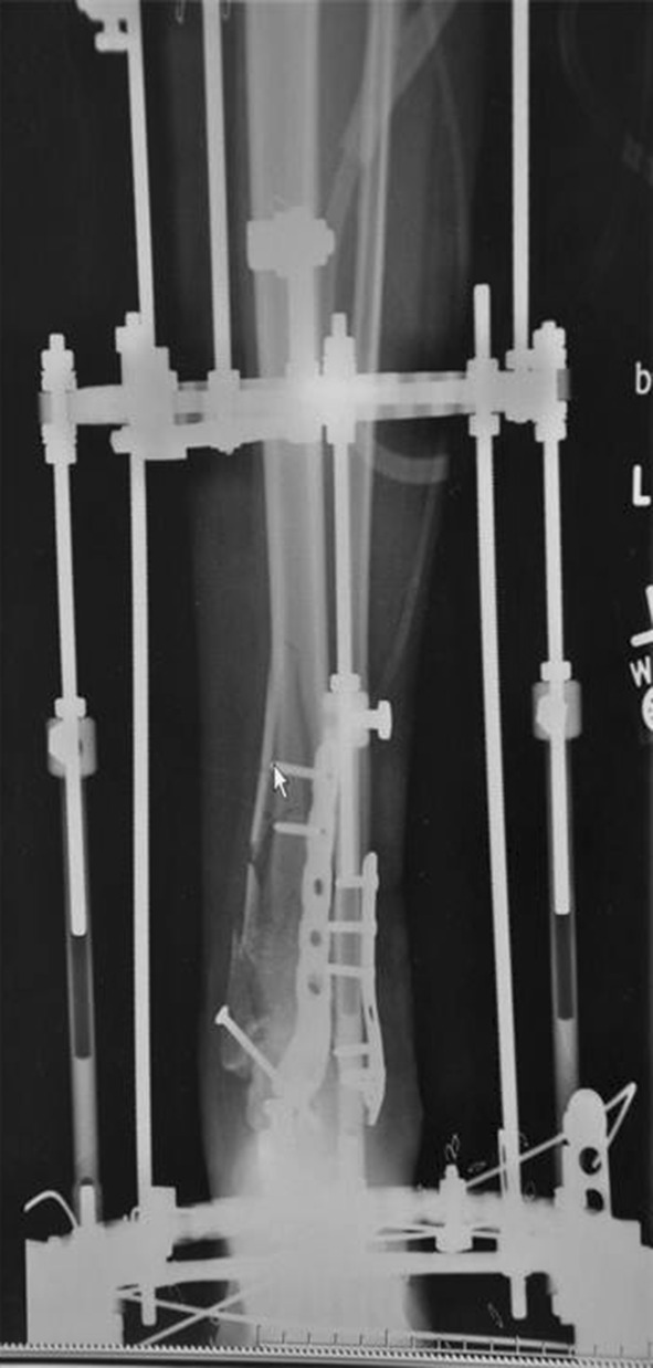
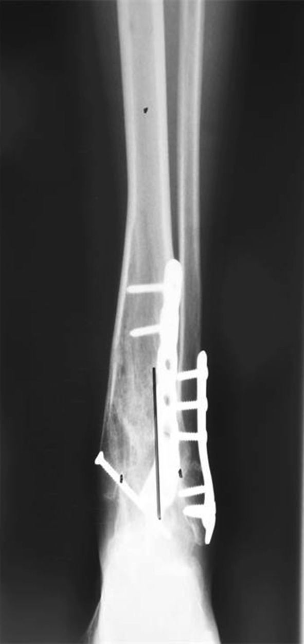
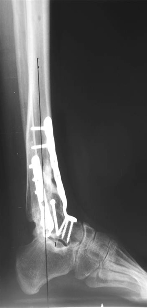
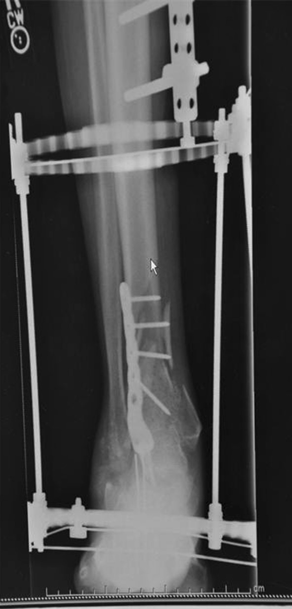
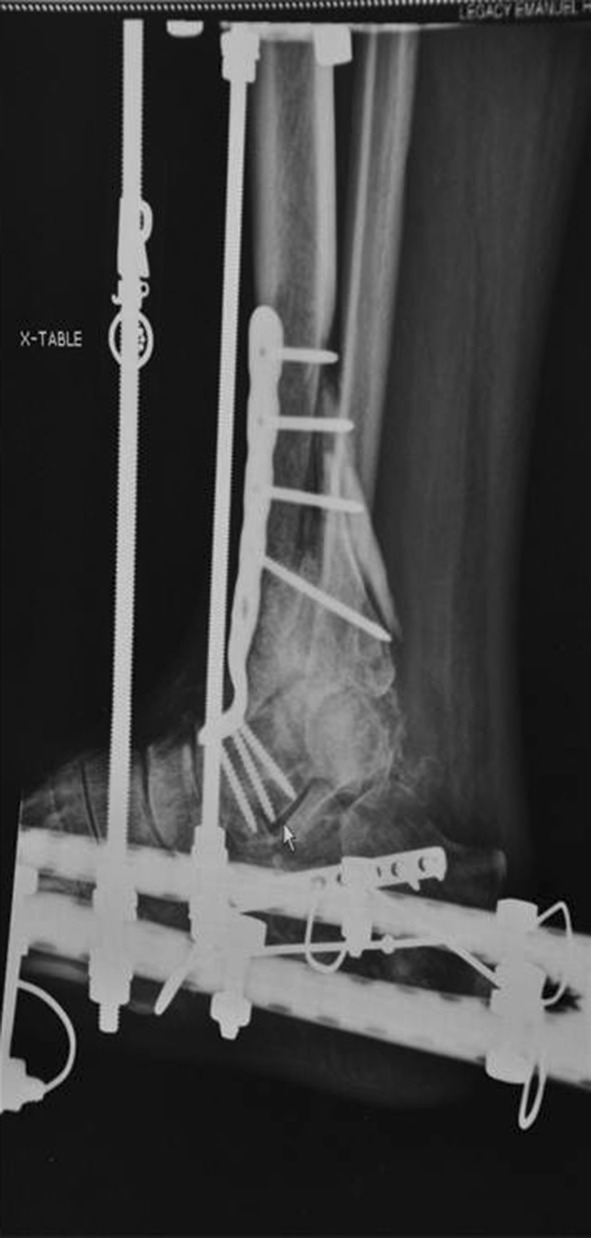
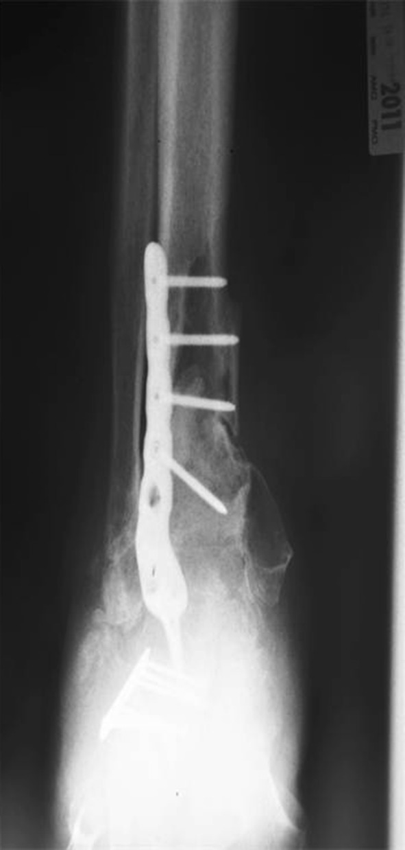
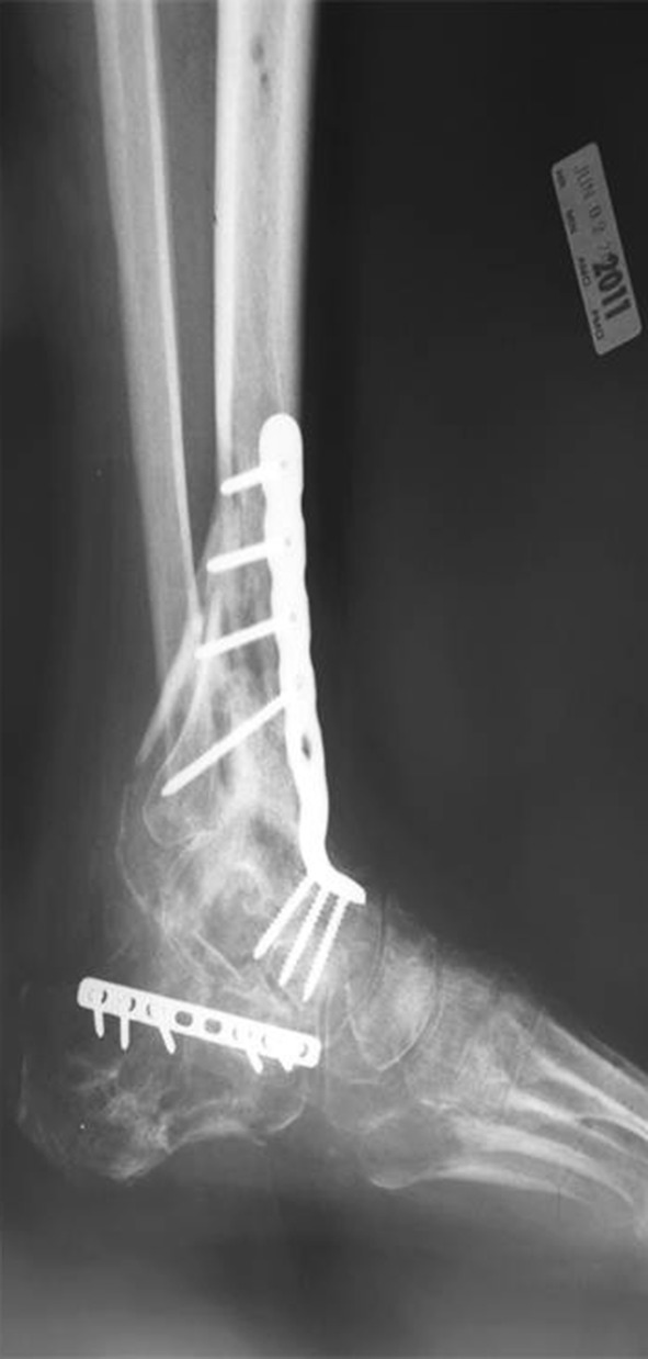
Methods: During a 2-year period, we performed open fracture reduction and internal fixation in 63 patients. Eleven patients (12 ankles) with severely comminuted high-energy tibial pilon fractures were retrospectively reviewed after surgical treatment with primary ankle arthrodesis and fracture reduction. Average patient age was 58 years, and minimum followup was 6 months (average, 14 months; range, 6-22 months). Anatomically designed anterior ankle arthrodesis plates were used in 10 ankles. Ring external fixation was used in nine ankles with concomitant tibia fracture or in instances requiring additional fixation. Clinical evaluation included chart review, interview, the AOFAS ankle-hindfoot score, and radiographic evaluation.

Results: All of the ankle arthrodeses healed at an average of 4.4 months (range, 3-5 months). One patient had a nonunion at the metaphyseal fracture, which healed with revision surgery. The average AOFAS ankle-hindfoot score was 83 with 88% having an excellent or good result. Radiographic and clinical analysis confirmed a plantigrade foot without malalignment. No patients required revision surgery for malunion.

Conclusions: Primary ankle arthrodesis combined with fracture reduction for the severely comminuted tibial pilon fracture reliably healed and restored acceptable function in this highly selective patient group. Ring external fixation may be a useful adjunct to internal fixation, and this concept should be further studied.

Level of evidence: Level IV, therapeutic study. See Guidelines for Authors for a complete description of levels of evidence.


Figures

Fig. 1
Fig. 1
Coronal CT scan cut demonstrates a high degree of tibial plafond comminution associated with severe articular cartilage damage, our primary indication for the inclusion of an ankle fusion with pilon fracture repair.
Fig. 2
Fig. 2
This is the injury AP radiograph of an open pilon fracture sustained resulting from a logging accident.
Fig. 3
Fig. 3
This AP radiograph is after initial spanning external fixation, open fracture management, and intramedullary fibular fixation.
Fig. 4
Fig. 4
This AP radiograph, after definitive fracture management and ankle arthrodesis, shows the use of both internal ankle arthrodesis plate fixation and ring external fixation.
Fig. 5
Fig. 5
This is the lateral radiograph of the same patient in Figure 4.
Fig. 6
Fig. 6
The AP radiograph shows a closed pilon fracture after spanning external fixation performed the day of injury.
Fig. 7
Fig. 7
The lateral radiograph more clearly depicts the plafond damage, which is better visualized on the CT scan shown in Figure 8.
Fig. 8
Fig. 8
This CT scan depicts plafond damage.
Fig. 9
Fig. 9
The AP radiograph is after surgical treatment, including fracture stabilization and ankle arthrodesis with an anatomically designed anterior fusion plate and ring external fixation.
Fig. 10
Fig. 10
The AP radiograph at followup demonstrates the radiographic measurements analyzed in this study.
Fig. 11
Fig. 11
Lateral followup radiograph demonstrates radiographic measurements.
Fig. 12
Fig. 12
The hindfoot alignment view of this patient with bilateral injuries shows the comparison with the contralateral limb, which was treated with traditional open reduction and internal fixation.
Fig. 13
Fig. 13
AP radiograph shows both the comminuted pilon fracture and calcaneal fracture sustained after a suicide attempt.
Fig. 14
Fig. 14
Lateral view shows a comminuted pilon fracture and calcaneal fracture from Figure 13.
Fig. 15
Fig. 15
AP radiograph shows a pilon fracture fixation and ankle arthrodesis performed through an anterior approach and the calcaneal fracture repair through a lateral approach. Ring external fixation provided additional fracture stability and allowed this patient with bilateral injuries to ambulate during the healing period.
Fig. 16
Fig. 16
Lateral view shows a pilon and calcaneal fracture from Figure 15.
Fig. 17
Fig. 17
AP radiograph after fracture healing shows restoration of limb alignment. This patient regained function and healed within 5 months after bilateral limb-threatening injuries without the need for additional reconstructive procedures.
Fig. 18
Fig. 18
Lateral view shows the patient in Figure 17.

Similar articles

Cited by

References

    1. Boraiah S, Kemp TJ, Erwteman A, Lucas PA, Asprinio DE. Outcome following open reduction and internal fixation of open pilon fractures. J Bone Joint Surg Am. 2010;92:346–352. doi: 10.2106/JBJS.H.01678. - DOI - PubMed
    1. Bozic V, Thordarson DB, Hertz J. Ankle fusion for definitive management of non-reconstructable pilon fractures. Foot Ankle Int. 2008;29:914–918. doi: 10.3113/FAI.2008.0914. - DOI - PubMed
    1. Davidovitch RI, Elkhechen RJ, Romo S, Walsh M, Egol KA. Open reduction with internal fixation versus limited internal fixation and external fixation for high grade pilon fractures (OTA type 43C) Foot Ankle Int. 2011;32:955–961. doi: 10.3113/FAI.2011.0955. - DOI - PubMed
    1. Feibel RJ, Uhthoff HK. [Primary Ilizarov ankle fusion for nonreconstructable tibial plafond fractures] [in German] Oper Orthop Traumatol. 2005;17:457–480. doi: 10.1007/s00064-005-1139-5. - DOI - PubMed
    1. Jansen H, Fenwick A, Doht S, Frey S, Meffert R. Clinical outcome and changes in gait pattern after pilon fractures. Int Orthop. 2013;37:51–58. doi: 10.1007/s00264-012-1716-1. - DOI - PMC - PubMed

MeSH terms