. 2014 Jul:45:128-41.
doi: 10.1016/j.psyneuen.2014.03.023. Epub 2014 Apr 12.

The neuroanatomical distribution of oxytocin receptor binding and mRNA in the male rhesus macaque (Macaca mulatta)

Affiliations

The neuroanatomical distribution of oxytocin receptor binding and mRNA in the male rhesus macaque (Macaca mulatta)

Sara M Freeman et al. Psychoneuroendocrinology. 2014 Jul.

Abstract

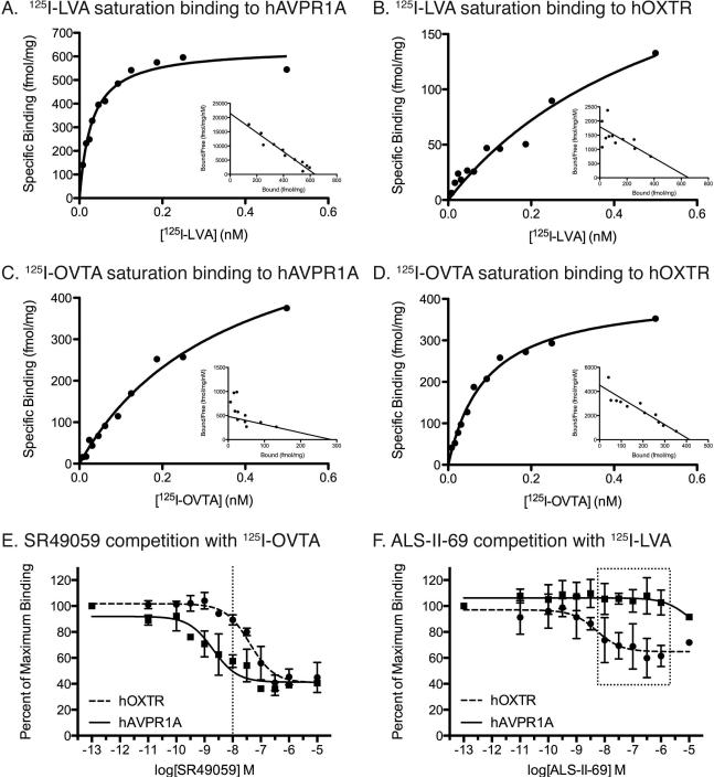
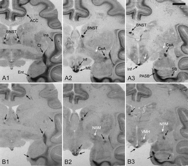
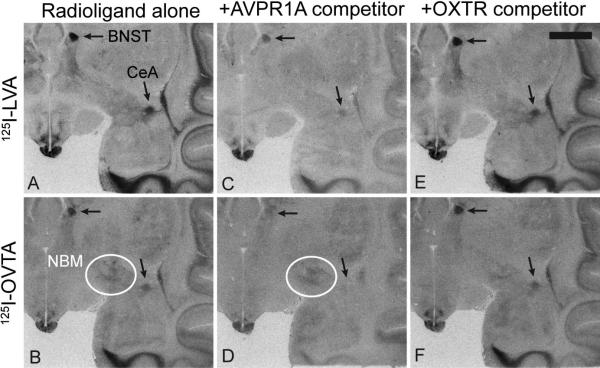
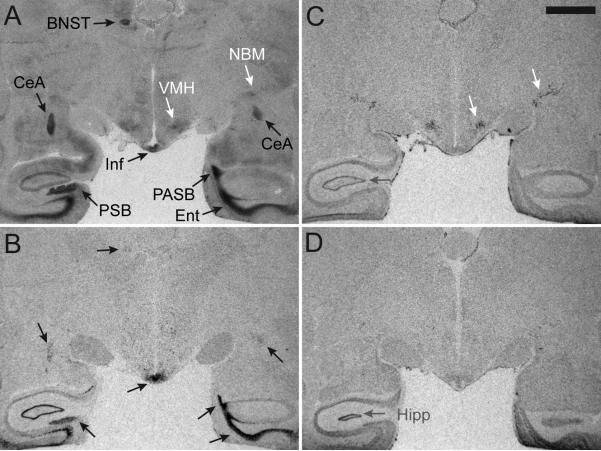
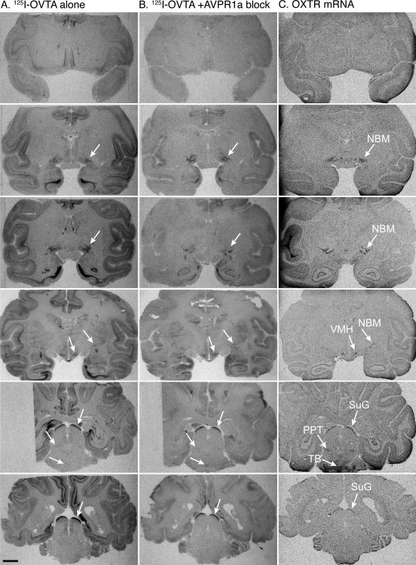
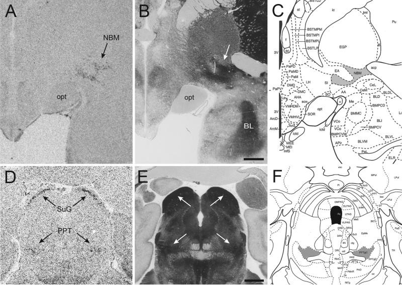
The rhesus macaque (Macaca mulatta) is an important primate model for social cognition, and recent studies have begun to explore the impact of oxytocin on social cognition and behavior. Macaques have great potential for elucidating the neural mechanisms by which oxytocin modulates social cognition, which has implications for oxytocin-based pharmacotherapies for psychiatric disorders such as autism and schizophrenia. Previous attempts to localize oxytocin receptors (OXTR) in the rhesus macaque brain have failed due to reduced selectivity of radioligands, which in primates bind to both OXTR and the structurally similar vasopressin 1a receptor (AVPR1A). We have developed a pharmacologically-informed competitive binding autoradiography protocol that selectively reveals OXTR and AVPR1A binding sites in primate brain sections. Using this protocol, we describe the neuroanatomical distribution of OXTR in the macaque. Finally, we use in situ hybridization to localize OXTR mRNA. Our results demonstrate that OXTR expression in the macaque brain is much more restricted than AVPR1A. OXTR is largely limited to the nucleus basalis of Meynert, pedunculopontine tegmental nucleus, the superficial gray layer of the superior colliculus, the trapezoid body, and the ventromedial hypothalamus. These regions are involved in a variety of functions relevant to social cognition, including modulating visual attention, processing auditory and multimodal sensory stimuli, and controlling orienting responses to visual stimuli. These results provide insights into the neural mechanisms by which oxytocin modulates social cognition and behavior in this species, which, like humans, uses vision and audition as the primary modalities for social communication.

Keywords: Autism; Neuroanatomy; Neuropeptide; Nonhuman primate; Oxytocin receptor.


Figures

Figure 1
Figure 1. Pharmacology results from ligand binding assays
A-D. Saturation binding curves and scatchard plots (insets) for the AVPR1A and OXTR radioligands binding to hAVPR1A (A,C) and hOXTR (B,D). E,F. Competition binding curves for competitor ligands. E. Competition curves for the AVPR1A ligand SR49059 showing that 10-8 M, or 10 nM, is the optimal concentration to occupy hAVPR1A without binding significantly to hOXTR (dotted line). F. Competition curves for the OXTR ligand ALS-II-69 showing that this ligand does not begin to occupy hAVPR1A until 10-5 M, or 10 μM. Thus, this ligand can be used at concentrations ranging to 1 μM (dotted box).
Figure 2
Figure 2. Radioligand binding to the rhesus macaque brain at three anatomical levels
The binding patterns produced by the AVPR1A radioligand, 125I-LVA (A), and the OXTR radioligand, 125I-OVTA (B), in rhesus monkey brain tissue sections at three anatomical levels (1, 2, 3), showing the overlap in binding by these two radioligands across brain regions. Black arrows indicate areas with overlapping binding between the two radioligands. White arrows highlight an example of two regions, the ventromedial hypothalamus and the nucleus basalis of Meynert, which has low levels of binding to 125I-OVTA but not 125I-LVA. Scale bar = 5mm. Abbreviations: ACC, anterior cingulate cortex; BNST, bed nucleus of stria terminalis; CeA, central amygdala; Cl, claustrum; Ent, entorhinal cortex; Inf, infundibulum; Ins, insular cortex; ME, median eminence; NBM, nucleus basalis of Meynert; PASB, parasubiculum; SB, subiculum; VMH, ventromedial hypothalamus
Figure 3
Figure 3. Representative results from competitive binding receptor autoradiography using 125I-LVA and 125I-OVTA
(A, B) Radioligand alone. (C, D) Radioligand coincubated with 10 nM of the AVPR1A competitor SR49059, showing that this compound is capable of competing off most of the signal from both radioligands, especially in the areas highlighted with arrows, the bed nucleus of stria terminalis (BNST) and the central amygdala (CeA). (E, F) Radioligand coincubated with 20 nM of the OXTR competitor ALS-II-69, showing little knockdown of 125I-OVTA or 125I-LVA compared to the radioligand alone condition. White circles in panels B and D indicate modest binding of 125I-OVTA to the nucleus basalis of Meynert (NBM), an area which does not show binding to 125I-LVA and does shows reduced binding in the presence of the OXTR competitor in panel F. Scale bar = 5mm.
Figure 4
Figure 4. Comparison of 125I-OVTA radioligand binding alone to in situ hybridization results
The binding pattern produced by 125I-OVTA alone (A) includes binding to both AVPR1A and OXTR, which can be parsed apart by comparing the radioligand binding densities to the in situ hybridization results for mRNA for these receptors. (B) An antisense probe for AVPR1A mRNA yields a binding pattern that matches areas of radioligand binding (black arrows, A,B). (C) An antisense probe for OXTR mRNA yields a binding pattern which allows the remaining lighter gray densities produced by 125I-OVTA to be interpreted as distinctly OXTR binding and not background (white arrows, A,C). (D) A sense probe designed as a negative control for in situ hybridization shows that the mRNA signal seen in the hippocampus and surrounding areas is background and not specific mRNA for OXTR (gray arrows, C,D). Scale bar = 5mm. Abbreviations: BNST, bed nucleus of stria terminalis; CeA, central amygdala; Ent, entorhinal cortex; Inf, infundibulum; NBM, nucleus basalis of Meynert; PASB, parasubiculum; PSB, presubiculum; VMH, ventromedial hypothalamus
Figure 5
Figure 5. OXTR distribution in the rhesus macaque brain
Receptor autoradiography results using 125I-OVTA alone (A) and 125I-OVTA plus 10 nM SR49059 (B) aligned with in situ hybridization results for OXTR mRNA (C) in six representative individuals at six different levels (rows). White arrows indicate areas of specific OXTR expression based on a visual comparison of the results from in situ hybridization (C) and competitive binding (B). Scale bar = 5mm. Abbreviations: III, oculomotor nucleus; NBM, nucleus basalis of Meynert; PPT, pedunculopontine tegmental nucleus; SuG, superficial gray layer of the superior colliculus; TB, trapezoid body; VMH, ventromedial hypothalamus; Sp5, spinal trigeminal nucleus
Figure 6
Figure 6. Comparison of OXTR mRNA distribution with acetylcholinesterase counterstained sections
Resulting OXTR mRNA at two anatomical levels (A, D) showing expression in the nucleus basalis of Meynert (NBM), the pedunculopontine tegmental nucleus (PPT), and the superficial gray layer of the superior colliculus (SuG). The adjacent acetylcholinesterase (AChE) counterstained sections (B, E) and adapted plates from the Paxinos rhesus brain atlas (C, F) show that OXTR expression is specific to the cholinergic NBM in the basal forebrain (gray area in panel C) and to the cholinergic subdivision of the PPT (“PPTgC”, gray area in panel F). Scale bars = 2mm.

References

    1. Acher R, Chauvet J, Chauvet MT. Man and the chimaera. Selective versus neutral oxytocin evolution. Adv. Exp. Med. Biol. 1995;395:615–627. - PubMed
    1. Ala Y, Morin D, Mahé E, Cotte N, Mouillac B, Jard S, Barberis C, Tribollet E, Dreifuss JJ, Sawyer WH, Wo NC, Chan WY, Kolodziejczyk AS, Cheng LL, Manning M. Properties of a new radioiodinated antagonist for human vasopressin V2 and V1a receptors. Eur. J. Pharmacol. 1997;331:285–293. - PubMed
    1. Andari E, Duhamel J-R, Zalla T, Herbrecht E, Leboyer M, Sirigu A. Promoting social behavior with oxytocin in high-functioning autism spectrum disorders. Proceedings of the National Academy of Sciences. 2010;107:4389–4394. - PMC - PubMed
    1. Bakermans-Kranenburg MJ, van IJzendoorn MH, Riem MME, Tops M, Alink LRA. Oxytocin decreases handgrip force in reaction to infant crying in females without harsh parenting experiences. Soc Cogn Affect Neurosci. 2012;7:951–957. - PMC - PubMed
    1. Bale TL, Davis AM, Auger AP, Dorsa DM, McCarthy MM. CNS region-specific oxytocin receptor expression: importance in regulation of anxiety and sex behavior. J. Neurosci. 2001;21:2546–2552. - PMC - PubMed

Publication types

LinkOut - more resources