

Powerful inner/outer controlled multi-target magnetic nanoparticle drug carrier prepared by liquid photo-immobilization
- PMID: 24845203
- PMCID: PMC4028896
- DOI: 10.1038/srep04990
Powerful inner/outer controlled multi-target magnetic nanoparticle drug carrier prepared by liquid photo-immobilization
Abstract
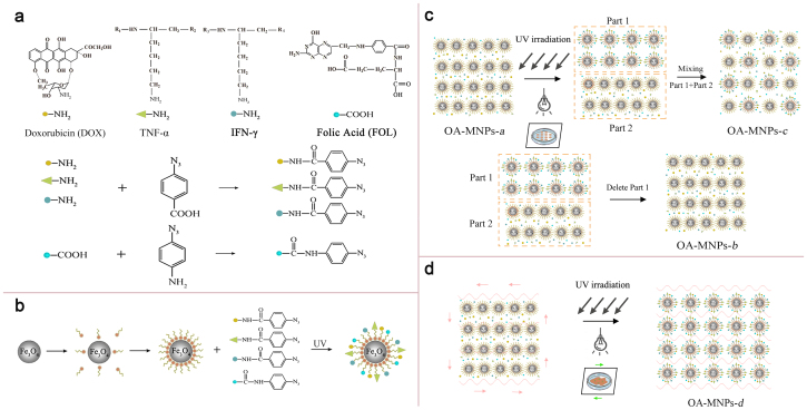
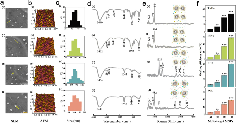
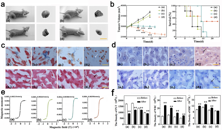
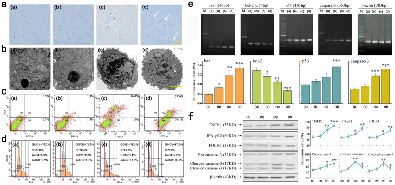
Nanomagnetic materials offer exciting avenues for advancing cancer therapies. Most researches have focused on efficient delivery of drugs in the body by incorporating various drug molecules onto the surface of nanomagnetic particles. The challenge is how to synthesize low toxic nanocarriers with multi-target drug loading. The cancer cell death mechanisms associated with those nanocarriers remain unclear either. Following the cell biology mechanisms, we develop a liquid photo-immobilization approach to attach doxorubicin, folic acid, tumor necrosis factor-α, and interferon-γ onto the oleic acid molecules coated Fe3O4 magnetic nanoparticles to prepare a kind of novel inner/outer controlled multi-target magnetic nanoparticle drug carrier. In this work, this approach is demonstrated by a variety of structural and biomedical characterizations, addressing the anti-cancer effects in vivo and in vitro on the HeLa, and it is highly efficient and powerful in treating cancer cells in a valuable programmed cell death mechanism for overcoming drug resistance.
Figures
Similar articles
-
Multi-functional nanocarriers based on iron oxide nanoparticles conjugated with doxorubicin, poly(ethylene glycol) and folic acid as theranostics for cancer therapy.Colloids Surf B Biointerfaces. 2018 Oct 1;170:529-537. doi: 10.1016/j.colsurfb.2018.06.051. Epub 2018 Jun 26. Colloids Surf B Biointerfaces. 2018. PMID: 29966906
-
Folate-mediated poly(3-hydroxybutyrate-co-3-hydroxyoctanoate) nanoparticles for targeting drug delivery.Eur J Pharm Biopharm. 2010 Sep;76(1):10-6. doi: 10.1016/j.ejpb.2010.05.005. Epub 2010 May 22. Eur J Pharm Biopharm. 2010. PMID: 20472060
-
Folic acid-conjugated polyethylene glycol-coated magnetic nanoparticles for doxorubicin delivery in cancer chemotherapy: Preparation, characterization and cytotoxicity on HeLa cell line.Hum Exp Toxicol. 2017 Aug;36(8):833-845. doi: 10.1177/0960327116672910. Epub 2016 Oct 10. Hum Exp Toxicol. 2017. PMID: 27758842
-
Redox-responsive mesoporous selenium delivery of doxorubicin targets MCF-7 cells and synergistically enhances its anti-tumor activity.Acta Biomater. 2017 May;54:294-306. doi: 10.1016/j.actbio.2017.02.042. Epub 2017 Mar 4. Acta Biomater. 2017. PMID: 28267598
-
An integrated targeting drug delivery system based on the hybridization of graphdiyne and MOFs for visualized cancer therapy.Nanoscale. 2019 Jun 20;11(24):11709-11718. doi: 10.1039/c9nr02017a. Nanoscale. 2019. PMID: 31180099
Cited by
-
Sui generis: gene therapy and delivery systems for the treatment of glioblastoma.Neuro Oncol. 2015 Mar;17 Suppl 2(Suppl 2):ii24-ii36. doi: 10.1093/neuonc/nou355. Neuro Oncol. 2015. PMID: 25746089 Free PMC article. Review.
-
Inhibition by Multifunctional Magnetic Nanoparticles Loaded with Alpha-Synuclein RNAi Plasmid in a Parkinson's Disease Model.Theranostics. 2017 Jan 1;7(2):344-356. doi: 10.7150/thno.16562. eCollection 2017. Theranostics. 2017. PMID: 28042339 Free PMC article.
-
Folate-conjugated nanoparticles as a potent therapeutic approach in targeted cancer therapy.Tumour Biol. 2015 Aug;36(8):5727-42. doi: 10.1007/s13277-015-3706-6. Epub 2015 Jul 5. Tumour Biol. 2015. PMID: 26142733 Review.
-
Development of Doxorubicin-Loaded Magnetic Silica-Pluronic F-127 Nanocarriers Conjugated with Transferrin for Treating Glioblastoma across the Blood-Brain Barrier Using an in Vitro Model.ACS Omega. 2018 Jul 31;3(7):8017-8026. doi: 10.1021/acsomega.8b00152. Epub 2018 Jul 18. ACS Omega. 2018. PMID: 30087932 Free PMC article.
-
Nanodelivery Systems Targeting Epidermal Growth Factor Receptors for Glioma Management.Pharmaceutics. 2020 Dec 10;12(12):1198. doi: 10.3390/pharmaceutics12121198. Pharmaceutics. 2020. PMID: 33321953 Free PMC article. Review.
References
-
- Dilnawaz F., Singh A., Mohanty C. & Sahoo S. K. Dual drug loaded superparamagnetic iron oxide nanoparticles for targeted cancer therapy. Biomaterials 31, 3694–3706 (2010). - PubMed
-
- Hua M. Y. et al. The effectiveness of a magnetic nanoparticle-based delivery system for BCNU in the treatment of gliomas. Biomaterials 32, 516–527 (2011). - PubMed
-
- Amstad E. et al. Triggered release from liposomes through magnetic actuation of iron oxide nanoparticle containing membranes. Nano Lett. 11, 1664–1670 (2011). - PubMed
Publication types
MeSH terms
Substances
LinkOut - more resources
Full Text Sources
Other Literature Sources
Molecular Biology Databases
