. 2014 Jul;10(7):558-66.
doi: 10.1038/nchembio.1528. Epub 2014 May 20.

Targeting the disordered C terminus of PTP1B with an allosteric inhibitor

Affiliations

Targeting the disordered C terminus of PTP1B with an allosteric inhibitor

Navasona Krishnan et al. Nat Chem Biol. 2014 Jul.

Abstract

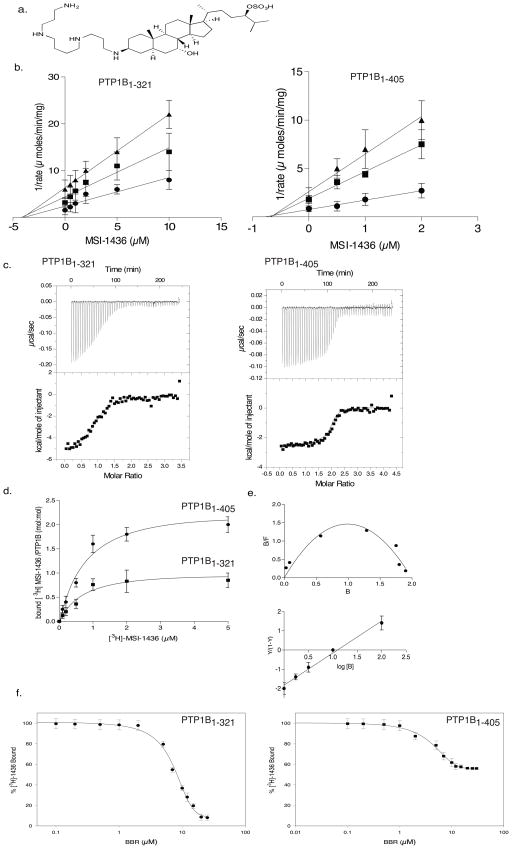
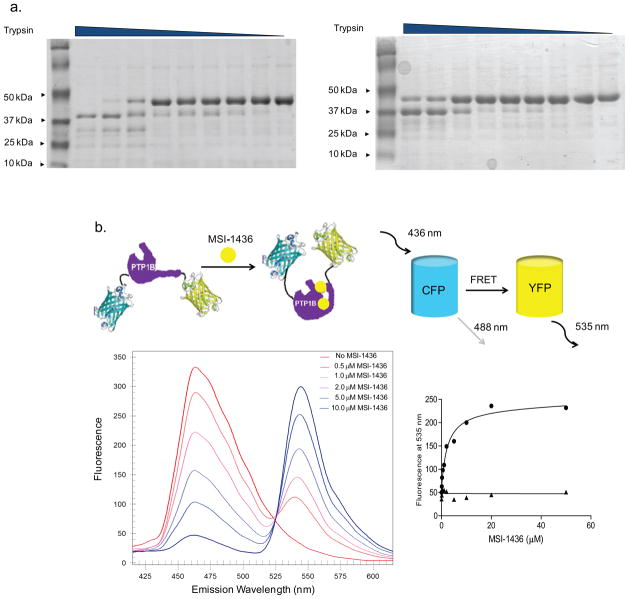
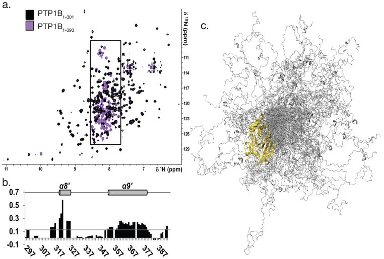
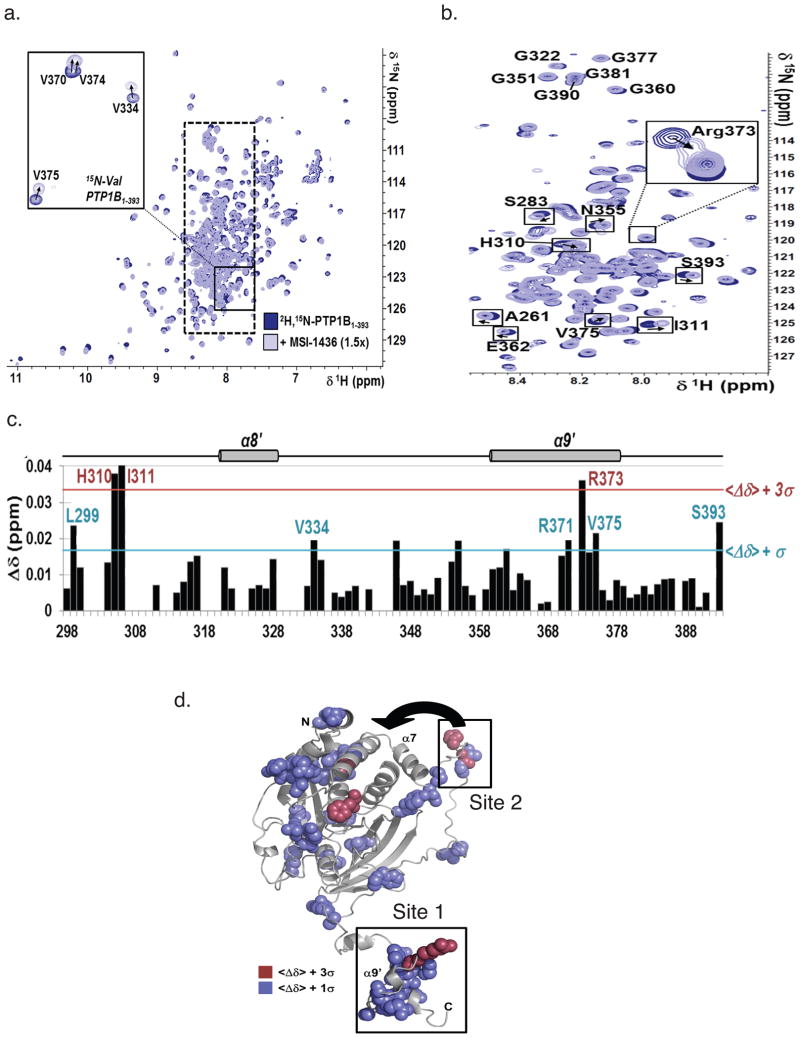
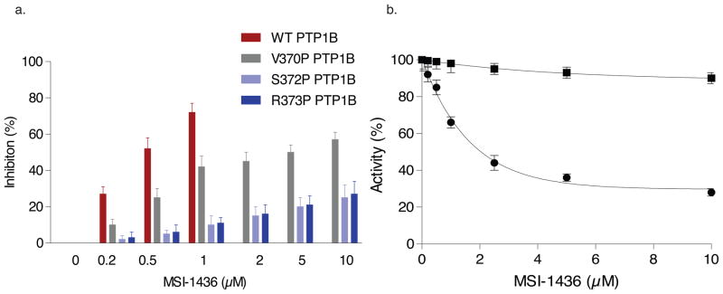
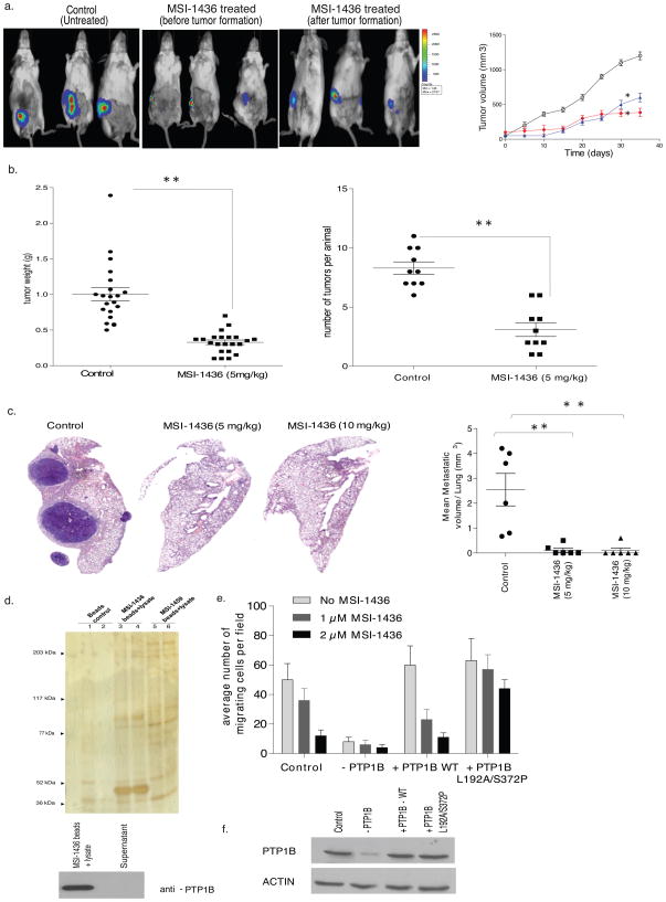
PTP1B, a validated therapeutic target for diabetes and obesity, has a critical positive role in HER2 signaling in breast tumorigenesis. Efforts to develop therapeutic inhibitors of PTP1B have been frustrated by the chemical properties of the active site. We define a new mechanism of allosteric inhibition that targets the C-terminal, noncatalytic segment of PTP1B. We present what is to our knowledge the first ensemble structure of PTP1B containing this intrinsically disordered segment, within which we identified a binding site for the small-molecule inhibitor MSI-1436. We demonstrate binding to a second site close to the catalytic domain, with cooperative effects between the two sites locking PTP1B in an inactive state. MSI-1436 antagonized HER2 signaling, inhibited tumorigenesis in xenografts and abrogated metastasis in the NDL2 mouse model of breast cancer, validating inhibition of PTP1B as a therapeutic strategy in breast cancer. This new approach to inhibition of PTP1B emphasizes the potential of disordered segments of proteins as specific binding sites for therapeutic small molecules.


Conflict of interest statement

COMPETING FINANCIAL INTERESTS

A company called DepYmed Inc was founded on 3 March 2014 with the primary purpose of taking MSI-1436 into the clinic in patients with HER2-positive breast cancer. It is owned 50-50 by Ohr Pharmaceuticals and Cold Spring Harbor Lab. Authors Krishnan and Tonks would be entitled to a share based upon the conditions of the Invention Agreement signed by all scientists at Cold Spring Harbor Lab.

Figures

Figure 1
Figure 1. MSI-1436 was a non-competitive inhibitor of PTP1B
a. Structure of MSI-1436/Trodusquemine. b. Dixon plot for PTP1B1-321 (left) and PTP1B1-405 (right) showing 1/rate versus varying concentrations of MSI-1436 at 2 μM (●), 5 μM (■) and10 μM (▲) substrate concentrations. The lines intersect on the x-axis to give a Ki of 4 μM for PTP1B1-321 and 0.6 μM for PTP1B1-405. Data are representative of three independent experiments. c. Isothermal titration calorimetry analysis. Binding thermograms for PTP1B1-321 (left), and PTP1B1-405 (right) are shown in the upper panels. Non-linear least square curves (lower panels) were fitted to enthalpies integrated from the individual titrations. The binding constants are 300 nM and 1 μM for PTP1B1-405 and 2 μM for PTP1B1-321. Data are representative of three independent experiments. d. Binding of radiolabelled MSI-1436. PTP1B1-405 (●) or PTP1B1-321 (■) (100 nM) was incubated with varying concentrations of [3H]-1436, PTP1B:[3H]-1436 complex was separated from unbound compound and bound radioactivity (cpm) was measured. The stoichiometry was found to be 1:1 for the PTP1B1-321:MSI-1436 and 1:2 for the PTP1B1-405: MSI-1436 complex. Data represent mean ± s.e.m from three independent experiments. e. Scatchard analysis of MSI-1436 binding to PTP1B1-405 (upper). B and F represent bound and free MSI-1436 (μmol). Hill plot (lower) where Y represents fraction of binding sites that are occupied by MSI-1436 on PTP1B1-405. The slope yields a Hill coefficient of 1.3 and Kd of 0.7 μM. Data are representative of three independent experiments. f. Displacement titrations. PTP1B1-321 (left) or PTP1B1-405 (right) (100 nM) was incubated with 5 μM tritiated MSI-1436 for 5 minutes in assay buffer. Following the incubation, varying concentrations of BBR (0–10 μM) were added for 30 minutes. The extent of MSI-1436 displaced by BBR was determined by measuring the residual [3H]-MSI-1436 bound to the enzyme. The experiment was repeated three times and error bars represent S.E.M.
Figure 2
Figure 2. MSI-1436 induced a conformational change in PTP1B
a. SDS-PAGE following limited proteolysis of PTP1B1-405 with trypsin in the absence (left) and presence (right) of MSI-1436. Purified PTP1B1-405 (2 μM) was incubated without (left) or with (right) MSI-1436 (5 μM) for 30 min with trypsin/PTP1B (mol/mol) ratio of 1:10, 0.5:10, 0.25:10, 0.125:10, 0.06:10,0.03:10, 0.01:10, 0.005:10, 0:10 from left to right, respectively. Reactions were terminated with SDS sample buffer, and the digested products were analyzed by 20% SDS-PAGE and stained by Coomassie blue. b. Schematic model of the PTP1B fusion protein tagged with CFP and YFP on the N- and C- terminus respectively, showing a conformational change induced by binding of the inhibitor (MSI-1436) that results in a change in FRET (upper panel). The effect of MSI-1436 binding to PTP1B1-405 was followed by FRET (lower left panel). Representative spectra from the experiment in which CFP-PTP1B1-405-YFP was titrated with MSI-1436, covering a concentration range of 0 – 10 μM revealed the inhibitor-induced decrease in CFP emission at 475 nm and concomitant increase in the YFP emission at 535 nm. The increase in YFP with addition of MSI- 1436 was plotted against the concentration of MSI-1436 to derive a Kd of 0.8 μM (lower right panel).
Figure 3
Figure 3. PTP1B residues 300-393 were flexible and predominantly disordered
a. Overlay of the 2D [1H,15N] TROSY spectra of PTP1B1-301 (black) and PTP1B1-393 (purple). Boxed region (7.5 - 8.5 ppm, 1H dimension) marks PTP1B residues 300-393. b. Secondary structure propensity (SSP) scores for PTP1B residues 300–393. A score of 1 indicates a fully populated α-helix, while a score of 0.5 indicates an α-helix that is ~50% populated in solution. Helices α8′ and α9′, which are 20 % populated, are denoted with a gray cylinder and labeled above. c. Representative ensemble of 100 conformers that are superimposed over PTP1B residues 1–284 (gold). PTP1B residues 285–398 (gray) sample a wide range of conformational space.
Figure 4
Figure 4. PTP1B residues were perturbed upon MSI-1436 binding.<
br>a. Overlay of the 2D [1H,15N] TROSY spectra of [2H,15N]-PTP1B1-393 with (light blue) and without (dark blue) MSI-1436 (1.5x molar excess). Inset: 2D [1H,15N] TROSY spectra of 15N-Val-PTP1B1-393 (same coloring scheme). Dashed box marks the region of the spectrum shown in b. b. Segment of the 2D [1H,15N] TROSY spectrum corresponding to PTP1B residues 300–393. c. CSP mapping of MSI-1436 binding to PTP1B residues 300–393. Colored lines indicate one (blue) and three (red) standard deviations from the mean,<Δδ>. d. CSPs mapped onto the structure of a single conformer of PTP1B1-393, using the same color scheme as in panel c. MSI-1436 binding site 1 and 2 are highlighted and the close proximity of binding site 2 to the exosite highlighted by an arrow.
Figure 5
Figure 5. Effect of mutations in the C-terminus of PTP1B on inhibition by MSI-1436<
br>a. The bar graph compares the extent of inhibition of PTP1B1-405 and three point mutants located in the NMR-detected helix α9′ (residues 360 – 379 in the C-terminus). The inhibition constants were ki 0.8 μM for PTP1B1-405 and 10 μM, 15 μM and 18 μM for V370P, S372P and R373P, respectively. Data represent mean ± s.e.m from three independent experiments. b. Introduction of two point mutations in PTP1B1-405, L192A and S372P, resulted in complete loss of inhibition by MSI-1436. Data represent mean ± s.e.m from three independent experiments.
Figure 6
Figure 6. Effect of the allosteric inhibitor of PTP1B, MSI-1436, in cell and animal models of HER2-positive breast cancer<
br>a. Effect MSI-1436 on tumor xenografts. BT474 cells (5×10⁶) were injected orthotopically into the mammary fat pad of SCID-beige mice. MSI-1436 (5 mg/kg) or saline (control) was injected intraperitoneally as soon as the surgery was performed or once the tumors reached ~100 mm3 in volume. Tumor volume was assessed with a digital caliper for control (open circles) and MSI-1436-treated. Quantitation of tumor volume, measured using a digital caliper, over time (Right panel) (Student t-test, p<0.01). Representative images of three independent experiments are shown. b. Distribution of tumor weight (left) and tumor number (right) in NDL2 animals treated with MSI-1436 (5 mg/kg) (●) or vehicle control (■) (Student t-test, p<0.001). Data are representative of three independent experiments with more than six animals in each experiment. c. Effect of MSI-1436 on lung metastases, monitored by histopathological analysis of hematoxylin and eosin-stained lung sections taken from the animals. Mice were treated with saline, or MSI-1436 at 5 mg/kg or 10 mg/kg. Each treatment group contained 6 animals. Left panels are representative images of lung sections from control and MSI-1436-treated animals subjected to H&E staining. The right panel is a quantitation of the whole lung obtained from all the animals in each group (control and MSI-1436-treated) (Student t-test, p<0.001). Data are representative of three independent experiments with more than six animals in each experiment. d. Tumor lysate was incubated with MSI-1436 immobilized to NHS-activated Sepharose beads for 30 minutes at 4 °C. The beads were washed and the samples were subjected to SDS-PAGE and silver staining (upper panel). Both beads and supernatant were subjected to immunoblotting using anti-PTP1B (FG6) antibody (lower panel) to demonstrate that PTP1B was cleared from the lysate by immobilized MSI-1436. Data are representative of three independent experiments. e. Migration of control and PTP1B-depleted MCF10-NeuNT cells, in the absence and presence of doxycycline, were compared to MCF10-NeuNT cells overexpressing wild type (WT) PTP1B or the double mutant, PTP1B L192A/S372P, in the presence of doxycycline and in the absence (pale grey bars) and presence of MSI-1436 (1 μM, grey bars and 2 μM dark grey bars). Data are representative of three independent experiments done in duplicates. f. PTP1B knock-down and re-expression of wild type (WT) and L192A/S372P mutant PTP1B in MCF10A-Neu-NT cells was analyzed by immunoblotting with anti-PTP1B (FG6) antibody. Data are representative of three independent experiments. (Full blot image Supplementary Fig. 20).

References

    1. Slamon DJ, et al. Studies of the HER-2/neu proto-oncogene in human breast and ovarian cancer. Science. 1989;244:707–12. - PubMed
    1. Tiwari RK, Borgen PI, Wong GY, Cordon-Cardo C, Osborne MP. HER-2/neu amplification and overexpression in primary human breast cancer is associated with early metastasis. Anticancer Research. 1992;12:419–25. - PubMed
    1. Engelman JA, Settleman J. Acquired resistance to tyrosine kinase inhibitors during cancer therapy. Current Opinion in Genetics & Development. 2008;18:73–9. - PubMed
    1. Rexer BN, Arteaga CL. Intrinsic and acquired resistance to HER2-targeted therapies in HER2 gene-amplified breast cancer: mechanisms and clinical implications. Critical Reviews in Oncogenesis. 2012;17:1–16. - PMC - PubMed
    1. Tonks NK, Diltz CD, Fischer EH. Purification of the major protein-tyrosine-phosphatases of human placenta. The Journal of Biological Chemistry. 1988;263:6722–30. - PubMed

Publication types

MeSH terms