

STAT6-mediated keratitis and blepharitis: a novel murine model of ocular atopic dermatitis
- PMID: 24845637
- PMCID: PMC4062401
- DOI: 10.1167/iovs.13-13685
STAT6-mediated keratitis and blepharitis: a novel murine model of ocular atopic dermatitis
Erratum in
-
Erratum.Invest Ophthalmol Vis Sci. 2020 Aug 3;61(10):42. doi: 10.1167/iovs.61.10.42. Invest Ophthalmol Vis Sci. 2020. PMID: 32832972 Free PMC article. No abstract available.
Abstract
Purpose: Atopic dermatitis (AD) is a common inflammatory disease that can affect the eye, resulting in ocular pathologies, including blepharitis, keratitis, and uveitis; however, the pathogenic mechanisms underlying the ocular manifestations of AD are not well understood.
Methods: In the present study, we characterized the ocular pathologies that develop in the Stat6VT mouse model of AD. We examined the cytokine profile of the eyelid lesions, measured the behavioral response, and documented the treatment response to topical steroids.
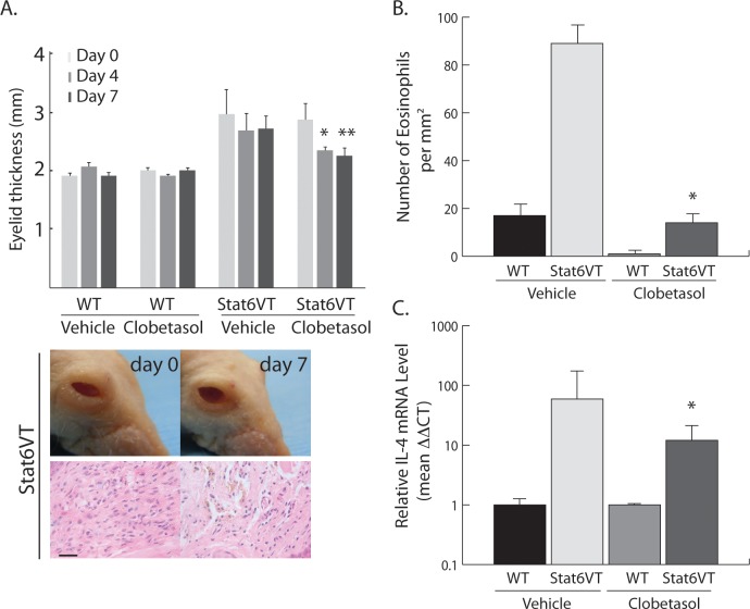
Results: Our results show that Stat6VT mice spontaneously developed blepharitis, keratitis, and uveitis similar to that observed in patients with AD. Histologic findings of allergic inflammation in affected eyelids in this model include the presence of a lymphocyte-predominant infiltrate and tissue eosinophilia in the dermis. Gene expression analysis of affected eyelid tissue by quantitative PCR revealed increased amounts of mRNAs for the Th2 cytokines IL-4, IL-5, and IL-13. In addition, increased eyelid scratching was seen in Stat6VT mice with blepharitis. Topical treatment with the corticosteroid clobetasol reduced eyelid inflammation, tissue eosinophilia, and Th2 cytokine expression.
Conclusions: The development of AD-like ocular pathologies in this model supports the idea that in humans, AD-associated disease of the eye may be driven by Th2-mediated inflammation and demonstrates that the Stat6VT mouse may be a useful system in which to further investigate pathogenesis of and treatment strategies for blepharitis and other ocular diseases that develop in association with AD.
Keywords: STAT6; atopic blepharitis; ocular atopic dermatitis.
Copyright 2014 The Association for Research in Vision and Ophthalmology, Inc.
Figures


Similar articles
-
Phenotyping acute and chronic atopic dermatitis-like lesions in Stat6VT mice identifies a role for IL-33 in disease pathogenesis.Arch Dermatol Res. 2018 Apr;310(3):197-207. doi: 10.1007/s00403-018-1807-y. Epub 2018 Jan 24. Arch Dermatol Res. 2018. PMID: 29368135 Free PMC article.
-
IL-4 is a critical determinant in the generation of allergic inflammation initiated by a constitutively active Stat6.J Immunol. 2008 Mar 1;180(5):3551-9. doi: 10.4049/jimmunol.180.5.3551. J Immunol. 2008. PMID: 18292582
-
Topical application of a vitamin D analogue exacerbates atopic dermatitis and induces the atopic dermatitis-like phenotype in Stat6VT mice.Pediatr Dermatol. 2013 Sep-Oct;30(5):574-8. doi: 10.1111/pde.12187. Epub 2013 Jul 25. Pediatr Dermatol. 2013. PMID: 23889122 Free PMC article.
-
[Ocular involvement in atopic dermatitis : Clinical aspects and therapy].Ophthalmologe. 2017 Jun;114(6):514-524. doi: 10.1007/s00347-017-0473-3. Ophthalmologe. 2017. PMID: 28283768 Review. German.
-
Bidirectional association between atopic dermatitis, conjunctivitis, and other ocular surface diseases: A systematic review and meta-analysis.J Am Acad Dermatol. 2021 Aug;85(2):453-461. doi: 10.1016/j.jaad.2020.11.037. Epub 2020 Nov 27. J Am Acad Dermatol. 2021. PMID: 33253849
Cited by
-
STAT6 and PARP Family Members in the Development of T Cell-dependent Allergic Inflammation.Immune Netw. 2016 Aug;16(4):201-10. doi: 10.4110/in.2016.16.4.201. Epub 2016 Aug 23. Immune Netw. 2016. PMID: 27574499 Free PMC article. Review.
-
Translational Animal Models of Atopic Dermatitis for Preclinical Studies.Yale J Biol Med. 2017 Sep 25;90(3):389-402. eCollection 2017 Sep. Yale J Biol Med. 2017. PMID: 28955179 Free PMC article. Review.
-
Quantitative assessment of redness in Asian patients with recurrent blepharitis: the utility of cross-polarized light.Front Med (Lausanne). 2025 Apr 30;12:1594764. doi: 10.3389/fmed.2025.1594764. eCollection 2025. Front Med (Lausanne). 2025. PMID: 40370733 Free PMC article.
-
The STAT6 inhibitor AS1517499 reduces the risk of asthma in mice with 2,4-dinitrochlorobenzene-induced atopic dermatitis by blocking the STAT6 signaling pathway.Allergy Asthma Clin Immunol. 2022 Feb 17;18(1):12. doi: 10.1186/s13223-022-00652-8. Allergy Asthma Clin Immunol. 2022. PMID: 35177102 Free PMC article.
-
Poly-ADP ribose polymerase-14 limits severity of allergic skin disease.Immunology. 2017 Nov;152(3):451-461. doi: 10.1111/imm.12782. Epub 2017 Jul 27. Immunology. 2017. PMID: 28653395 Free PMC article.
References
-
- Leung DY, Bieber T. Atopic dermatitis. Lancet. 2003; 361: 151–160 - PubMed
-
- Carmi E, Defossez-Tribout C, Ganry O, et al. Ocular complications of atopic dermatitis in children. Acta Derm Venereol. 2006; 86: 515–517 - PubMed
-
- Ebihara N, Funaki T, Matsuda H, Okumura K, Murakami A, Ra C. Corneal abnormalities in the NC/Nga mouse: an atopic dermatitis model. Cornea. 2008; 27: 923–929 - PubMed
-
- Matsuo T, Saito H, Matsuo N. Cataract and aqueous flare levels in patients with atopic dermatitis. Am J Ophthalmol. 1997; 124: 36–39 - PubMed
-
- Iijima Y, Wagai K, Matsuura Y, Ueda M, Miyazaki I. Retinal detachment with breaks in the pars plicata of the ciliary body. Am J Ophthalmol. 1989; 108: 349–355 - PubMed
Publication types
MeSH terms
Substances
Grants and funding
LinkOut - more resources
Full Text Sources
Other Literature Sources
Molecular Biology Databases
Research Materials
Miscellaneous
