. 2014 May 20;55(7):4186-98.
doi: 10.1167/iovs.13-13217.

Relationship between foveal cone specialization and pit morphology in albinism

Affiliations

Relationship between foveal cone specialization and pit morphology in albinism

Melissa A Wilk et al. Invest Ophthalmol Vis Sci. .

Abstract

Purpose: Albinism is associated with disrupted foveal development, though intersubject variability is becoming appreciated. We sought to quantify this variability, and examine the relationship between foveal cone specialization and pit morphology in patients with a clinical diagnosis of albinism.

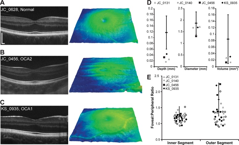
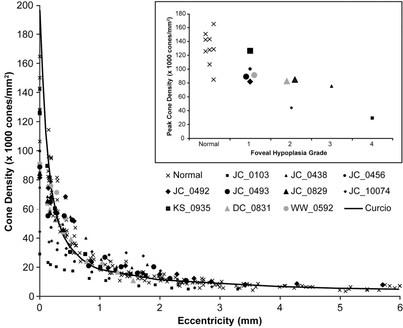
Methods: We recruited 32 subjects with a clinical diagnosis of albinism. DNA was obtained from 25 subjects, and known albinism genes were analyzed for mutations. Relative inner and outer segment (IS and OS) lengthening (fovea-to-perifovea ratio) was determined from manually segmented spectral domain-optical coherence tomography (SD-OCT) B-scans. Foveal pit morphology was quantified for eight subjects from macular SD-OCT volumes. Ten subjects underwent imaging with adaptive optics scanning light ophthalmoscopy (AOSLO), and cone density was measured.

Results: We found mutations in 22 of 25 subjects, including five novel mutations. All subjects lacked complete excavation of inner retinal layers at the fovea, though four subjects had foveal pits with normal diameter and/or volume. Peak cone density and OS lengthening were variable and overlapped with that observed in normal controls. A fifth hyper-reflective band was observed in the outer retina on SD-OCT in the majority of the subjects with albinism.

Conclusions: Foveal cone specialization and pit morphology vary greatly in albinism. Normal cone packing was observed in the absence of a foveal pit, suggesting a pit is not required for packing to occur. The degree to which retinal anatomy correlates with genotype or visual function remains unclear, and future examination of larger patient groups will provide important insight on this issue.

Keywords: adaptive optics; albinism; foveal development; foveal morphology.


Figures

Figure 1
Figure 1
Variability in foveal morphology in albinism. The SD-OCT horizontal line scans through the expected foveal location are shown for all subjects with albinism, grouped by clinical diagnosis (phenotype). Yellow, OA1; blue, OCA1; green, OCA2; orange, OCA4; and gray, OCA with unspecified type. Scale bar: 200 μm for all subjects.
Figure 2
Figure 2
Foveal morphology in albinism can overlap with normal values. (AC) Left panel is a high-resolution SD-OCT horizontal line scan image acquired through the fovea. Scale bar: 200 μm for all subjects. Right panel shows macular thickness maps centered on the fovea. (A) Subject JC_0628 represents a control subject with a shallow foveal pit with incomplete excavation of the inner retinal layers. (B, C) Subjects JC_0456 (OCA2) and KS_0935 (OCA1) are subjects with albinism who have well-defined foveal pits. (D, E) Open symbols represent the subjects with no genetic mutations identified, filled gray symbols represent the subjects with one mutation identified, filled black symbols represent subjects with 2 or more mutations or hemizygous mutations, and circles represent subjects with pit metrics outside 2 SDs of the normal mean or no measurable pit. (D) Quantification of foveal pit depth, diameter, and volume. The black bar represents the mean of 64 control subjects, and the error bars represent ±2 SDs. Four subjects with albinism had at least one aspect of their foveal pit morphology (depth, diameter, and/or volume) within 2 SDs of the normal mean. (E) The distribution of IS and OS ratios for all 32 subjects with albinism relative to 167 control subjects (the black bar is the previously published mean of 167 control subjects, and the error bars represent ±2 SDs9). No differences in IS or OS lengthening were seen between subjects with no mutations and those with 1 or more mutations (P = 0.37 for IS and P = 0.34 for OS; Mann-Whitney U test).
Figure 3
Figure 3
Visualizing foveal cone packing in albinism. Montages of the central retina surrounding the location of peak cone density (*) are shown using a logarithmic display for 10 subjects with albinism and two normal controls. Qualitatively, it can be seen that cone density decreases (larger, more coarsely packed cells) moving away from the location of peak density for all subjects except JC_0103, whose cone mosaic appears to have larger cones and uniform packing across the central retina. No significant differences in peak cone density were seen between subjects with one TYR mutation and those with two or more TYR mutations (P = 0.86, Wilcoxon rank sum test). Scale bar: 50 μm for all subjects.
Figure 4
Figure 4
Quantifying foveal cone packing in albinism. Cone density is plotted over a range of eccentricities (0 being the location of peak cone density) for 10 subjects with albinism, nine control subjects (x), and normative data from Curcio et al. (black line). For the subjects with albinism, filled black symbols represent subjects with 2 or more mutations or hemizygous mutations, filled gray symbols represent subjects with 1 mutation. Though some subjects with albinism appear to have lower absolute peak cone densities, all show a prominent increase in cone density at the fovea, with the exception of JC_0103 and JC_10074. Inset: Peak cone density versus degree of foveal hypoplasia using the grading scheme of Thomas et al. Subjects with more normal foveal morphology (i.e., lower grade of foveal hypoplasia) tend to have higher peak cone density.
Figure 5
Figure 5
Altered appearance of the outer retinal hyper-reflective SD-OCT bands in albinism. (AD) Top shows a logarithmic high-resolution horizontal line scan. Arrows indicate the nomenclature of Spaide and Curcio given to the outer retinal hyper-reflective bands: orange, ELM; blue, EZ; green, IZ; purple, RPE and BrM. Scale bars = 200 μm. White boxes (263 × 345 μm) delineate the areas of each image that are analyzed in the bottom. The bottom shows an LRP analysis for the central five pixels of the image excerpts (linear format) to the left of each plot. The gray line is the LRP for the logarithmic image, and the black line is the LRP for the linear image. Faint ELM bands are lost in the linear LRP, but the presence of a fifth hyper-reflective band is illustrated clearly in the linear LRP. Arrows in the bottom are color-matched to the image in the top to show the respective peaks for each hyper-reflective band. (A) Subject JC_0677 is a representative normal subject with a total of four outer retinal hyper-reflective bands at foveal and perifoveal locations. (B) Subject JC_1212 is one of seven control subjects (of 64 analyzed control subjects) with a fifth hyper-reflective band (denoted by the second purple arrow). (C, D) Representative subjects with albinism (JC_10073 and JC_0438) also display the presence of a fifth hyper-reflective band in the outer retina. The presence of a fifth band is more prevalent in subjects with albinism than in the normal population (P < 0.0001, Fisher's exact test).

Comment in

References

    1. King RA, Pietsch J, Fryer JP, et al. Tyrosinase gene mutations in oculocutaneous albinism 1 (OCA1): definition of the phenotype. Hum Genet. 2003; 113: 502–513 - PubMed
    1. Oetting WS, King RA. Molecular basis of albinism: mutations and polymorphisms of pigmentation genes associated with albinism. Hum Mutat. 1999; 13: 99–115 - PubMed
    1. Newton JM, Cohen-Barak O, Hagiwara N, et al. Mutations in the human orthologue of the mouse underwhite gene (uw) underlie a new form of oculocutaneous albinism, OCA4. Am J Hum Genet. 2001; 69: 981–988 - PMC - PubMed
    1. Oetting WS, Summers CG, King RA. Albinism and the associated ocular defects. Metab Pediatr Syst Ophthalmol. 1994; 17: 5–9 - PubMed
    1. Summers CG. Vision in albinism. Trans Am Ophthalmol Soc. 1996; 94: 1095–1155 - PMC - PubMed

Publication types

MeSH terms

LinkOut - more resources