Review
. 2014 May 19;24(10):R495-501.
doi: 10.1016/j.cub.2014.04.027.

Stress sensitivity and mechanotransduction during heart development

Affiliations
Review

Stress sensitivity and mechanotransduction during heart development

Stephanie Majkut et al. Curr Biol. .

Abstract

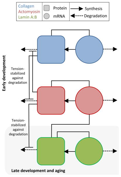
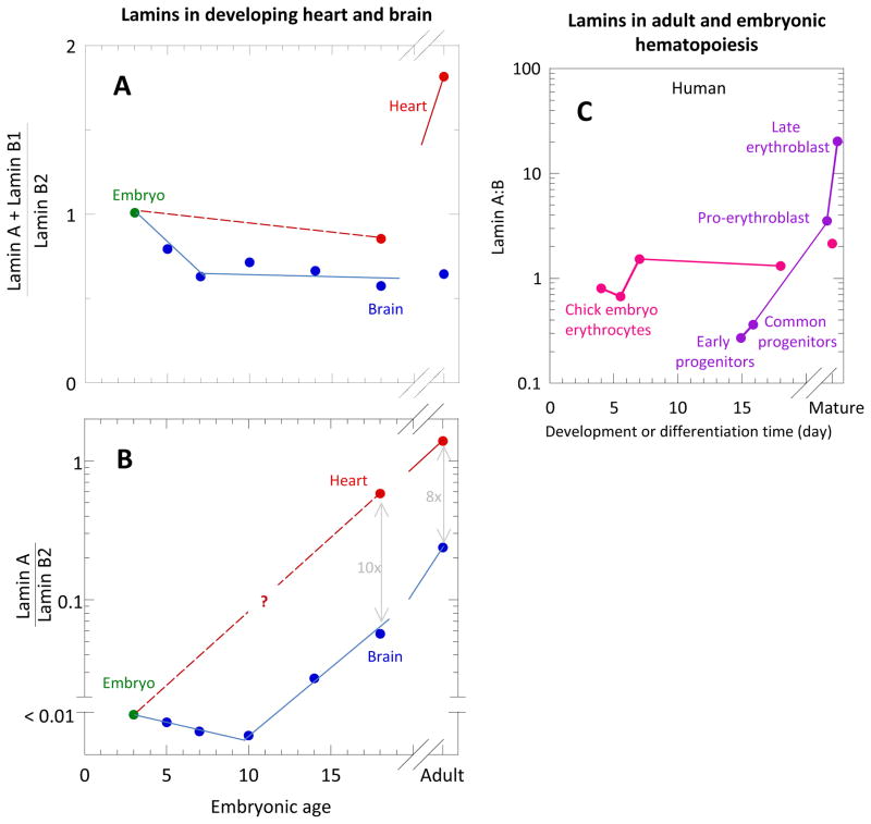
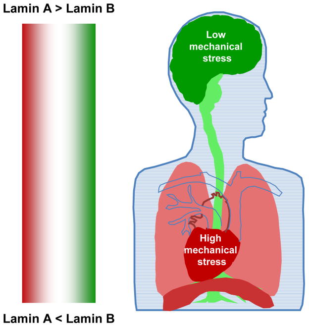
Early in embryogenesis, the heart begins its rhythmic contractions as a tube that helps perfuse the nascent vasculature, but the embryonic heart soon changes shape and mechanical properties, like many other developing organs. A key question in the field is whether stresses in development impact the underlying gene circuits and, if so, how? Here, we attempt to address this question as we review the mechanical maturation of heart - and, to a limited extent, lung and blood - with a focus on a few key abundant structural proteins whose expression dynamics have been suggested to be directly sensitive to mechanical stress. In heart maturation, proliferating fibroblasts deposit increasing amounts of collagenous matrix in parallel with cardiomyocytes expressing more sarcomeric proteins that increase the contractile stress and strength of the tissue, which in turn pumps more blood at higher stress throughout the developing vasculature. Feedback of beating cardiomyocytes on the expression of matrix by fibroblasts seems a reasonable model, with both synthesis and turnover of matrix and contractile elements achieving a suitable balance. Based on emerging evidence for coiled-coil biopolymers that are tension-stabilized against degradation, a minimal network model of a dynamic cell-matrix interaction is proposed. This same concept is extended to nuclear mechanics as regulated by stress on the nuclear structural proteins called lamins, which are examined in part because of the prominence of mutations in these coiled-coil proteins in diseases of the heart, amongst other organs/tissues. Variations in lamin levels during development and across adult tissues are to some extent known and appear to correlate with extracellular matrix mechanics, which we illustrate across heart, lung, and blood development. The formal perspective here on the mechanochemistry of tissue development and homeostasis could provide a useful framework for 'big data' quantitative biology, particularly of stress-sensitive differentiation, maturation, and disease processes.


Figures

Fig. 1
Fig. 1. Cardiomyocytes and Fibroblasts create a balance between contractile ability and ECM abundance during development
(A) Schema illustrating concept of how such a balance could be struck. Early in development, cardiomyocytes are relatively small with unorganized and relatively spare myofibril content. Cardiac fibroblasts feel strain from passive and active contraction of surrounding cells (orange arrows) propagated through the ECM and cell-cell adhesions (yellow arrows) prompting them to divide and produce ECM in a strain and growth-factor responsive manner. The increased ECM due to increased CF population prompts increased production of myofibril proteins and encourages myofibril organization, which in turn increases contractile strain on the cardiac fibroblasts. We propose that fibroblast population growth is at least in part limited by stiffness conferred significantly by collagenous matrix production, leading to an ultimate steady state of cardiomyocyte to CF volume fractions in normal adult tissue. (B) Tissue stiffness of embryonic chick heart and brain tissue was measured to increase throughout embryonic development in a way that is paralleled with both collagen-I and cardiac myosin-II expression. (C) Half-lives of collagens (dark blue) and collagen-binding integrins (light blue), actomyosin contractility (red), and nuclear Lamin mRNAs and proteins measured coincidently in NIH3T3 mouse fibroblasts. Half-lives are fairly constant within functional groups suggesting similar dynamics within groups.
Fig. 2
Fig. 2. Network model of interplay between ECM, actomyosin, and nuclear lamina genetic modules in cardiac tissue development
Collagen and myosin modules are modeled as simple genetic regulatory network in which the protein is formed proportionally to the amount of mRNA, mRNA is produced at a protein-dependentrate, and both proteins and mRNA degrade in relation to the applied tension that stabilizes the protein networks against degradation. The nuclear lamin module can be coupled in turn to the myosin module with the same mutually applied tension stabilization against degradation. This coupling is an example of the types of possible testable implications of the mechanical interactions between the protein structural networks.
Fig. 3
Fig. 3. Lamin levels in developing heart and brain
[41, 47]. (A) Total variable lamins A plusB1 normalized by constant lamin-B2 in brain (blue) and heart (red). In brain, this total remained relatively constant; in heart, this total increases from late embryogenesis to adult levels. (B) Lamin-A to lamin-B2 for brain (blue) and heart (red). In brain, the variable lamins is dominated by lamin-B1 throughout the development and aging, although, while the ratio of lamin-A:B was negligible before E10, it then increases to a non-negligible level adulthood. In heart, measurements are more sparse and missing during early embryogenesis. However, by late embryogenesis and into adulthood, lamin-A is the major variable isoform and constitutes much more of the heart cell nuclear lamina than in the brain. (C) Lamin-A:B for embryonic chick erythrocytes as measured by Lehner et al. (1987) compared to the same for adult mouse hematopoietic cells as measured by Shin et al. (2013) (inset).
Fig. 4
Fig. 4. Schema of adult tissue Lamin A:B stoichiometries
Summary figure of the lamin-A:B stoichiometries of the heart (high, red), brain (low, green) and the intermediate Lung, with heterogeneous lung tissue, diaphragm muscle (high), and nerves(low).

References

    1. Majkut SF, Idema T, Swift J, Krieger C, Liu A, Discher D. Heart stiffening in early embryos parallels matrix and myosin levels to optimize beating. Current Biology. 2013 - PMC - PubMed
    1. McCain M, Parker K. Mechanotransduction: the role of mechanical stress, myocyte shape, and cytoskeletal architectre on cardiac form and function. Pflugers Arch - Eur J Physiol. 2011;462:89–104. - PubMed
    1. Majkut S, Discher D. Cardiomyocytes from late embryos and neonates do optimal work and striate best on substrates with tissue-level elasticity: metris and mathematics. Biomechanics and modeling in mechanobiology. 2012;11(8):1219–1225. - PMC - PubMed
    1. Chung C, Pruitt B, Heilshorn S. Spontaneous cardiomyocyte differentiation of mouse embryoid bodies regulated by hydrogel rosslin density. Biomaterials Science. 2013;2013(1):1082–1090. - PMC - PubMed
    1. Young J, Engler A. Hydrogels with time-dependent material properties enhance cardiomyocyte differentiation in vitro. Biomaterials. 2011;32(4):1002–1009. - PMC - PubMed

LinkOut - more resources