Review
. 2014 Jun;13(6):547-57.
doi: 10.1038/nmat3937.

Materials as stem cell regulators

Affiliations
Review

Materials as stem cell regulators

William L Murphy et al. Nat Mater. 2014 Jun.

Erratum in

  • Nat Mater. 2014 Jul;13(7):756

Abstract

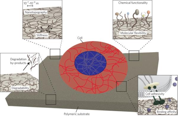
The stem cell/material interface is a complex, dynamic microenvironment in which the cell and the material cooperatively dictate one another's fate: the cell by remodelling its surroundings, and the material through its inherent properties (such as adhesivity, stiffness, nanostructure or degradability). Stem cells in contact with materials are able to sense their properties, integrate cues via signal propagation and ultimately translate parallel signalling information into cell fate decisions. However, discovering the mechanisms by which stem cells respond to inherent material characteristics is challenging because of the highly complex, multicomponent signalling milieu present in the stem cell environment. In this Review, we discuss recent evidence that shows that inherent material properties may be engineered to dictate stem cell fate decisions, and overview a subset of the operative signal transduction mechanisms that have begun to emerge. Further developments in stem cell engineering and mechanotransduction are poised to have substantial implications for stem cell biology and regenerative medicine.


Figures

Figure 1
Figure 1. Inherent material properties
Stem cell fate decisions can be affected by properties inherent to materials (exemplified by a two-dimensional polymeric substrate in this schematic) near the cell/material interface, such as nanotopography, stiffness (pictured as force vectors), chemical functionality (represented by coloured beads), molecular flexibility (indicated by the vertical strands sticking out of the substrate), the adhesivity of cells to the material (exemplified by ligand binding to the transmembrane receptor integrin), its binding affinity for soluble factors (pictured as blue spheres), its cell-mediated degradability and its degradation by-products.
Figure 2
Figure 2. stiffness, nanotopography and chemical functionality influence the behaviour of human mesenchymal stem cells
a, The modulus of poly(acrylamide) substrates influences lineage-specific (neurogenic, myogenic or osteogenic) differentiation, as indicated by immunostaining for the appropriate markers (β3-tubulin, MyoD and CBFα1, respectively, shown in green; cell nucleus in blue). Scale bars, 5 μm. b, Substrates with asymmetrically organized nanopits (top row) stimulate osteogenesis (middle and bottom rows), as indicated by immunostaining for bone-specific extracellular-matrix proteins (osteopontin and osteocalcin, green). c, Poly(ethylene glycol) (PEG) substrates modified with 50 mM of simple functional groups (insets) influence gene expression associated with chondrogenesis (top), osteogenesis (middle) and adipogenesis (bottom), as indicated by the normalized expression of appropriate markers (aggrecan, CBFα1 and PPARG, respectively) at days 0 (black bars), 4 (white bars) and 10 (grey bars) of culture. Gene expression was normalized by the expression of β-actin in cells cultured on PEG. Error bars, standard deviation. Asterisks denote statistical significance with respect to PEG (p < 0.05). Figures reproduced with permission from: a, ref. , © 2006 Elsevier; b, ref. , 2007 NPG; c, ref. , 2008 NPG.
Figure 3
Figure 3. Cell–material interactions established at the outset but evolving during the course of cell culture regulate the behaviour of mesenchymal stem cells (MSCs)
a, Substrates patterned with fibronectin in the shape of circles or holly leaves of the same area control human MSC (hMSC) shape on adhesion and spreading (left; colours from blue (low) to red (high) represent the levels of myosin IIa immunofluorescence). In turn, cell shape influences their fate after exposure to mixed media (right). Error bars, standard deviation of n samples. b, Cells encapsulated in an alginate hydrogel actively cluster cell-adhesion ligands covalently linked to the hydrogel, whose stiffness dictates the ability of the cells to exert traction forces and cluster the ligands. After 1 week of culture in hydrogels of different stiffness but identical ligand density, mouse MSCs differentiated into fat (bone) for hydrogels with low (high) stiffness, as indicated by the staining of cryosectioned samples. Scale bars, 100 μm (yellow) and 20 μm (white). c, differences in hydrogel degradability create either a permissive (left) or a restrictive (right) environment that leads hMSCs to undergo osteogenesis or adipogenesis, respectively, as indicated by three-dimensional traction-force microscopy images (top) of hMSCs (grey) within hyaluronic acid hydrogels with embedded beads (punctate spots throughout; bead displacement, a proxy for hydrogel degradability, is indicated by the colour map), as well as by alkaline phosphatase staining (purple; top bright-field images in bottom panels), and osteocalcin (green; bottom bright-field image in left bottom panel) and fatty acid-binding protein (red; bottom bright-field image in right bottom panel) immunostaining and associated quantification of stained cells (bar graphs). Error bars, standard error of the mean. Scale bars, 10 μm (top), 25 μm (bottom, (ii)) and 20 μm (bottom, (iii)). Figures reproduced with permission from: a, ref. , © 2010 NAS; b, ref. , 2010 NPG; c, ref. , 2013 NPG.
Figure 4
Figure 4. ‘Give and take’ at the stem cell/material interface
Materials can give signals to cells in the form of degradation by-products, here exemplified by the translocation of material-released by-products to the cell's cytosol through a transmembrane channel (left), and also take signals from cells through molecular sequestering, as in the binding and unbinding of a growth factor (GF) from a material-associated ligand (right).
Figure 5
Figure 5. Autocatalytic processes in stem cell culture
Human mesenchymal stem cells (hMSCs) secrete key inductive growth factors (dots) during proliferation (for instance, autologous production of fibroblast growth factor (FGF); left) and differentiation (for example, the production of transforming growth factor-β (TGF-β) or bone morphogenetic protein (BMP), which can induce differentiation into cartilage or bone tissue, respectively; right), and these factors can be harnessed by material-mediated sequestering.
Figure 6
Figure 6. Materials-based signalling mechanisms within cells
The inherent material properties of an extracellular matrix (ECM) can vary (as indicated by the height of the horizontal bar at the top) and result in cellular responses (specified at the bottom) that are mediated by signals from the ECM: (1) actin–myosin contraction, (2) focal adhesion (FA) signalling, (3) stretch-activated-channel (SAC)-induced ion changes, (4) nuclear-associated-protein signalling and chromatin unfolding. There are tissue- and stem cell-specific transcription factor (TF) responses for both sets of properties. The question mark signifies that it is unclear whether the perinuclear and linking proteins (pink lines) such as LINK and SUN1/2 attach to the actin cytoskeleton.

References

    1. Engler AJ, Sen S, Sweeney HL, Discher DE. Matrix elasticity directs stem cell lineage specification. Cell. 2006;126:677–689. - PubMed
    1. Lee J, Abdeen AA, Zhang D, Kilian KA. Directing stem cell fate on hydrogel substrates by controlling cell geometry, matrix mechanics and adhesion ligand composition. Biomaterials. 2013;34:8140–8148. - PubMed
    1. Dalby MJ, et al. The control of human mesenchymal cell differentiation using nanoscale symmetry and disorder. Nature Mater. 2007;6:997–1003. - PubMed
    1. McMurray RJ, et al. Nanoscale surfaces for the long-term maintenance of mesenchymal stem cell phenotype and multipotency. Nature Mater. 2011;10:637–644. - PubMed
    1. Yim EK, Darling EM, Kulangara K, Guilak F, Leong KW. Nanotopography-induced changes in focal adhesions, cytoskeletal organization, and mechanical properties of human mesenchymal stem cells. Biomaterials. 2010;31:1299–1306. - PMC - PubMed

Publication types

Substances