. 2014 Aug;3(4):835-44.
doi: 10.1002/cam4.262. Epub 2014 May 21.

FGFR3 expression in primary and metastatic urothelial carcinoma of the bladder

Affiliations

FGFR3 expression in primary and metastatic urothelial carcinoma of the bladder

Elizabeth A Guancial et al. Cancer Med. 2014 Aug.

Abstract

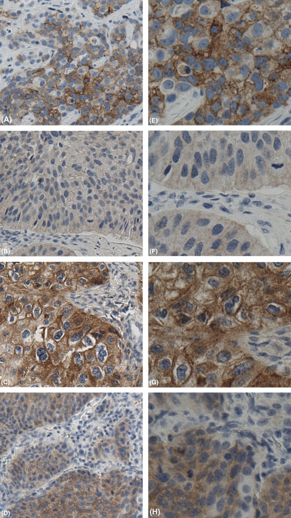
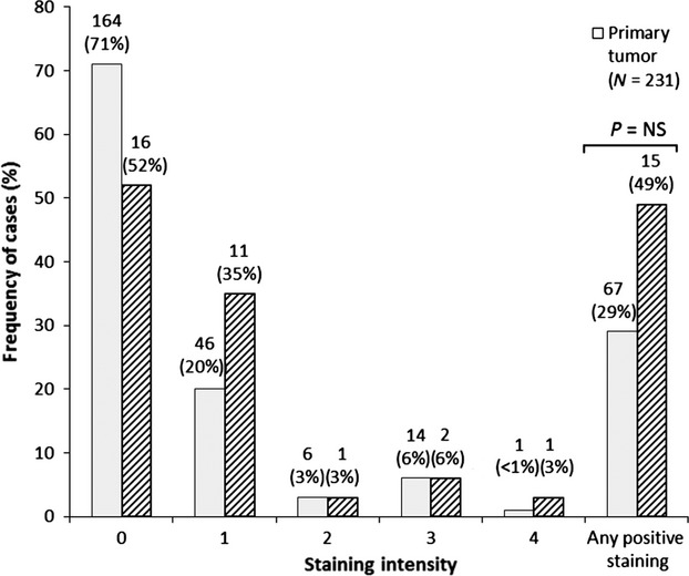
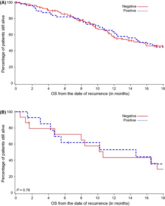
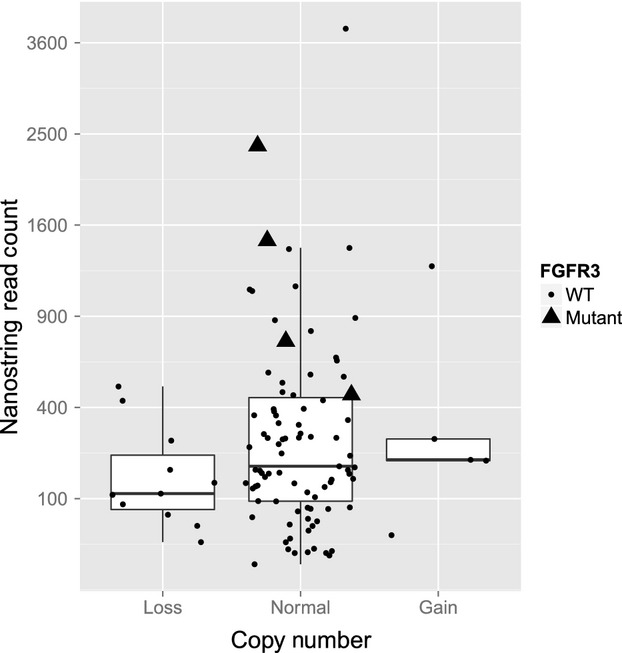
While fibroblast growth factor receptor 3 (FGFR3) is frequently mutated or overexpressed in nonmuscle-invasive urothelial carcinoma (UC), the prevalence of FGFR3 protein expression and mutation remains unknown in muscle-invasive disease. FGFR3 protein and mRNA expression, mutational status, and copy number variation were retrospectively analyzed in 231 patients with formalin-fixed paraffin-embedded primary UCs, 33 metastases, and 14 paired primary and metastatic tumors using the following methods: immunohistochemistry, NanoString nCounterTM, OncoMap or Affymetrix OncoScanTM array, and Gain and Loss of Analysis of DNA and Genomic Identification of Significant Targets in Cancer software. FGFR3 immunohistochemistry staining was present in 29% of primary UCs and 49% of metastases and did not impact overall survival (P = 0.89, primary tumors; P = 0.78, metastases). FGFR3 mutations were observed in 2% of primary tumors and 9% of metastases. Mutant tumors expressed higher levels of FGFR3 mRNA than wild-type tumors (P < 0.001). FGFR3 copy number gain and loss were rare events in primary and metastatic tumors (0.8% each; 3.0% and 12.3%, respectively). FGFR3 immunohistochemistry staining is present in one third of primary muscle-invasive UCs and half of metastases, while FGFR3 mutations and copy number changes are relatively uncommon.

Keywords: Biomarker; FGFR3; bladder cancer; metastatic urothelial carcinoma; muscle-invasive urothelial carcinoma; targeted therapy.


Figures

Figure 1
Figure 1
FGFR3 immunostaining in muscle-invasive UC of the bladder with metastatic phenotype. (A and E) Low- and high-power (20× and 40×) FGFR3 expression in invasive urothelial carcinoma, graded as moderate with both cytoplasmic and membranous staining. (B and F) Low- and high-power (20× and 40×) FGFR3 expression in metastatic urothelial carcinoma, graded as weak with cytoplasmic staining evident. (C and G) Low- and high-power (20× and 40×) FGFR3 expression in metastatic urothelial carcinoma, graded as strong, with prominent cytoplasmic and membranous staining. (D and H) Low- and high-power (20× and 40×) FGFR3 expression in metastatic urothelial carcinoma, graded as moderate, with prominent cytoplasmic staining.
Figure 2
Figure 2
Immunohistochemical staining of FGFR3 in primary and metastatic tumors. Negative staining = 0, weak = 1, weak–moderate = 2, moderate = 3, strong = 4. *P = NS by Fisher's exact test (two tailed) between negative and any positive staining.
Figure 3
Figure 3
OS and FGFR3 staining in primary and metastatic tumors. (A) FGFR3 staining was not associated with a difference in OS in the primary tumor cohort (= 0.89). (B) FGFR3 staining was not associated with differences in OS from time of disease recurrence in the metastatic tumor cohort (= 0.78).
Figure 4
Figure 4
FGFR3 copy number and mRNA expression for FGFR3 mutant and wild-type (WT) tumors. The Nanostring read counts for all patients with loss, gain, or normal FGFR3 copy numbers are visualized with dots for WT tumors and triangles for FGFR3 mutants. In addition, the distributions of read counts in the three categories are visualized with box plots.

References

    1. Tomlinson DC, Lamont FR, Shnyder SD. Knowles MA. Fibroblast growth factor receptor 1 promotes proliferation and survival via activation of the mitogen-activated protein kinase pathway in bladder cancer. Cancer Res. 2009;69:4613–4620. - PMC - PubMed
    1. Barbisan F, Santinelli A, Mazzucchelli R, Lopez-Beltran A, Cheng L, Scarpelli M, et al. Strong immunohistochemical expression of fibroblast growth factor receptor 3, superficial staining pattern of cytokeratin 20, and low proliferative activity define those papillary urothelial neoplasms of low malignant potential that do not recur. Cancer. 2008;112:636–644. - PubMed
    1. Tomlinson DC, Baldo O, Harnden P. Knowles MA. FGFR3 protein expression and its relationship to mutation status and prognostic variables in bladder cancer. J. Pathol. 2007;213:91–98. - PMC - PubMed
    1. Reis PP, Waldron L, Goswami RS, Xu W, Xuan Y, Perez-Ordonez B, et al. mRNA transcript quantification in archival samples using multiplexed, color-coded probes. BMC Biotechnol. 2011;11:46–56. - PMC - PubMed
    1. Kim WJ, Kim EJ, Kim SK, Kim YJ, Ha YS, Jeong P, et al. Predictive value of progression-related gene classifier in primary non-muscle invasive bladder cancer. Mol. Cancer. 2010;9:3. - PMC - PubMed

Publication types

MeSH terms

Substances