. 2014 May 20;9(5):e97851.
doi: 10.1371/journal.pone.0097851. eCollection 2014.

Mice deficient in transmembrane prostatic acid phosphatase display increased GABAergic transmission and neurological alterations

Affiliations

Mice deficient in transmembrane prostatic acid phosphatase display increased GABAergic transmission and neurological alterations

Heidi O Nousiainen et al. PLoS One. .

Abstract

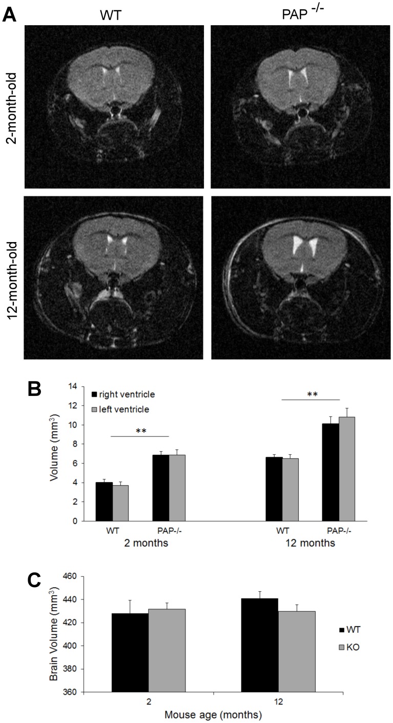
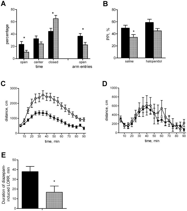
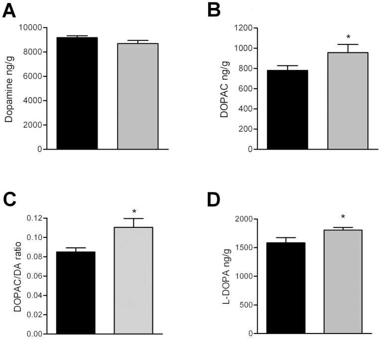
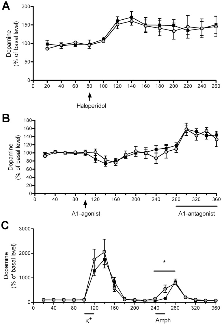
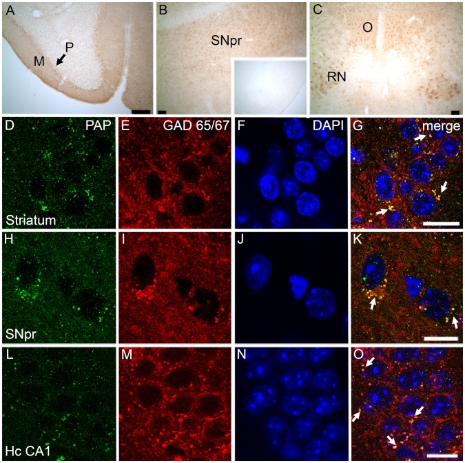
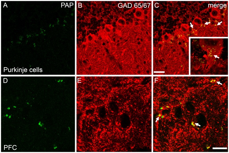
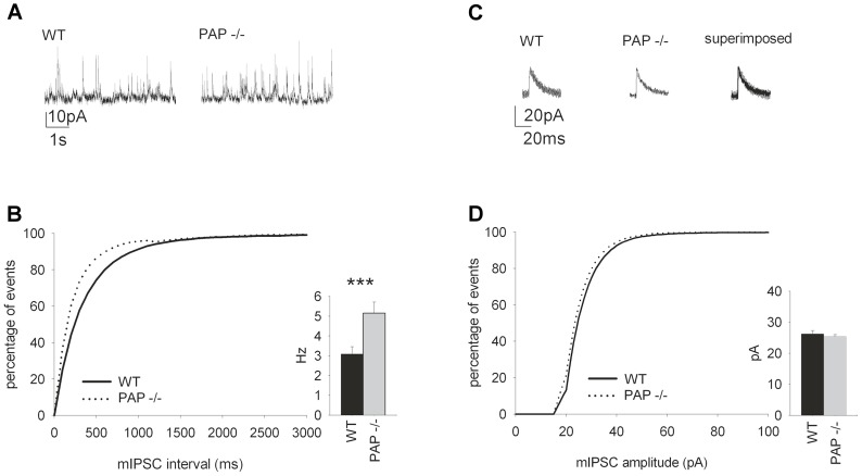
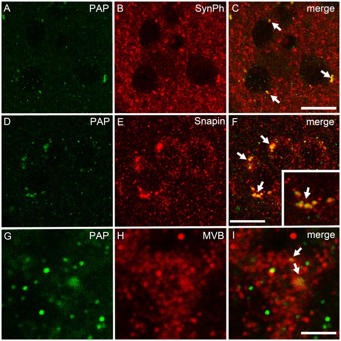
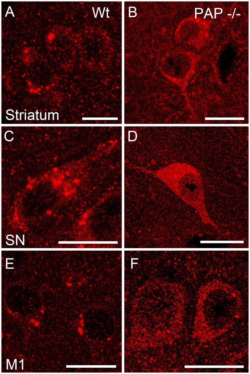
Prostatic acid phosphatase (PAP), the first diagnostic marker and present therapeutic target for prostate cancer, modulates nociception at the dorsal root ganglia (DRG), but its function in the central nervous system has remained unknown. We studied expression and function of TMPAP (the transmembrane isoform of PAP) in the brain by utilizing mice deficient in TMPAP (PAP-/- mice). Here we report that TMPAP is expressed in a subpopulation of cerebral GABAergic neurons, and mice deficient in TMPAP show multiple behavioral and neurochemical features linked to hyperdopaminergic dysregulation and altered GABAergic transmission. In addition to increased anxiety, disturbed prepulse inhibition, increased synthesis of striatal dopamine, and augmented response to amphetamine, PAP-deficient mice have enlarged lateral ventricles, reduced diazepam-induced loss of righting reflex, and increased GABAergic tone in the hippocampus. TMPAP in the mouse brain is localized presynaptically, and colocalized with SNARE-associated protein snapin, a protein involved in synaptic vesicle docking and fusion, and PAP-deficient mice display altered subcellular distribution of snapin. We have previously shown TMPAP to reside in prostatic exosomes and we propose that TMPAP is involved in the control of GABAergic tone in the brain also through exocytosis, and that PAP deficiency produces a distinct neurological phenotype.


Conflict of interest statement

Competing Interests: The authors have declared that no competing interests exist.

Figures

Figure 1
Figure 1. Lateral ventricles volume is enlarged in PAP−/− mice.
Lateral ventricles (right and left ventricles) volume is significantly larger (**p<0.01) in both young and old PAP−/− mice compared to corresponding WT mice. (A) T2-weighted images from young (2 months) and old (12 months) WT and PAP−/− mice. Plot of (B) lateral ventricle volumes and (C) total brain size for WT and PAP−/− mice. The data is expressed as mean±S.E.M.
Figure 2
Figure 2. PAP−/− mice show increased anxiety, disrupted PPI, augmented locomotor response to amphetamine and increased tolerance to diazepam-induced loss of righting reflex.
(A) PAP−/− mice spent less time in open arms of the elevated plus maze, and more time in closed arms than WT mice. The ratio of open arm entries was lower in PAP−/− mice. (B) PAP−/− mice have reduced PPI of the acoustic startle reflex. PPI was enhanced by treatment with haloperidol. (C) Amphetamine-induced locomotor activity was significantly increased in PAP−/− mice compared to WT mice (p = 0.0001), whereas the effect of MK-801 (D) was similar between the genotypes. (E) PAP−/− mice displayed shorter duration of loss of righting reflex after treatment with high dose of diazepam. The data is expressed as mean±S.E.M.; black bars/squares represent WT, grey bars/dots PAP−/− mice; *p<0.05 between the genotypes.
Figure 3
Figure 3. Dopamine synthesis is augmented in the striatum of PAP−/− mice.
(A) Tissue levels of DA are similar in WT and PAP−/− mice. (B) Level of the principal metabolite of DA, DOPAC, is elevated in the PAP−/− mice, and also the DOPAC/DA ratio is elevated (C). (D) Accumulation of L-DOPA is greater in PAP−/− mice than WT mice 30 min after administration of a blocker of L-amino acid decarboxylase, indicating increased DA synthesis. The data is expressed as mean±S.E.M.; black bars represent WT, grey bars PAP−/− mice; *p<0.05, two tailed t-test (monoamines) or repeated measures ANOVA (microdialysis).
Figure 4
Figure 4. Characterization of dopaminergic transmission in PAP−/− mice by microdialysis.
(A) There is no difference in haloperidol (0.2 mg/kg) -induced dopamine release between PAP−/− and WT mice. (B) Adenosine A1-agonist GR79236X (1 mg/kg) and A1-antagonist 8-CPT (300 µM in dialysis fluid) significantly decreased and increased the release of dopamine, respectively (repeated measures ANOVA) but there was no difference in the magnitude of response between the genotypes. (C) There is no significant difference in potassium–induced DA release between the genotypes, but amphetamine induces DA release significantly faster. The data is expressed as mean±S.E.M.; black squares represent WT, grey dots PAP−/− mice.
Figure 5
Figure 5. TMPAP is expressed in the mouse brain and colocalizes with GABAergic marker, GAD 65/67.
Representative confocal images depict intense TMPAP (brown color) expression in molecular cell layer (M) and Purkinje cells (P) of cerebellum (Panel A), in substantia nigra pars reticulata (SNpr; Panel B), in red nucleus (RN; Panel C) and in oculomotor nucleus (O; Panel C). Small picture in Panel B depicts the TMPAP staining of the substantia nigra in PAP−/− mouse. TMPAP (green) was colocalized with GABAergic marker (red) in medium spiny neurons of striatum (Panels D-G, yellow color and white arrows indicating the colocalization) and in SNpr (Panels H–K). Colocalization was evident also in GABAergic neurons of hippocampus CA1 (Panels L–O). DAPI (blue color) was used as a nuclear marker. Scale bars are 500 µm in Panels A–C, and 10 µm in panels D–O.
Figure 6
Figure 6. The colocalization of PAP (green) and GAD65/67 (red) was seen in several areas of brain.
In cerebral Purkinje cells (A–C), strong colocalization was seen especially in the axon hillock of the neuron (small picture in C; yellow color and white arrows depicting the colocalization). Similarly, PAP was present in GABAergic neurons in prefrontal cortex (PFC; infralimbic cortex) (D–F). Scale bars are 10 µm.
Figure 7
Figure 7. The frequency of spontaneous hippocampal mIPSCs is increased in PAP−/− mice.
(A) Sample traces of mIPSC from WT and PAP−/− mice (P14–P18). (B) The mIPSC frequency in the PAP−/− mice was higher (p = 0.003) compared to WT mice. The graph shows the cumulative distribution of the events. (C) Sample traces from 8 averaged mIPSCs from PAP−/− and WT mice. (D) There were no differences in the amplitude in mIPSCs recorded from WT and PAP−/− mice. The graph shows the cumulative distribution of the amplitudes.
Figure 8
Figure 8. Immunofluorescent colocalization stainings of TMPAP and synaptic vesicle associated proteins.
TMPAP (green) is colocalized with a presynaptic marker, synaptophysin (red) (A–C; yellow color and white arrows depicting the colocalization). PAP was seen in vesicle-like structures that had strong colocalization with Snapin (D–F). Small picture is a magnification from panel C, depicting the colocalization. Moreover, largest PAP-immunoreactive structures had a colocalization with multivesicular bodies (MVB, red; G–I). All pictures are from striatum. Scale bars are 10 µm in A–C and G–I, and 3 µm in D–F.
Figure 9
Figure 9. Immunostaining of Snapin in various brain areas of WT and PAP−/− mice.
In the PAP−/− mouse, Snapin (red) is localized more diffusely in the cell soma (B, D, F). Cellular localization of Snapin in WT mouse is more vesicular-like (A, C, E). Scale bar is 10 µm in all figures. M1 - primary motor cortex; SN - substantia nigra.

References

    1. Quintero IB, Araujo CL, Pulkka AE, Wirkkala RS, Herrala AM, et al. (2007) Prostatic acid phosphatase is not a prostate specific target. Cancer Res 67: 6549–6554. - PubMed
    1. Vihko P (1979) Human prostatic acid phosphatases: Purification of a minor enzyme and comparisons of the enzymes. Invest Urol 16: 349–352. - PubMed
    1. Quintero IB, Herrala AM, Araujo CL, Pulkka AE, Hautaniemi S, et al. (2013) Transmembrane prostatic acid phosphatase (TMPAP) interacts with snapin and deficient mice develop prostate adenocarcinoma. PLoS One 8: e73072. - PMC - PubMed
    1. Südhof TC (2013) Neurotransmitter release: The last millisecond in the life of a synaptic vesicle. Neuron 80: 675–690. - PMC - PubMed
    1. Dickman DK, Tong A, Davis GW (2012) Snapin is critical for presynaptic homeostatic plasticity. J Neurosci 32: 8716–8724. - PMC - PubMed

Publication types

MeSH terms