Clinical Trial
. 2014 May 20;9(5):e96703.
doi: 10.1371/journal.pone.0096703. eCollection 2014.

Serum amyloid A induces NLRP-3-mediated IL-1β secretion in neutrophils

Affiliations
Clinical Trial

Serum amyloid A induces NLRP-3-mediated IL-1β secretion in neutrophils

Kiyoshi Migita et al. PLoS One. .

Abstract

Background/aims: Serum amyloid A (SAA) is an acute phase reactant with significant immunological activities, including effects on cytokine synthesis and neutrophil chemotaxis. Neutrophils can also release cytokines with proinflammatory properties. IL-1β is a key proinflammatory cytokine, the secretion of which is controlled by inflammasome. We investigated the proinflammatory effects of SAA in vitro in relation to the NLRP3 inflammasome in neutrophils.

Methodology/principal findings: Human neutrophils isolated form healthy subjects were stimulated with serum amyloid A (SAA). The cellular supernatants were analyzed by western blot using anti-IL-1β or anti-caspase-1 antibodies. IL-1β or Nod-like receptor family, pyrin domain containing 3 (NLRP3) mRNA expressions were analyzed by real-time PCR or reverse transcription-PCR (RT-PCR) method. SAA stimulation induced pro-IL-1β mRNA expression in neutrophils. Furthermore, SAA engaged the caspase-1-activating inflammasome, resulting in the production of active IL-1β. SAA-induced pro-IL-1β expression was marginally suppressed by the Syk specific inhibitor, R406, and SAA-induced pro-IL-1β processing in neutrophils was prevented by R406. Furthermore, SAA-induced NLRP3 mRNA expression was completely blocked by R406. Analysis of intracellular signaling revealed that SAA stimulation activated the tyrosine kinase Syk and mitogen-activated protein kinase (MAPK).

Conclusions/significance: These results demonstrate that the innate neutrophil immune response against SAA involves a two-step activation process: an initial signal promoting expression of pro-IL-1β and a second signal involving Syk-dependent activation of the NLRP3 inflammasome and caspase-1, allowing processing of pro-IL-1β and secretion of mature IL-1β.


Conflict of interest statement

Competing Interests: The authors have declared that no competing interests exist.

Figures

Figure 1
Figure 1. SAA induces the transcription of pro-IL-1β in human neutrophils.
Neutrophils were incubated with SAA (5 µg/ml) for the indicated periods. The cells were harvested and analyzed for IL-1β and GAPDH mRNA levels by real-time PCR. Values represent the mean ± SD of three independent experiments.
Figure 2
Figure 2. SAA induces mature IL-1β synthesis from neutrophils.
A Neutrophils were stimulated with the indicated concentrations of SAA for 8-1β production using ELISA. Values represent the mean ± SD of two independent experiments. *p<0.001 compared to SAA-untreated neutrophils. B Neutrophils were stimulated with the indicated concentrations of SAA for 8 hr. After stimulation, supernatants were analyzed by western blot analysis for the presence of mature IL-1β. Three experiments were performed using different neutrophils and a representative result is shown.
Figure 3
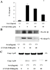
Figure 3. SAA-induced IL-1β processing is dependent on caspase-1.
A Neutrophils were stimulated with SAA (5 µg/ml) in the presence or absence of Z-YVAD-FMK for 8 h. After stimulation, supernatants were analyzed for IL-1β production using ELISA. Values represent the mean ± SD of two independent experiments. *p<0.001 compared to SAA-stimulated neutrophils. B Neutrophils were stimulated with SAA (5 µg/ml) in the presence or absence of Z-YVAD-FMK for 8 h. Supernatants were analyzed by western blot analysis for the presence of mature IL-1β. C Neutrophils were stimulated with SAA (5 µg/ml) in the presence or absence of Z-YVAD-FMK for 8 h. Culture supernatants (SN) and cellular lysates (CL) were analyzed by immunoblot using anti-caspase-1 Ab. Caspase-1 (p20, cleaved subunit; p45, precursor). Two experiments were performed using different neutrophils and a representative result is shown.
Figure 4
Figure 4. Effects of R406 on the transcription of pro-IL-1β in SAA-stimulated neutrophils.
Neutrophils were pretreated or untreated with the indicated concentrations of R406 for 1(5 µg/ml) for 4 hr. The cells were harvested and analyzed for IL-1β and GAPDH mRNA levels by real-time PCR. Values represent the mean ± SD of three independent experiments. *p<0.005 compared to SAA-stimulated neutrophils.
Figure 5
Figure 5. Effects of R406 on SAA-induced IL-1β processing in neutrophils.
A Neutrophils were pretreated or untreated with the indicated concentrations of R406 for 1(5 µg/ml) for 8 hr. After stimulation, supernatants were analyzed by western blot analysis for the presence of mature IL-1β. Three experiments were performed using different neutrophils and a representative result is shown. B Neutrophils were pretreated or untreated with the indicated concentrations of R406 for 1 hr. The cells were stimulated with SAA (5 µg/ml) for 8 hr. After stimulation, culture supernatants (SN) and cellular lysates (CL) were analyzed by immunoblot using anti-caspase-1 Ab. Caspase-1 (p20, cleaved subunit; p45, precursor). Three experiments were performed using different neutrophils and a representative result is shown.
Figure 6
Figure 6. Effects of R406 on the transcription of NLRP3 in SAA-stimulated neutrophils.
A Neutrophils were pretreated or untreated with the indicated concentrations of R406 for 1(5 µg/ml) for the indicated periods. The cells were harvested and analyzed for NLRP3 mRNA by RT-PCR. B Neutrophils were pretreated or untreated with the indicated concentrations of R406 for 1 hr. The cells were stimulated with SAA (5 µg/ml) for 4 hr. The cells were harvested and analyzed for NLRP3 mRNA by RT-PCR. Three experiments were performed using different neutrophils and a representative result is shown.
Figure 7
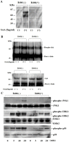
Figure 7. Effects of R406 on the protein phosphorylation in SAA-stimulated neutrophils.
A Quiescent neutrophils pretreated or untreated with the indicated concentrations of R406 for 1(5 µg/ml) for 20 min. Cellular lysates were subjected to western blotting using phosphotyrosine-specific antibody, 4G10. Three experiments were performed using different neutrophils and a representative result is shown. B Quiescent neutrophils pretreated or untreated with the indicated concentrations of R406 for 1 hr. The cells were stimulated with SAA (5 µg/ml) for 20 min. Syk was immunoprecipitated from each lysates and the immunoprecipitates were subjected to western blotting using phosphotyrosine-specific antibody, 4G10 or anti-Syk antibody. Each lane shows Syk precipitated from 107 cells. Three experiments were performed using different neutrophils and a representative result is shown. C Quiescent neutrophils pretreated or untreated with the indicated concentrations of R406 for 1 hr. The cells were stimulated with SAA (5 µg/ml). Cellular lysates were subjected to western blotting using phospho-specific or pan antibodies against ERK1/2, p38 and JNK1. Three experiments were performed using different neutrophils and a representative result is shown.
Figure 8
Figure 8. Effects of R406 on NF-κB p65 phosphorylation in SAA-stimulated neutrophils.
Neutrophils pretreated with or without R406 were stimulated with SAA (5 µg/ml) for 20 min. Cellular lysates were analyzed by western blotting using anti-phospho-specific p65 (A) or anti-p65 (B) antibodies. Two experiments were performed using different neutrophils and a representative result is shown.

References

    1. Uhlar CM, Whitehead AS (1999) Serum amyloid A, the major vertebrate acute-phase reactant. Eur J Biochem 265: 501–23. - PubMed
    1. Cunnane G, Whitehead AS (1999) Amyloid precursors and amyloidosis in rheumatoid arthritis. Baillieres Best Pract Res Clin Rheumatol 13: 615–28. - PubMed
    1. Eklund KK, Niemi K, Kovanen PT (2012) Immune functions of serum amyloid A. Crit Rev Immunol. 32: 335–48. - PubMed
    1. Niemi K, Teirilä L, Lappalainen J, Rajamäki K, Baumann MH, et al. (2011) Serum amyloid A activates the NLRP3 inflammasome via P2×7 receptor and a cathepsin B-sensitive pathway. J Immunol 186: 6119–28. - PubMed
    1. Ather JL, Ckless K, Martin R, Foley KL, Suratt BT, et al. (2011) Serum amyloid A activates the NLRP3 inflammasome and promotes Th17 allergic asthma in mice. J Immunol 187: 64–73. - PMC - PubMed

Publication types

MeSH terms

Substances

LinkOut - more resources