. 2014 Sep;44(9):2659-66.
doi: 10.1002/eji.201344134. Epub 2014 Jun 17.

NK cell-extrinsic IL-18 signaling is required for efficient NK-cell activation by vaccinia virus

Affiliations

NK cell-extrinsic IL-18 signaling is required for efficient NK-cell activation by vaccinia virus

Joshua D Brandstadter et al. Eur J Immunol. 2014 Sep.

Abstract

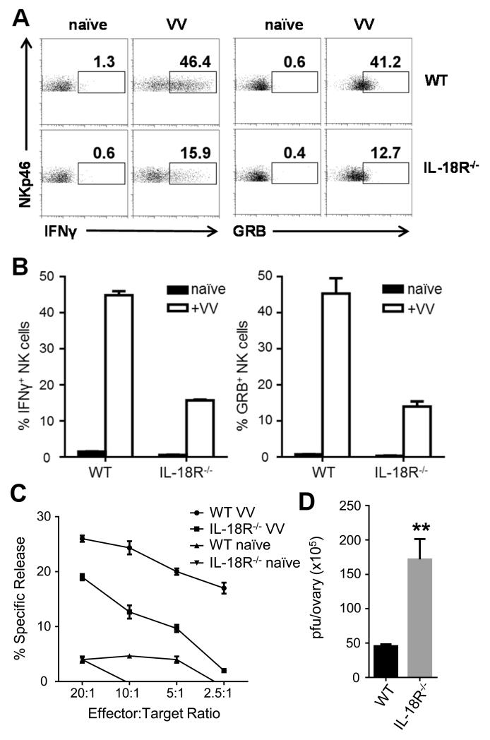
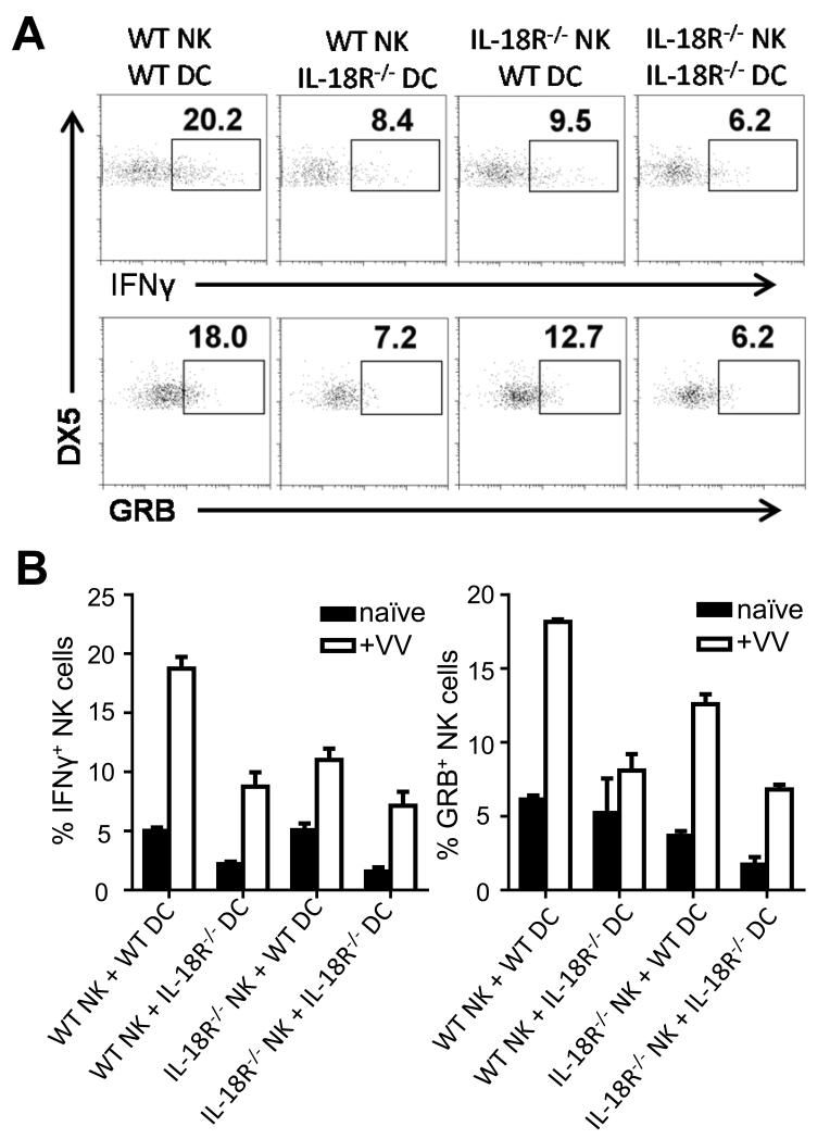
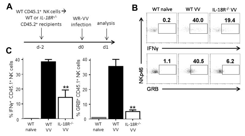
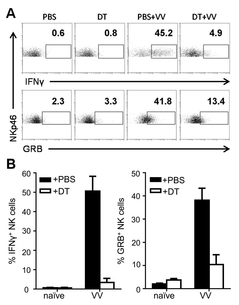
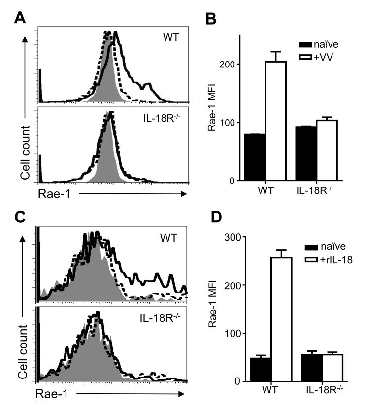
NK cells are important for the control of vaccinia virus (VV) in vivo. Recent studies have shown that multiple pathways are required for effective activation of NK cells. These include both TLR-dependent and -independent pathways, as well as the NKG2D activating receptor that recognizes host stress-induced NKG2D ligands. However, it remains largely unknown what controls the upregulation of NKG2D ligands in response to VV infection. In this study using C57BL/6 mice, we first showed that IL-18 is critical for NK-cell activation and viral clearance. We then demonstrated that IL-18 signaling on both NK cells and DCs is required for efficient NK-cell activation upon VV infection in vitro. We further showed in vivo that efficient NK-cell activation in response to VV is dependent on DCs and IL-18 signaling in non-NK cells, suggesting an essential role for NK cell-extrinsic IL-18 signaling in NK-cell activation. Mechanistically, IL-18 signaling in DCs promotes expression of Rae-1, an NKG2D ligand. Collectively, our data reveal a previously unrecognized role for NK cell-extrinsic IL-18 signaling in NK-cell activation through upregulation of NKG2D ligands. These observations may provide insights into the design of effective NK-cell-based therapies for viral infections and cancer.

Keywords: IL-18; NK cells; NKG2D; Rae-1; Vaccinia virus.


Figures

FIGURE 1
FIGURE 1
IL-18 is required for efficient NK cell activation and VV clearance. WT and IL-18R−/− C57BL/6 mice were infected by i.p. injection of 5 × 10⁶ pfu VV or left uninfected (naïve). (A) 24 h after infection, splenocytes were assayed for IFN-γ and GRB production. Representative FACS plots showing the percentage of IFN-γ and GRB-positive NKp46+CD3-NK cells are shown. (B) The mean percentages ±s.e.m. of IFN-γ and GRB-positive NK cells (n=3 mice per group) are shown. Interaction term for two-way ANOVA is p<0.01 for IFN-γ and GRB. Data is representative of three independent experiments. (C) 48 h after infection, splenocytes were enriched for DX5+ cells and NK cell lytic activity was assayed on YAC-1 target cells by a standard 4-hour chromium release assay at different effector:target ratios. The mean percentages ±s.e.m. of specific lysis are indicated (n=3 per group). ANCOVA comparing infected WT and IL-18R−/− mice shows p<0.05. Data is representative of two independent experiments. (D) 48 h after infection, ovaries of female mice were harvested for measurement of viral load by plaque assay using TK-143B cells. Data represents the mean viral titer ±s.e.m. as pfu per ovary (n=4 per group). Data is representative of two independent experiments. ** signifies a p-value <0.01 on an unpaired student’s t test.
FIGURE 2
FIGURE 2
IL-18 signaling on both NK cells and DCs is required for NK cell activation to VV in vitro. WT or IL-18R−/− NK cells were co-cultured with WT or IL-18R−/− bone-marrow derived CD11c+ DCs and stimulated with VV or left uninfected (naïve) for 18 h. (A) The percentage of IFN-γ and GRB-positive DX5+CD3 NK cells are shown on representative FACS plots. (B) The mean percentages ±s.e.m. of IFN-γ and GRB-positive DX5+CD3 NK cells are shown (n=3 wells per condition). Data is representative of three independent experiments. Interaction term for two-way ANOVA is p<0.01 for both IFN-γ and GRB.
FIGURE 3
FIGURE 3
NK cell-extrinsic IL-18 signaling is critical for NK cell activation in vivo. (A) Five to 10 × 105 DX5+CD3 NK cells from the spleens of CD45.1+ WT C57BL/6 mice were purified by cell sorting and injected intravenously into WT or IL-18R−/− recipients, which are both CD45.2+. Two days after cell transfer, recipient mice were infected with 5 × 10⁶pfu VV i.p. or left uninfected (naïve). 24 h after infection, spleens were assayed for IFN-γ and GRB production by donor (CD45.1+) NK cells. (B) The percentages of IFN-γ and GRB-positive NKp46+CD3 NK cells from the WT CD45.1+ donor NK cells are shown on representative FACS plots with recipient genotype and stimulation indicated above. (C) The mean percentages ±s.e.m. of IFN-γ and GRB-positive WT donor NKp46+CD3 NK cells are shown (n=2 infected mice). Interaction term for one-way ANOVA is p<0.01 for IFN-γ and GRB production. Data is representative of four independent experiments. ** signifies a p-value <0.05 on post-hoc unpaired student’s t test comparing WT donor NK cell activation between infected WT and IL-18R−/− recipients.
FIGURE 4
FIGURE 4
Dendritic cells are required for NK cell activation to VV in vivo. CD11c-DTR+ transgenic mice were treated with diphtheria toxin (DT) and the next day were infected i.p. with 5 × 10⁶pfu VV or left uninfected (naïve). Control mice were injected with PBS. 24 h after infection, splenocytes were assayed for IFN-γ and GRB production. In the naïve mice, CD11c+ DCs, but not NK cells or total splenocytes, were depleted in mice carrying the transgene (data not shown). (A) Representative FACS plots with the percentages of IFN-γ and GRB-positive NKp46+CD3 NK cells are shown. (B) The mean percentages ± s.e.m. of IFN-γ and GRB-positive NKp46+CD3 NK cells (n=2 mice per group). Interaction term for two-way ANOVA is p<0.01 for IFN-γ and GRB. Data is representative of two independent experiments.
FIGURE 5
FIGURE 5
IL-18 signaling on DCs is required for Rae-1 upregulation to VV in vivo. WT or IL-18R−/− mice were infected with 5 × 10⁶pfu VV i.p. or left uninfected (naïve). 24 h after infection, splenocytes were assayed for Rae-1 surface expression on CD11c+CD11b+B220 cDCs. (A) Histograms compare Rae-1 expression on cDCs from infected (thick line) and naïve (dotted line) WT (top panel) and IL-18R−/− (bottom panel) mice. Shaded histogram indicates isotype control. (B) The mean fluorescent intensity ±s.e.m. of Rae-1 on cDCs is shown (n=3 infected mice). Data is representative of three independent experiments. Interaction term for two-way ANOVA is p<0.01. (C) Histograms compare Rae-1 expression on WT (top panel) and IL-18R−/− (bottom panel) bone marrow-derived DCs cultured with 5 ng/mL rIL-18 (thick line) or left untreated (dotted line) for four days. Shaded histogram indicates isotype control (D) The mean fluorescent intensity ±s.e.m. of Rae-1 on bone-marrow derived DCs is shown (n=3 wells). Data is representative of three independent experiments. Interaction term for two-way ANOVA is p<0.0001.

Similar articles

Cited by

References

    1. Lee SH, Miyagi T, Biron CA. Keeping NK cells in highly regulated antiviral warfare. Trends Immunol. 2007;28:252–259. - PubMed
    1. Lanier LL. Up on the tightrope: natural killer cell activation and inhibition. Nat Immunol. 2008;9:495–502. - PMC - PubMed
    1. Bukowski JF, Woda BA, Habu S, Okumura K, Welsh RM. Natural killer cell depletion enhances virus synthesis and virus-induced hepatitis in vivo. J Immunol. 1983;131:1531–1538. - PubMed
    1. Natuk RJ, Welsh RM. Accumulation and chemotaxis of natural killer/large granular lymphocytes at sites of virus replication. J Immunol. 1987;138:877–883. - PubMed
    1. Martinez J, Huang X, Yang Y. Direct action of type I IFN on NK cells is required for their activation in response to vaccinia viral infection in vivo. J Immunol. 2008;180:1592–1597. - PubMed

Publication types

MeSH terms

Substances