

Fatty foci within the heart diagnosed with ECG-gated multi-slice computed tomography: frequency and morphology
- PMID: 24846568
- PMCID: PMC4043540
- DOI: 10.12659/MSM.890271
Fatty foci within the heart diagnosed with ECG-gated multi-slice computed tomography: frequency and morphology
Abstract
Background: The purpose of our study was to analyze the frequency of focal fatty replacement (FR) of the heart, as well as the distribution and detailed morphology of FR in a large group of patients referred to multi-slice computed tomography with ECG-gating examinations (ECG-MSCT) for various clinical reasons.
Material and methods: The ECG-MSCT examinations of 1830 consecutive patients were analyzed. The examinations were performed using 8-row (1015 patients) and 64-row (815 patients) MSCT, in pre- and post-contrast scanning. We analyzed the morphology of FR, the dimensions and densities of changes, as well as the morphology and localization of FR with regard to clinical diagnosis.
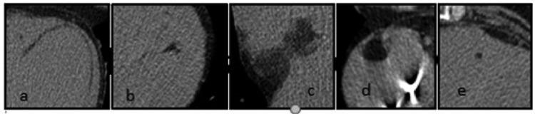
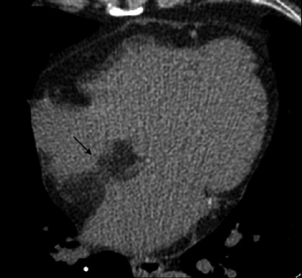
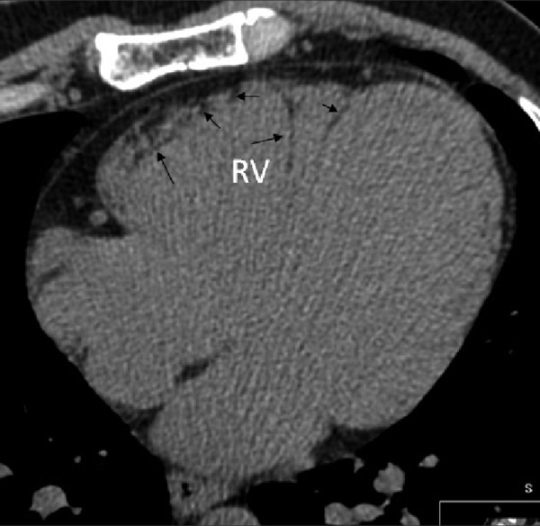
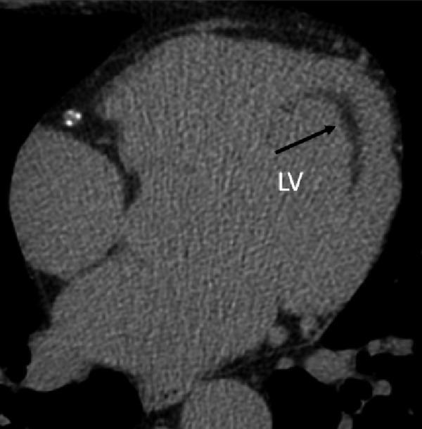
Results: 204 subjects (11.1%) had FR within the heart (113 men; 91 women; mean age 57.8 years); 66% of fatty foci were seen only in the native scanning. The distribution of the fat was: right ventricle (RV) 31.9%, left ventricle (LV) 21.5%, biventricular 39.7%, interventricular or atrial septum 5.9%, and atria 1%. In the RV, fat was localized mainly in the papillary muscles, while in the LV fat was mainly subendocardial (p<0.001). The morphology of the fat was: linear 61.6%, oval 14.8%, punctuate 10.6%, irregular 10.2%, and bilobular 2.8%. Fat was primarily located subendocardially in the LV in patients after myocardial infarction. In patients with suspected coronary artery disease, it was mainly observed subpericardially in the RV and in papillary muscles (p<0.001).
Conclusions: The incidental frequency of FR within the heart in patients diagnosed with the ECG-MSCT examinations is about 11%. Pre-contrast scanning is the most valuable for FR assessment.
Figures
References
-
- Kiès P, Bootsma M, Bax J, et al. Arrhythmogenic right ventricular dysplasia/cardiomyopathy: screening, diagnosis, and treatment. Heart Rhythm. 2006;3:225–34. - PubMed
-
- McKenna WJ, Thiene G, Nava A, et al. Diagnosis of arrhythmogenic riht ventricular dysplasia/cardiomyopathy. Task Force of the Working Group Myocardial and Pericardial Disease of the European Society of Cardiology and of the Scientific Council an Cardiomyopathies of the International Society and Federation of Cardiology. Br Heart J. 1994;71:215–18. - PMC - PubMed
-
- Baroldi G, Silver MD, De Maria R, et al. Lipomatous metaplasia in left ventricular scar. Can J Cardiol. 1997;13:65–71. - PubMed
-
- Beltrami AP, Urbanek K, Kajstura J, et al. Evidence that human cardiac myocytes divide after myocardial infarction. N Engl J Med. 2001;344:1750–57. - PubMed
-
- Basso C, Barbazza R, Thiene G. Lipomatous hypertrophy of the atrial septum. Circulation. 1998;97:1423. - PubMed
MeSH terms
Substances
LinkOut - more resources
Full Text Sources
Other Literature Sources
