. 2014 Sep;171(18):4273-88.
doi: 10.1111/bph.12785.

Buprenorphine signalling is compromised at the N40D polymorphism of the human μ opioid receptor in vitro

Affiliations

Buprenorphine signalling is compromised at the N40D polymorphism of the human μ opioid receptor in vitro

Alisa Knapman et al. Br J Pharmacol. 2014 Sep.

Abstract

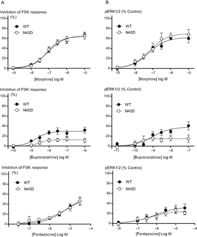
Background and purpose: There is significant variation in individual response to opioid drugs, which may result in inappropriate opioid therapy. Polymorphisms of the μ opioid receptor (MOP receptor) may contribute to individual variation in opioid response by affecting receptor function, and the effect may be ligand-specific. We sought to determine functional differences in MOP receptor signalling at several signalling pathways using a range of structurally distinct opioid ligands in cells expressing wild-type MOP receptors (MOPr-WT) and the commonly occurring MOP receptor variant, N40D.

Experimental approach: MOPr-WT and MOPr-N40D were stably expressed in CHO cells and in AtT-20 cells. Assays of AC inhibition and ERK1/2 phosphorylation were performed on CHO cells, and assays of K activation were performed on AtT-20 cells. Signalling profiles for each ligand were compared between variants.

Key results: Buprenorphine efficacy was reduced by over 50% at MOPr-N40D for AC inhibition and ERK1/2 phosphorylation. Buprenorphine potency was reduced threefold at MOPr-N40D for K channel activation. Pentazocine efficacy was reduced by 50% for G-protein-gated inwardly rectifying K channel activation at MOPr-N40D. No other differences were observed for any other ligands tested.

Conclusions and implications: The N40D variant is present in 10-50% of the population. Buprenorphine is a commonly prescribed opioid analgesic, and many individuals do not respond to buprenorphine therapy. This study demonstrates that buprenorphine signalling to several effectors via the N40D variant of MOP receptors is impaired, and this may have important consequences in a clinical setting for individuals carrying the N40D allele.


Figures

Figure 1
Figure 1
Saturation binding curve of [3H]-DAMGO in intact CHO-MOPr-WT and CHO-MOPr-N40D cells, 24 h after induction of receptor expression with tetracycline. Radioligand binding was carried out as described in the Methods. No significant difference in Bmax or KD was observed between cells expressing MOPr-WT or MOPr-N40D (P > 0.05). Each point represents the mean ± SEM of triplicate determinations from a single experiment. The assay was repeated three times.
Figure 2
Figure 2
DAMGO inhibits AC and activates ERK1/2 in CHO cells expressing MOPr-WT or MOPr-N40D. AC inhibition and levels of ERK1/2 phosphorylation were determined as described in the Methods. (A) Traces showing changes in fluorescent signal following application of 300 nM FSK + vehicle (HBSS) to MOPr-WT, and 300 nM FSK + 1 μM DAMGO to MOPr-WT and MOPr-N40D cells. Drugs were added at 120 s. Changes in raw fluorescent units (RFU) are normalized to predrug values. (B) DAMGO inhibited FSK-stimulated AC hyperpolarization of MOPr-WT or MOPr-N40D to a similar degree and with a similar potency (P > 0.05). (C) DAMGO stimulated ERK1/2 phosphorylation in cells expressing MOPr-WT or MOPr-N40D to a similar degree and with a similar potency (P > 0.05). Maximum ERK1/2 phosphorylation via 100 nM PMA was used as a control for pERK1/2 experiments. Data represent the mean ± SEM of pooled data from five to six independent determinations performed in duplicate.
Figure 3
Figure 3
Endogenous opioids inhibit AC and activate ERK1/2 in CHO cells expressing MOPr-WT or MOPr-N40D. AC inhibition and levels of ERK phosphorylation were determined as described in the Methods. (A) β-endorphin, endomorphins-1 and -2, and met-enkephalin inhibited FSK-stimulated AC hyperpolarization of MOPr-WT or MOPr-N40D to a similar degree and with a similar potency (P > 0.05). (B) β-endorphin, endomorphi-1 and -2, and met-enkephalin stimulated ERK1/2 phosphorylation in cells expressing MOPr-WT or MOPr-N40D to a similar degree and with a similar potency (P > 0.05). Maximum ERK1/2 phosphorylation via 100 nM PMA was used as a control for pERK1/2 experiments. Data represent the mean ± SEM of pooled data from five to six independent determinations performed in duplicate.
Figure 4
Figure 4
Buprenorphine inhibits AC and activates ERK1/2 less effectively in CHO cells expressing MOPr-N40D. AC inhibition and levels of ERK phosphorylation were determined as described in the Methods. (A) Buprenorphine Emax for inhibition of FSK-stimulated AC hyperpolarization was decreased from 35 ± 6 % in CHO-MOPr-WT to 16 ± 4% in CHO-MOPr-N40D (P < 0.05). Morphine and pentazocine inhibited AC hyperpolarization to a similar degree and with similar potency in CHO cells expressing MOPr-WT and MOPr-N40D (P > 0.05). (B) Buprenorphine Emax for stimulation of ERK1/2 phosphorylation was decreased from 35 ± 7 % in CHO-MOPr-WT to 14 ± 6% in CHO-MOPr-N40D (P < 0.05). Morphine and pentazocine stimulated ERK1/2 phosphorylation to a similar degree and with similar potency in CHO cells expressing MOPr-WT and MOPr-N40D (P > 0.05). Maximum ERK1/2 phosphorylation via 100 nM PMA was used as a control for pERK1/2 experiments. Data represent the mean ± SEM of pooled data from five to six independent determinations performed in duplicate.
Figure 5
Figure 5
Fentanyl, oxycodone and methadone inhibit AC and activate ERK1/2 in CHO cells expressing MOPr-WT or MOPr-N40D. AC inhibition and levels of ERK phosphorylation were determined as described in the Methods. (A) Fentanyl, oxycodone and methadone inhibited FSK-stimulated AC hyperpolarization of MOPr-WT or MOPr-N40D to a similar degree and with a similar potency (P > 0.05). (B) Fentanyl, oxycodone and methadone stimulated ERK1/2 phosphorylation in cells expressing MOPr-WT or MOPr-N40D to a similar degree and with a similar potency (P > 0.05). Maximum ERK1/2 phosphorylation via 100 nM PMA was used as a control for pERK1/2 experiments. Data represent the mean ± SEM of pooled data from five to six independent determinations performed in duplicate.
Figure 6
Figure 6
DAMGO causes membrane hyperpolarization in AtT-20 cells expressing MOPr-WT. Raw trace showing decrease in fluorescent signal following application of vehicle (HBSS), 3 nM DAMGO, 300 nM DAMGO or 3 μM buprenorphine to AtT20 cells expressing MOPr-WT, corresponding to membrane hyperpolarization from GIRK activation. The Y-axis is raw fluorescent units (RFU). Drugs were added for the duration of the bar. The traces are representative of at least six individual experiments, each performed in duplicate.
Figure 7
Figure 7
Buprenorphine is less potent at activating GIRK in AtT20 cells expressing MOPr-N40D than in those expressing MOPr-WT. GIRK activation was determined as described in the Methods. (A) Buprenorphine activated GIRK channels to a similar degree in AtT20 cells expressing MOPr-WT and MOPr-N40D, but with a threefold lower pEC50, from 7.0 ± 0.1 in AtT20-MOPr-WT to 6.7 ± 0.1 in AtT20-MOPr-N40D (P < 0.05). (B) Pentazocine activated GIRK channels with 50% lower efficacy at MOPr-N40D, Emax was decreased from 8 ± 1% in AtT20-MOPr-WT to 4 ± 1% in AtT20-MOPr-N40D (P < 0.05). (C) DAMGO, morphine, β-endorphin and methadone activated GIRK channels to a similar degree and with similar potency in AtT-20 cells expressing MOPr-WT and MOPr-N40D (P > 0.05). Data represent the mean ± SEM of pooled data from five to six independent determinations performed in duplicate.

References

    1. Abrol R, Kim SK, Bray JK, Trzaskowski B, Goddard WA., 3rd Conformational ensemble view of G protein-coupled receptors and the effect of mutations and ligand binding. Methods Enzymol. 2013;520:31–48. - PubMed
    1. Alexander SP, Benson HE, Faccenda E, Pawson AJ, Sharman JL, Spedding M, et al. The concise guide to PHARMACOLOGY 2013/14: G protein-coupled receptors. Br J Pharmacol. 2013;170:1459–1581. - PMC - PubMed
    1. Allouche S, Polastron J, Hasbi A, Homburger V, Jauzac P. Differential G-protein activation by alkaloid and peptide opioid agonists in the human neuroblastoma cell line SK-N-BE. Biochem J. 1999;342:71–78. - PMC - PubMed
    1. Atwood BK, Lopez J, Wager-Miller J, Mackie K, Straiker A. Expression G protein-coupled receptors and related proteins in HEK293, AtT20, BV2, and N18 cell lines as revealed by microarray analysis. BMC Genomics. 2011;12:14. - PMC - PubMed
    1. Befort K, Filliol D, Decaillot FM, Gaveriaux-Ruff C, Hoehe MR, Kieffer BL. A single nucleotide polymorphic mutation in the human mu-opioid receptor severely impairs receptor signaling. J Biol Chem. 2001;276:3130–3137. - PubMed

Publication types

MeSH terms

Substances