Review
. 2014 Jun;78(2):199-230.
doi: 10.1128/MMBR.00055-13.

The bicomponent pore-forming leucocidins of Staphylococcus aureus

Affiliations
Review

The bicomponent pore-forming leucocidins of Staphylococcus aureus

Francis Alonzo 3rd et al. Microbiol Mol Biol Rev. 2014 Jun.

Abstract

The ability to produce water-soluble proteins with the capacity to oligomerize and form pores within cellular lipid bilayers is a trait conserved among nearly all forms of life, including humans, single-celled eukaryotes, and numerous bacterial species. In bacteria, some of the most notable pore-forming molecules are protein toxins that interact with mammalian cell membranes to promote lysis, deliver effectors, and modulate cellular homeostasis. Of the bacterial species capable of producing pore-forming toxic molecules, the Gram-positive pathogen Staphylococcus aureus is one of the most notorious. S. aureus can produce seven different pore-forming protein toxins, all of which are believed to play a unique role in promoting the ability of the organism to cause disease in humans and other mammals. The most diverse of these pore-forming toxins, in terms of both functional activity and global representation within S. aureus clinical isolates, are the bicomponent leucocidins. From the first description of their activity on host immune cells over 100 years ago to the detailed investigations of their biochemical function today, the leucocidins remain at the forefront of S. aureus pathogenesis research initiatives. Study of their mode of action is of immediate interest in the realm of therapeutic agent design as well as for studies of bacterial pathogenesis. This review provides an updated perspective on our understanding of the S. aureus leucocidins and their function, specificity, and potential as therapeutic targets.


Figures

FIG 1
FIG 1
Current model of leucocidin pore formation. Leucocidin pore formation is believed to occur in a stepwise fashion that begins with toxin recognition of cellular receptors on the surface of target host cells. On most host cells, the “S” subunit recognizes a proteinaceous receptor (either a chemokine receptor [LukED and PVL] or an integrin [LukAB/HG]) to facilitate high-affinity binding to the cell surface (1). The S subunit then recognizes and recruits the “F” subunit (2), leading to dimerization on the host cell surface (3). Dimerization is followed by oligomer formation (4). Toxin oligomers assemble into an octameric prepore structure containing alternating S and F subunits. Following oligomerization, a major structural change occurs in the stem domains of the S and F subunits, leading to membrane insertion and the formation of a β-barrel pore that spans the host cell lipid bilayer (5).
FIG 2
FIG 2
Morphological changes associated with leucocidin-mediated killing of immune cells. (A) Light and fluorescence microscopy images of murine phagocytic leukocytes (macrophages and neutrophils) and light microscopy (LM) images of the human T cell line HUT-R5 (cell line overexpressing CCR5) after exposure to a 90% lethal dose of LukED (5 μg/ml). Characteristic membrane halos and expansion of cellular nuclei are seen, along with increased permeability to ethidium bromide (EtBr) (red), an indicator of pore formation and membrane damage. Arrows point to characteristic cellular morphology changes upon leucocidin intoxication. (B) Electron microscopy images of the human PMN-like cell line PMN-HL60 after exposure to S. aureus supernatant containing a 100% lethal dose of LukAB/HG (>2.5 μg/ml). All intoxications and microscopic image acquisition were conducted as previously described (47, 97, 227).
FIG 3
FIG 3
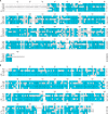
Amino acid sequence alignment of mature leucocidins without their signal peptide. Amino acid sequence comparisons were generated by ClustalW alignment using Lasergene MegAlign Pro software (DNASTAR). Identical amino acids are shown in blue, and divergent residues are shown in white. (A) Alignment of the leucocidin S subunits HlgA, HlgC, LukA, LukE, LukM, and LukS-PV. Notable distinctions are the unique N- and C-terminal extensions that are present in LukA/H but absent from all other toxins. (B) Alignment of the leucocidin F subunits HlgB, LukB/G, LukD, LukF′-PV, and LukF-PV.
FIG 4
FIG 4
Genome organization of the S. aureus leucocidins. Shown is a schematic representation of the S. aureus leucocidin genetic loci within the genome of the sequenced USA300 strain FPR3757 (GenBank accession number NC_007793.1) (blue arrows, hlgACB; green arrows, lukED; red arrows, lukAB [lukHG]; purple arrows, lukSF-PV) or the sequenced genome of bovine isolate ED133 (GenBank accession number NC_017337.1) (yellow arrows, lukMF′). Numbers to the right and left indicate the nucleotide base positions of the indicated region designated in the genome repository of the National Center for Biotechnology Information (NCBI). Vertical lines with arrowheads demarcate the location of prophage insertions (ϕSa3, ϕSa2/ϕSLT, and ϕSa1/ϕPV83) within the S. aureus genome. Flanking genes upstream and downstream of the respective leucocidins are supplied with either their designated nomenclature or their gene number. The S. aureus pathogenicity island vSaβ (the site where lukED is located) is indicated with branching arrows above the lukED-containing region.
FIG 5
FIG 5
Leucocidin structural features. (A) Crystal structure of the monomeric F subunit of gamma-hemolysin (HlgB) (178). Structural information was acquired from the Protein Data Bank (PDB) (accession number 1LKF), and the major structural domains were colored by using PyMOL software. Blue, amino latch; green, β-sandwich; orange, stem domain; red, rim domain. (B) Crystal structure of the HlgAB octamer (190). The S subunit (HlgA) is in cyan, while the F subunit (HlgB) is in red. The stem, rim, and cap as well as the β-barrel pore are shown. Structural information for the HlgAB octamer was acquired from the PDB (accession number 3B07), and the major structural domains were colored by using PyMOL software.
FIG 6
FIG 6
Sublytic effects of S. aureus leucocidins. Sublytic activities of leucocidins have been investigated primarily for PVL and gamma-hemolysin. Some sublytic functions are shown. (1) Priming of PMNs through the engagement of cellular receptors and other mechanisms yet to be defined that lead to increased reactive oxygen species formation, enhanced granule exocytosis, robust phagocytosis, and increased bactericidal activity of host neutrophils. (2) Induction of the NLRP3 inflammasome and subsequent IL-1β release mediated by potassium efflux from the cytosol due to pore formation. (3) Stimulation of immune cell chemotaxis and NF-κB activation as a result of calcium influx. The subsequent activation of cellular kinases leads to IκB phosphorylation and targeted degradation, followed by translocation of NF-κB to the nucleus and induction of proinflammatory gene expression. (4) Engagement of Toll-like receptors (TLR2 and TLR4) to stimulate the same canonical NF-κB activation pathway described above (3). (5) Activation of apoptosis via mitochondrial disruption potentially caused by pore formation.
FIG 7
FIG 7
Leucocidin gene regulation. The molecular details of leucocidin gene regulation have not been extensively defined, but a number of master regulators and external signals provide major inputs into their altered gene expression in various environments. Some major regulatory inputs are shown. (1) The Agr quorum-sensing system is activated upon reaching a high bacterial density, leading to AgrA-dependent activation of the P3 promoter, which encodes the regulatory RNA, RNAIII. RNAIII negatively regulates the translation of the leucocidin repressor Rot, leading to increased leucocidin production. (2) The global regulator SarA indirectly facilitates leucocidin expression by positively regulating the expression of the P3 promoter, leading to a similar repression of Rot translation and increased leucocidin production. (3) Rot is believed to bind directly to leucocidin promoters to inhibit toxin gene expression. (4) The SaeRS two-component system recognizes external stimuli from the environment, leading to direct binding of SaeR to leucocidin promoters and subsequent enhancement of gene expression. (5) Other environmental stimuli positively and negatively regulate leucocidin gene expression, although the precise stimuli and their mechanism(s) of activation/repression are yet to be defined.
None
None

References

    1. David MZ, Daum RS. 2010. Community-associated methicillin-resistant Staphylococcus aureus: epidemiology and clinical consequences of an emerging epidemic. Clin. Microbiol. Rev. 23:616–687. 10.1128/CMR.00081-09 - DOI - PMC - PubMed
    1. Klevens RM, Morrison MA, Nadle J, Petit S, Gershman K, Ray S, Harrison LH, Lynfield R, Dumyati G, Townes JM, Craig AS, Zell ER, Fosheim GE, McDougal LK, Carey RB, Fridkin SK, Active Bacterial Core Surveillance MRSA Investigators 2007. Invasive methicillin-resistant Staphylococcus aureus infections in the United States. JAMA 298:1763–1771. 10.1001/jama.298.15.1763 - DOI - PubMed
    1. Klevens RM, Edwards JR, Tenover FC, McDonald LC, Horan T, Gaynes R, National Nosocomial Infections Surveillance System 2006. Changes in the epidemiology of methicillin-resistant Staphylococcus aureus in intensive care units in US hospitals, 1992-2003. Clin. Infect. Dis. 42:389–391. 10.1086/499367 - DOI - PubMed
    1. Liu C, Bayer A, Cosgrove SE, Daum RS, Fridkin SK, Gorwitz RJ, Kaplan SL, Karchmer AW, Levine DP, Murray BE, Ryback MJ, Talan DA, Chambers HF. 2011. Clinical practice guidelines by the Infectious Diseases Society of America for the treatment of methicillin-resistant Staphylococcus aureus infections in adults and children: executive summary. Clin. Infect. Dis. 52:285–292. 10.1093/cid/cir034 - DOI - PubMed
    1. Stefani S, Chung DR, Lindsay JA, Friedrich AW, Kearns AM, Westh H, Mackenzie FM. 2012. Methicillin-resistant Staphylococcus aureus (MRSA): global epidemiology and harmonisation of typing methods. Int. J. Antimicrob. Agents 39:273–282. 10.1016/j.ijantimicag.2011.09.030 - DOI - PubMed

Publication types

MeSH terms

LinkOut - more resources