. 2015 Apr;10(4):474-85.
doi: 10.1093/scan/nsu073. Epub 2014 May 19.

Childhood physical abuse predicts stressor-evoked activity within central visceral control regions

Affiliations

Childhood physical abuse predicts stressor-evoked activity within central visceral control regions

Layla Banihashemi et al. Soc Cogn Affect Neurosci. 2015 Apr.

Abstract

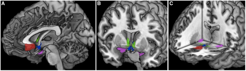
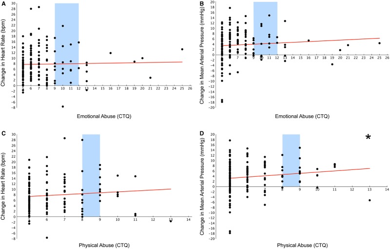
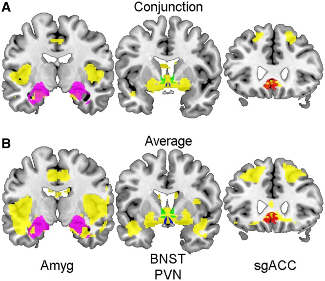
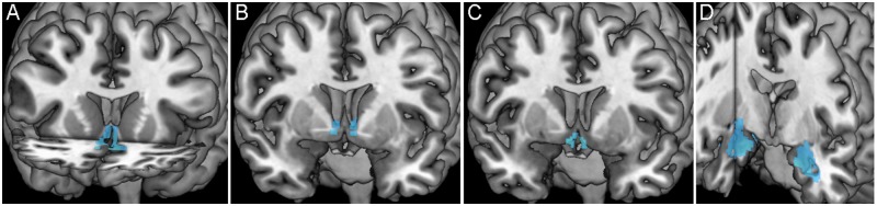
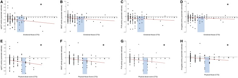
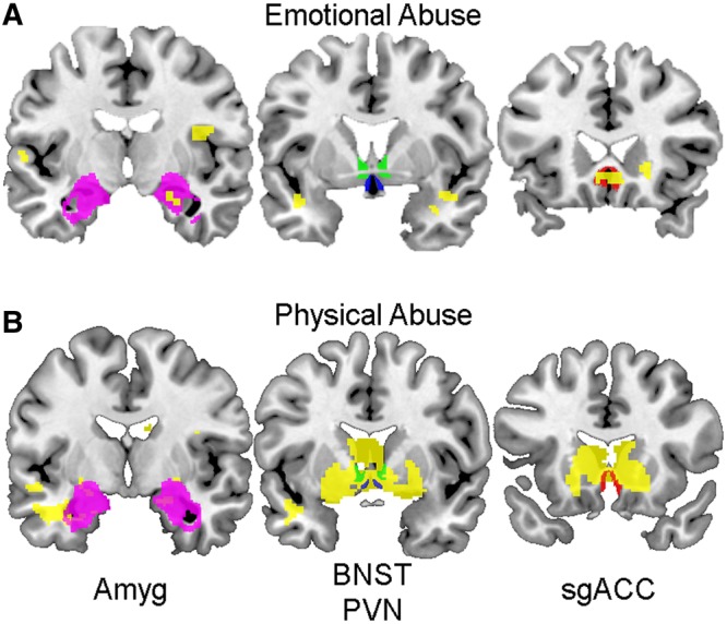
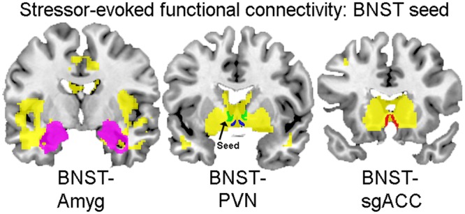
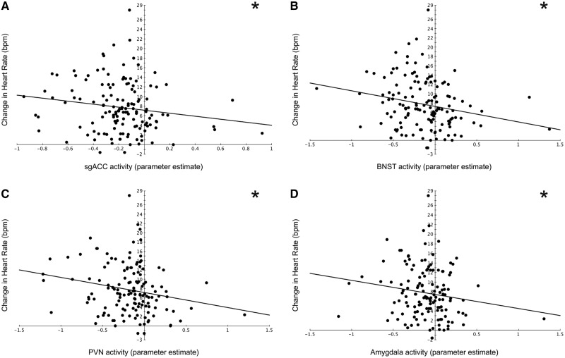
Early life experience differentially shapes later stress reactivity, as evidenced by both animal and human studies. However, early experience-related changes in the function of central visceral neural circuits that control stress responses have not been well characterized, particularly in humans. The paraventricular nucleus of the hypothalamus (PVN), bed nucleus of the stria terminalis (BNST), amygdala (Amyg) and subgenual anterior cingulate cortex (sgACC) form a core visceral stress-responsive circuit. The goal of this study is to examine how childhood emotional and physical abuse relates to adulthood stressor-evoked activity within these visceral brain regions. To evoke acute states of mental stress, participants (n = 155) performed functional magnetic resonance imaging (fMRI)-adapted versions of the multi-source interference task (MSIT) and the Stroop task with simultaneous monitoring of mean arterial pressure (MAP) and heart rate. Regression analyses revealed that childhood physical abuse correlated positively with stressor-evoked changes in MAP, and negatively with unbiased, a priori extractions of fMRI blood-oxygen level-dependent signal change values within the sgACC, BNST, PVN and Amyg (n = 138). Abuse-related changes in the function of visceral neural circuits may reflect neurobiological vulnerability to adverse health outcomes conferred by early adversity.

Keywords: abuse; amygdala; bed nucleus of the stria terminalis; hypothalamus; stress.


Figures

Fig. 1
Fig. 1
ROI masks. Images showing sagittal (A), coronal (B) and axial (C) views of all four ROIs: sgACC (in red), BNST (in green), PVN (in blue) and Amyg (in magenta). The sgACC, BNST and PVN ROI masks were hand-drawn using MRIcron, and the Amyg mask was derived from the SPM anatomy toolbox. (See Table 2 for ROI characteristics).
Fig. 2
Fig. 2
Abuse and stressor-evoked cardiovascular reactivity. Scatterplots displaying the relationship between emotional (A and B) and physical (C and D) abuse and cardiovascular reactivity [i.e. the change in HR (bpm = beats per minute; A and C) or MAP (mmHg = millimeters of mercury; B and D) from baseline to the incongruent, or stress-inducing condition of the tasks]. Childhood physical abuse significantly predicts the stressor-evoked change in MAP in adulthood. Blue bars indicate the range considered low to moderate abuse (A–D). All scatterplots show raw, uncorrected data. *P < 0.05.
Fig. 3
Fig. 3
Stressor tasks main effects maps. Coronal sections (posterior to anterior from left to right) displaying significant voxels (in yellow) derived from a conjunction analysis of both the MSIT and the Stroop task (A), and significant task-related activity (in yellow) averaged across both tasks (B). ROIs are overlying main effects maps (A and B): Amyg (left, in magenta), BNST (middle, in green), PVN (middle, in blue) and sgACC (right, in red). Areas of significant conjunction between both tasks and significant task-related activity overlap with all four ROIs.
Fig. 4
Fig. 4
Averaged stressor-evoked deactivation within each ROI. Images display significant stressor-evoked deactivation (in blue) within each ROI [sgACC (A, in coronal and axial planes), BNST (B, coronal plane), PVN (C, coronal plane) and Amyg (D, coronal and sagittal planes)] from which parameter estimates were extracted for subsequent analyses.
Fig. 5
Fig. 5
Abuse and stressor-evoked activity within ROIs. Scatterplots displaying the relationship between emotional (A–D) and physical (E–H) abuse and stressor-evoked activity within each ROI. Childhood emotional abuse significantly predicts stressor-evoked activity within the sgACC (A) and the Amyg (D) but not the BNST (B) or PVN (C). Childhood physical abuse significantly predicts the stressor-evoked change in activity within all four ROIs: sgACC (E), BNST (F), PVN (G) and Amyg (H). Blue bars indicate the range considered low to moderate abuse (A–H). All scatterplots show raw, uncorrected data. *P < 0.05.
Fig. 6
Fig. 6
Whole-brain regressions of abuse and stressor-evoked activity. Coronal sections (posterior to anterior from left to right) displaying voxels that significantly describe the relationship between emotional (A) and physical (B) abuse with stressor-evoked activity (in yellow). ROIs are overlaid to display the extent of overlap between ROIs and significant voxels: Amyg (left, in magenta), BNST (middle, in green), PVN (middle, in blue) and sgACC (right, in red). Whole-brain voxel-wise regression revealed that emotional abuse predicts activity within both the sgACC and Amyg, but not the BNST and PVN (A). Also, whole-brain voxel-wise regression revealed that physical abuse predicts activity located within all four ROIs (B).
Fig. 7
Fig. 7
PPI main-effects map with BNST seed. Coronal sections (posterior to anterior from left to right) displaying stressor-evoked functional connectivity with the BNST seed. Voxels that are positively correlated with BNST seed activity are displayed in yellow. ROIs are overlaid to demonstrate the extent of their overlap with positively correlated voxels: Amyg (left, in magenta), BNST (middle, in green), PVN (middle, in blue) and sgACC (right, in red).
Fig. 8
Fig. 8
Stressor-evoked ROI activity and HR reactivity. Scatterplots displaying the relationship between stressor-evoked activity within each ROI and the change in HR (bpm = beats per minute) from baseline to the incongruent, or stress-inducing condition of the tasks. Stressor-evoked activity within each ROI [sgACC (A), BNST (B), PVN (C) and the Amyg (D)] predicts HR reactivity. All scatterplots show raw, uncorrected data. *P < 0.05

References

    1. Abrahám IM, Kovacs KJ. Postnatal handling alters the activation of stress-related neuronal circuitries. European Journal of Neuroscience. 2000;12:3003–14. - PubMed
    1. Amunts K, Kedo O, Kindler M, et al. Cytoarchitectonic mapping of the human amygdala, hippocampal region and entorhinal cortex: intersubject variability and probability maps. Anatomy and embryology. 2005;210(5):343–52. - PubMed
    1. Aston-Jones G, Delfs JM, Druhan J, Zhu Y. The bed nucleus of the stria terminalis: a target site for noradrenergic actions in opiate withdrawal. Annals of the New York Academy of Sciences. 1999;877:486–98. - PubMed
    1. Banihashemi L, Rinaman L. Repeated brief postnatal maternal separation enhances hypothalamic gastric autonomic circuits in juvenile rats. Neuroscience. 2010;165(1):265–77. - PMC - PubMed
    1. Banihashemi L, O'Neill EJ, Rinaman L. Central neural responses to restraint stress are altered in rats with an early life history of repeated brief maternal separation. Neuroscience. 2011;192:413–28. - PMC - PubMed

Publication types

MeSH terms