. 2014 May 20;7(326):ra47.
doi: 10.1126/scisignal.2005070.

Targeting poly(ADP-ribose) polymerase and the c-Myb-regulated DNA damage response pathway in castration-resistant prostate cancer

Affiliations

Targeting poly(ADP-ribose) polymerase and the c-Myb-regulated DNA damage response pathway in castration-resistant prostate cancer

Likun Li et al. Sci Signal. .

Abstract

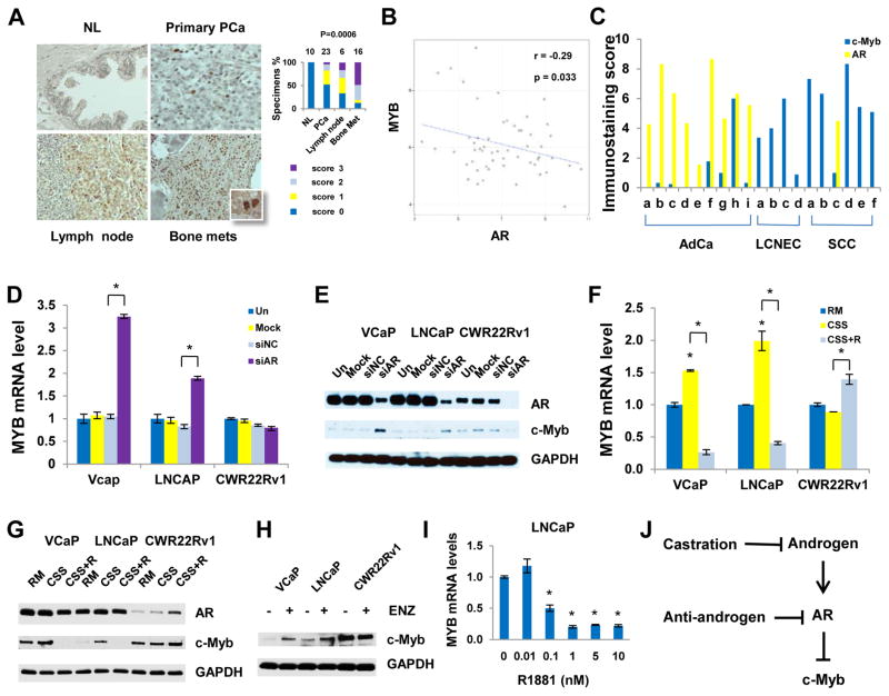
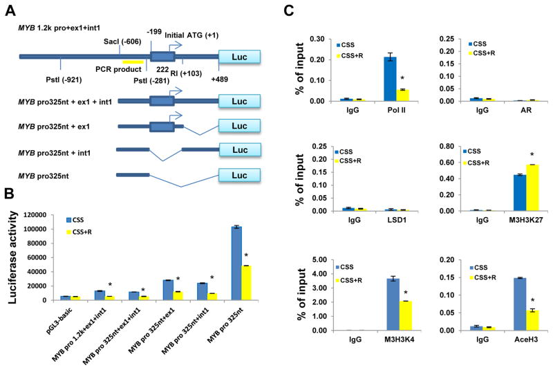
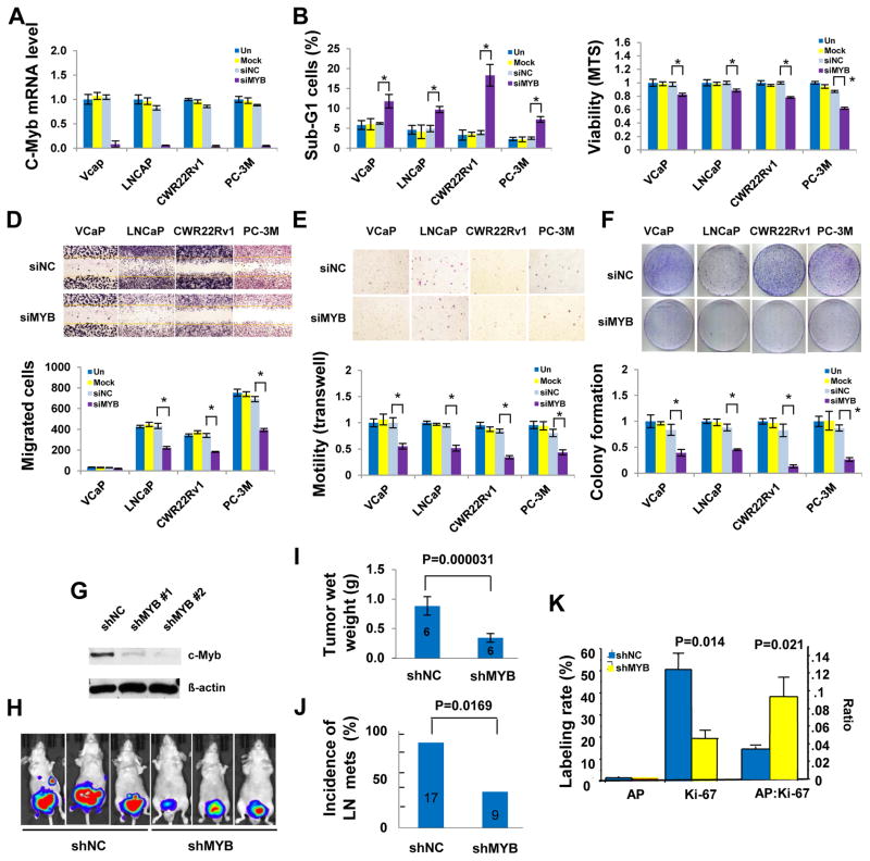
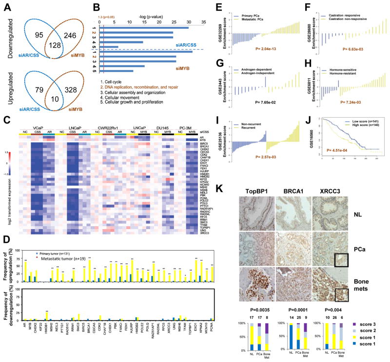
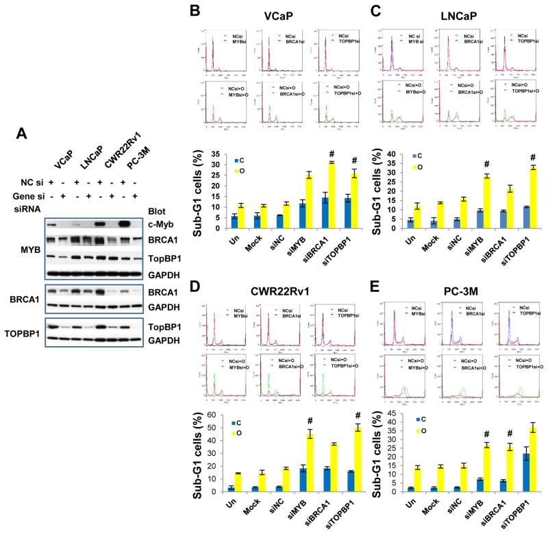
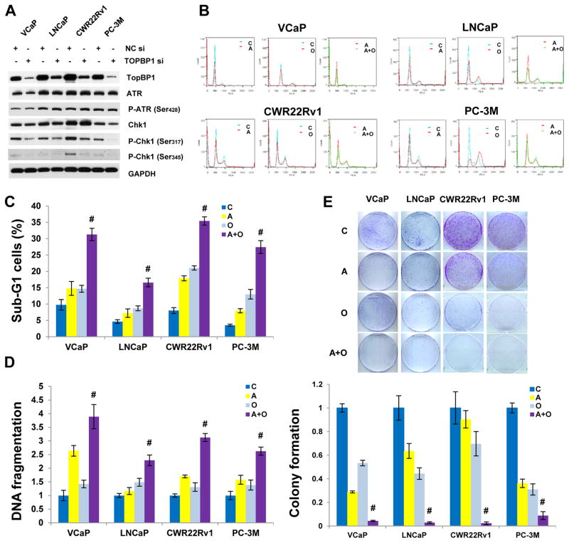
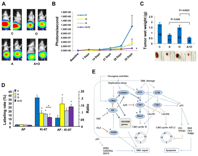
Androgen deprivation is the standard treatment for advanced prostate cancer (PCa), but most patients ultimately develop resistance and tumor recurrence. We found that MYB is transcriptionally activated by androgen deprivation therapy or genetic silencing of the androgen receptor (AR). MYB silencing inhibited PCa growth in culture and xenografts in mice. Microarray data revealed that c-Myb and AR shared a subset of target genes that encode DNA damage response (DDR) proteins, suggesting that c-Myb may supplant AR as the dominant regulator of their common DDR target genes in AR inhibition-resistant or AR-negative PCa. Gene signatures including AR, MYB, and their common DDR-associated target genes positively correlated with metastasis, castration resistance, tumor recurrence, and decreased survival in PCa patients. In culture and in xenograft-bearing mice, a combination strategy involving the knockdown of MYB, BRCA1, or TOPBP1 or the abrogation of cell cycle checkpoint arrest with AZD7762, an inhibitor of the checkpoint kinase Chk1, increased the cytotoxicity of the poly[adenosine 5'-diphosphate (ADP)-ribose] polymerase (PARP) inhibitor olaparib in PCa cells. Our results reveal new mechanism-based therapeutic approaches for PCa by targeting PARP and the DDR pathway involving c-Myb, TopBP1, ataxia telangiectasia mutated- and Rad3-related (ATR), and Chk1.


Conflict of interest statement

Competing interests: The authors declare no competing financial interest.

Figures

Fig. 1
Fig. 1. Activation of c-Myb in CRPC and under experimental conditions that impair AR signaling
(A) IHC analysis for expression of c-Myb in human PCa tissues. The numbers on the top of bar graphs denotes the number of specimens. P values were derived using Kruskal-Wallis rank testing. (B) Correlation analysis of MYB and AR mRNA expression in a published data set (GSE32269). (C) Human PCa xenograft tissue microarray. AdCa, adenocarcinoma; LCNEC, large cell neuroendocrine carcinoma; SCC, small cell prostate carcinoma. X axis labeling represent individual xenograft specimens. The information regarding patient derived xenografts is summarized in Table S2. (D and E) PCa cells were untreated, mock transfected or transfected with 20 nM siAR or negative control siRNA (siNC) for 48 hours. (D) qRT-PCR analysis (E) Western blotting analysis. (F and G) PCa cells were grown in regular medium (RM), charcoal-stripped serum medium (CSS) or CSS plus 10 nM R1881 (CSS+R) for 48 hours. (F) qRT-PCR analysis. (G) Western blotting analysis. (H) Western blotting analysis showing c-Myb abundances after the treatment of 1 μM enzalutamide (ENZ) for 48 hours. (I) qRT-PCR analysis 48 hours after R1818 treatment. (J) Summary of AR regulation of c-Myb. *P < 0.05.
Fig. 2
Fig. 2. AR transcriptionally suppresses MYB through modification of chromatin architecture on MYB promoter
(A) The structural illustration of different MYB promoter luciferase reporters. (B) Luciferase reporter assays. LNCaP cells were transfected with MYB promoter reporters or control pGL3-basic empty vector and β-gal–expressing vector (internal control) for 24 h, followed by treatment with CSS or CSS plus 10 nM R1881 (CSS+R) for 24 h. (C) ChIP assays. LNCaP cells were treated with or without 10 nM R1881 in CSS medium for 24 h prior to ChIP assays. The data are expressed as percentage of input. Pol II, RNA polymerase II; LSD1, lysine-specific demethylase 1; M3H3K27, trimethyl-histone 3 lysine 27; M3H3K4, trimethyl-histone 3 lysine 4; AceH3, acetyl-histone 3 lysine 27. *P < 0.05.
Fig. 3
Fig. 3. c-Myb silencing inhibits PCa Growth in vitro and in vivo
(A) qRT-PCR analysisfor validation of MYB siRNA efficiency. MYB silencing led to increased sub-G-1 cells in flow cytometry analysis (B), reduced cell proliferation and survival in MTS assay (C), fewer migrated cells in wound healing assay (D) and in Boyden chamber assay (E), and decreased colony growth (F) of PCa cells. Top panels of DF arerepresentative images and bottom panels are average of data from triplicate experiments. (G to K) PC-3M orthotopic xenograft model. (G) Western blotting analysis of c-Myb expression in input PC-3M cells. (H) Representative bioluminescent images (day 21). (I) The tumor wet weights in shMYB mice were significantly reduced compared with those in the shNC 28 days after cancer cell injection. (J) shMYB significantly reduced incidence of nymph node metastasis. (K) Apoptosis (TUNEL), cell proliferation (Ki67 staining) and ratio of apoptosis to cell proliferation (AP: Ki67) were compared between the shNC (n=6) and the shMYB tumors (n=5). The data are presented as mean ± standard errors. The numbers in the bars indicate total number of mice. *P < 0.05 unless indicated otherwise.
Fig. 4
Fig. 4. Integrated analysis of AR siRNA/CSS and MYB siRNA responsive genes and Correlation analysis of the DDR gene signature
(A) Venn diagram showing genes targeted by siAR or CSS, siMYB, or both. (B) Ingenuity Pathway Analysis gene ontology enrichment analysis for siAR or CSS and MYB siRNA negatively regulated top five biologic processes. (C) Heat map for DDR genes negatively regulated by siAR or CSS and siMYB. (D) mRNA expression of AR, MYB, and their DDR target genes in primary and metastatic PCa by the analysis of online data (www.cbioportal.org/public-portal) by Taylor et al. (27). Statistical analysis was performed using chi-square analysis. (EJ) Correlation analysis of the DDR gene signature. (E) Primary PCa versus metastatic PCa. (F–H) Androgen-dependent versus androgen-independent. (I) Non-recurrence versus recurrence. (J) Kaplan-Meier curves that compare the survival of patients that have a higher-than-median signature score (red) with those that have a lower-than-median score (blue). (K) Upregulation of DDR genes TOPBP1, BRCA1, and XRCC3 in PCa bone metastases. Normal human prostate (NL), primary PCa, or bone metastasis (bone mets) tissues were evaluated by IHC. The percentages of each type of specimen with a given immunostaining score were compared. P values were derived using Kruskal-Wallis rank testing.
Fig. 5
Fig. 5. DDR gene silencing synergizes with PARP inhibition to increase cytotoxicity to PCa cells
(A) Western blotting analysis showing efficiency of MYB, BRCA1 and TOPBP1 siRNAs. Note MYB gene silencing led to downregulation of BRCA1 and TopBP1. (B–E), Cell cycle analysis. PCa cells were untreated, mock transfected or transfected with specific siRNAs (20 nM) and grown for 24 h, and then followed by the treatment of PARP inhibitor olaparib (OLA; 10 μM) for 48 h. Top panels are representative cell cycle profiles. Bottom bar graphs are average of triplicates. C, vehicle control; O, olaparib; Data are presented as mean ± standard deviation. # indicates synergistic effect. Synergy quantification was summarized in Supplementary Table S4.
Fig. 6
Fig. 6. Chk1 inhibition synergizes with PARP inhibition to increase cytotoxicity and DNA fragmentation and suppress cologenic growth in PCa cells
(A) Effect of TopBP1 on ATR-Chk1 signaling pathway. (B) Representative cell cycle profiles. (C) Cell cycle analysis. PCa cells were treated with Chk1 inhibitor AZD7762 (200 nM) or PARP inhibitor olaparib (10 μM for VCaP and LNCaP and 5 μM for CWR22Rv1 and PC-3M) for 48 h. C, vehicle control (DMSO); A, AZD7762; O, olaparib; A+O, AZD7762+olaparib. (D) DNA fragmentation assay using a Cell Death ELISA Kit (Roche, Mannheim, Germany). Experimental conditions are the same as cell cycle analysis. (E) Colony assay. The data are presented as mean ± standard deviation in (C) and (D), as mean ± standard error in (E). # indicates synergistic effect.
Fig. 7
Fig. 7. Chk1 inhibition synergizes with PARP inhibition to suppress xenograft tumor growth in vivo
C, vehicle control; A, AZD7762; O, olaparib; A+O, AZD7762+olaparib. (A) Representative luminescence images. (B) Quantitative luminescence data showing xenograft tumor growth. Photon signals in combination group were significantly smaller than those in AZD alone (P= 0.0459), but not statistically significant compared to OLA alone due to bigger signal variation in OLA group. (C) Tumor wet weight. Numbers in the bar graph are number of mice in each group. Error bars in (B and C) are standard errors. (D) IHC analysis of apoptosis (TUNEL), cell proliferation (Ki67 staining) and ratio of apoptosis to cell proliferation (AP: Ki67) in the tumors treated with C (n= 8), O (n= 8), A (n= 7) or A+O (n= 7). *: P values <0.05. (E) Proposed AR and c-Myb coregulated DDR signaling pathway. In AR-positive, androgen-sensitive prostate cancer, AR and c-Myb coregulate TOPBP1, BRCA1 and other their common DDR target genes, promoting ATR-Chk1 signaling and DNA repair. In AR negative PCa or upon ADT or the impairment of AR signaling, MYB is derepressed and predominately in charge of DDR gene regulation. * indicates druggable therapeutic targets. PARP and Chk1 are two important DDR gene targets.

References

    1. Heinlein CA, Chang C. Androgen receptor in prostate cancer. Endocr Rev. 2004;25:276–308. - PubMed
    1. Karantanos T, Corn PG, Thompson TC. Prostate cancer progression after androgen deprivation therapy: mechanisms of castrate resistance and novel therapeutic approaches. Oncogene. 2013 - PMC - PubMed
    1. Cai C, He HH, Chen S, Coleman I, Wang H, Fang Z, Nelson PS, Liu XS, Brown M, Balk SP. Androgen receptor gene expression in prostate cancer is directly suppressed by the androgen receptor through recruitment of lysine-specific demethylase 1. Cancer Cell. 2011;20:457–471. - PMC - PubMed
    1. Yuan X, Balk SP. Mechanisms mediating androgen receptor reactivation after castration. Urol Oncol. 2009;27:36–41. - PMC - PubMed
    1. Tzelepi V, Zhang J, Lu JF, Kleb B, Wu G, Wan X, Hoang A, Efstathiou E, Sircar K, Navone NM, Troncoso P, Liang S, Logothetis CJ, Maity SN, Aparicio AM. Modeling a lethal prostate cancer variant with small-cell carcinoma features. Clinical Cancer Research. 2012;18:666–677. - PMC - PubMed

Publication types

MeSH terms

Associated data