

The extrahepatic role of TFR2 in iron homeostasis
- PMID: 24847265
- PMCID: PMC4019842
- DOI: 10.3389/fphar.2014.00093
The extrahepatic role of TFR2 in iron homeostasis
Abstract
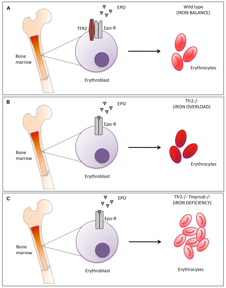
Transferrin receptor 2 (TFR2), a protein homologous to the cell iron importer TFR1, is expressed in the liver and erythroid cells and is reported to bind diferric transferrin, although at lower affinity than TFR1. TFR2 gene is mutated in type 3 hemochromatosis, a disorder characterized by iron overload and inability to upregulate hepcidin in response to iron. Liver TFR2 is considered a sensor of diferric transferrin, possibly in a complex with hemochromatosis protein. In erythroid cells TFR2 is a partner of erythropoietin receptor (EPOR) and stabilizes the receptor on the cell surface. However, Tfr2 null mice as well as TFR2 hemochromatosis patients do not show defective erythropoiesis and tolerate repeated phlebotomy. The iron deficient Tfr2-Tmprss6 double knock out mice have higher red cells count and more severe microcytosis than the liver-specific Tfr2 and Tmprss6 double knock out mice. TFR2 in the bone marrow might be a sensor of iron deficiency that protects against excessive microcytosis in a way that involves EPOR, although the mechanisms remain to be worked out.
Keywords: hemochromatosis; hepcidin; iron deficiency; iron metabolism; transferrin; transferrin receptors.
Figures
References
-
- Camaschella C., Roetto A. (2011). “TFR2-related hereditary hemochromatosis,” in GeneReviews eds Pagon R. A., Adam M. P., Bird T. D., Dolan C. R., Fong C. T., Stephens K. (Seattle, WA)
Publication types
LinkOut - more resources
Full Text Sources
Other Literature Sources