Review
. 2014 May 5:5:94.
doi: 10.3389/fphar.2014.00094. eCollection 2014.

Atherogenesis and iron: from epidemiology to cellular level

Affiliations
Review

Atherogenesis and iron: from epidemiology to cellular level

Francesca Vinchi et al. Front Pharmacol. .

Abstract

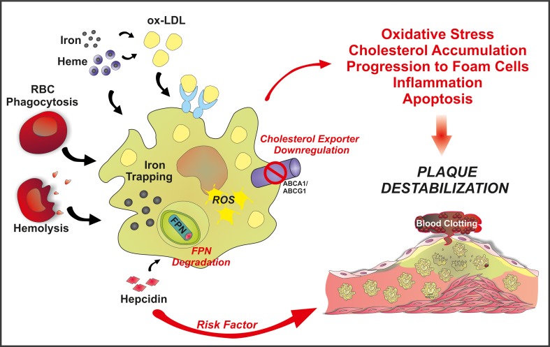
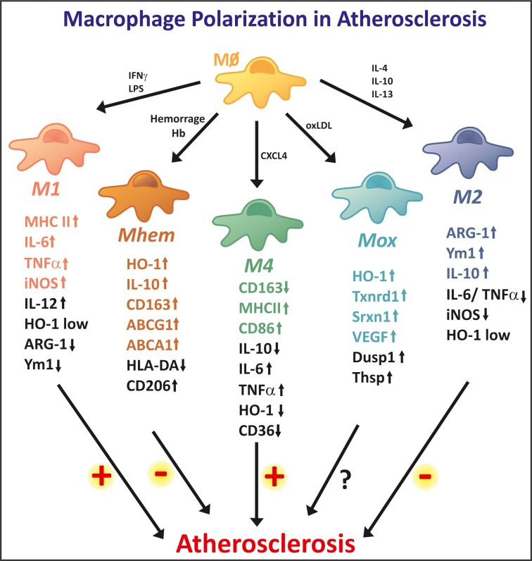
Iron accumulates in human atherosclerotic lesions but whether it is a cause or simply a downstream consequence of the atheroma formation has been an open question for decades. According to the so called "iron hypothesis," iron is believed to be detrimental for the cardiovascular system, thus promoting atherosclerosis development and progression. Iron, in its catalytically active form, can participate in the generation of reactive oxygen species and induce lipid-peroxidation, triggering endothelial activation, smooth muscle cell proliferation and macrophage activation; all of these processes are considered to be proatherogenic. On the other hand, the observation that hemochromatotic patients, affected by life-long iron overload, do not show any increased incidence of atherosclerosis is perceived as the most convincing evidence against the "iron hypothesis." Epidemiological studies and data from animal models provided conflicting evidences about the role of iron in atherogenesis. Therefore, more careful studies are needed in which issues like the source and the compartmentalization of iron will be addressed. This review article summarizes what we have learnt about iron and atherosclerosis from epidemiological studies, animal models and cellular systems and highlights the rather contributory than innocent role of iron in atherogenesis.

Keywords: LDL; atherosclerosis; heme; hemoglobin; intraplaque hemorrhage; iron; macrophages; oxidative stress.


Figures

Figure 1
Figure 1
A role for iron in atherosclerotic lesions. Iron accumulates in the plaque either as inorganic or hemoglobin-bound iron. Inorganic iron in part derives from circulating transferrin (Tf)-bound iron and non-transferrin-bound iron (NTBI). NTBI is generated in chronic iron overload conditions, such as in iron-loading anemia, hereditary hemochromatosis or secondary hemochromatosis due to blood transfusions. Circulating NTBI is accessible to many cell types in the atherosclerotic plaque: endothelial cells, monocytes/macrophages, and vascular smooth muscle cells (VSMCs). Hemoglobin- and heme-derived iron can access the plaque upon intravascular hemolysis and intraplaque hemorrhage, affecting endothelial cells and macrophages. Hemoglobin (Hb), heme and iron promote endothelial activation, by enhancing adhesion molecule expression. As a consequence, monocyte recruitment is expected to be increased. Circulating iron and Hb oxidize LDLs, thus enhancing subendothelial LDL retention and macrophage progression to foam cells. Iron also affects VSMC proliferation and migration into the lesion, favoring plaque progression.
Figure 2
Figure 2
Schematic overview of the “refined iron hypothesis”: a role for macrophage-retained iron in atherosclerosis. Iron can accumulate in macrophages as inorganic iron and Hb-iron, upon erytrophagocytosis or hemolysis. Once stored in the cell, iron can be made available to the bloodstream via FPN-mediated export. According to the refined iron hypothesis, high hepcidin levels are considered a risk factor for plaque progression and destabilization. Hepcidin is known to bind to FPN, thus promoting its degradation and blocking iron export. This increases intracellular ROS levels and decreases cholesterol efflux. As a result, the oxidative status alters and LDL accumulation occurs, promoting foam cell formation, inflammation and eventually plaque instability.
Figure 3
Figure 3
High macrophage plasticity in atherosclerosis. In the atherosclerotic plaque, macrophages differentiate into different phenotypes. The two extreme phenotypes are represented by M1 and M2 macrophages. M1 macrophages show strong pro-inflammatory properties, thus potentially being involved in lesion progression. M1 macrophages show high expression levels of iNOS, MHCII, and inflammatory cytokines, such as IL-6 and TNF-a. M2 macrophages are considered anti-inflammatory and are involved in tissue repair and remodeling. M2 specific markers are Arginase 1, Ym1, and IL-10. The M2 phenotype is reported as anti-atherogenic. In addition, several other macrophage phenotypes are observed in the atherosclerotic plaque. Mhem macrophages originate as a consequence of intraplaque hemorrhage and are endowed with high Hb handling ability. These anti-atherogenic macrophages express high levels of the heme-degrading enzyme HO-1 and the Hp-Hb scavenger receptor CD163. Additionally, Mheme macrophages express the cholesterol exporters ABCA1 and ABCG1, thus efficiently activating reverse cholesterol efflux. Mox macrophages are generated upon oxidized phospholipid stimulation. They show anti-oxidant properties, as they express genes involved in the anti-oxidant responses such as HO-1, Txnrd1, and Srxn1. Their potentially athero-protective effect still needs to be demonstrated. M4 macrophages differentiate in response to the chemo-attractant CXCL4, thus showing pro-inflammatory and pro-atherogenic effects. These macrophages express low levels of CD163 and high levels of MHCII and CD86. M1 and M4 macrophages promote, while M2 and Mhem macrophages counteract foam cell formation, thus having opposite effect on atherosclerosis progression.

References

    1. Aessopos A., Farmakis D., Tsironi M., Diamanti-Kandarakis E., Matzourani M., Fragodimiri C., et al. (2007). Endothelial function and arterial stiffness in sickle-thalassemia patients. Atherosclerosis 191, 427–432 10.1016/j.atherosclerosis.2006.04.015 - DOI - PubMed
    1. Ahluwalia N., Genoux A., Ferrieres J., Perret B., Carayol M., Drouet L., et al. (2010). Iron status is associated with carotid atherosclerotic plaques in middle-aged adults. J. Nutr. 140, 812–816 10.3945/jn.109.110353 - DOI - PMC - PubMed
    1. Alayash A. I. (2011). Haptoglobin: old protein with new functions. Clin. Chim. Acta 412, 493–498 10.1016/j.cca.2010.12.011 - DOI - PubMed
    1. Araujo J. A., Romano E. L., Brito B. E., Parthe V., Romano M., Bracho M., et al. (1995). Iron overload augments the development of atherosclerotic lesions in rabbits. Arterioscler. Thromb. Vasc. Biol. 15, 1172–1180 10.1161/01.ATV.15.8.1172 - DOI - PubMed
    1. Bach F. H. (2005). Heme oxygenase-1: a therapeutic amplification funnel. FASEB J. 19, 1216–1219 10.1096/fj.04-3485cmt - DOI - PubMed

LinkOut - more resources