. 2014 Apr 28:11:35.
doi: 10.1186/1742-9994-11-35. eCollection 2014.

Organization and metamorphic remodeling of the nervous system in juveniles of Phoronopsis harmeri (Phoronida): insights into evolution of the bilaterian nervous system

Affiliations

Organization and metamorphic remodeling of the nervous system in juveniles of Phoronopsis harmeri (Phoronida): insights into evolution of the bilaterian nervous system

Elena N Temereva et al. Front Zool. .

Abstract

Background: Metamorphic remodeling of the nervous system and its organization in juvenile may shed light on early steps of evolution and can be used as an important criterion for establishing the relationships among large groups of animals. The protostomian affiliation of phoronids does not still have certain morphological and embryological proofs. In addition, the relationship of phoronids and other former "lophophorates" is still uncertain. The resolving of these conflicts requires detailed information from poorly investigated members of phoronids, such as Phoronopsis harmeri.

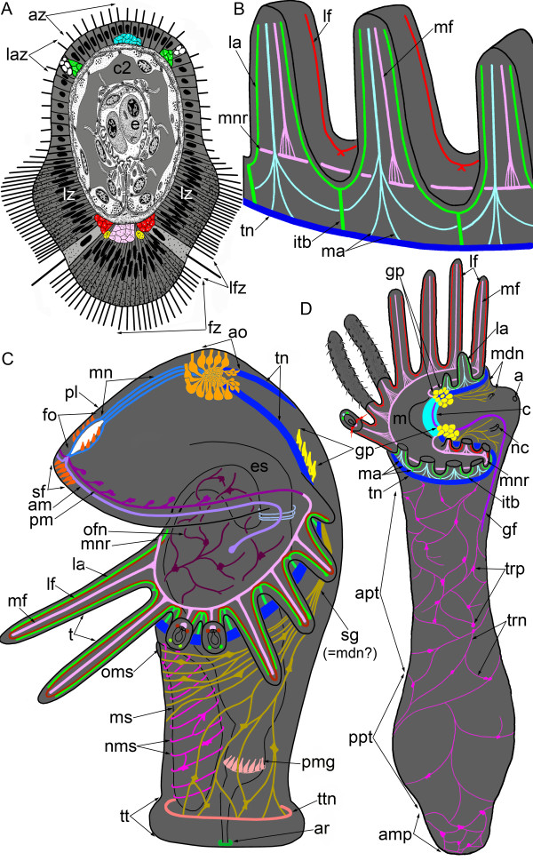
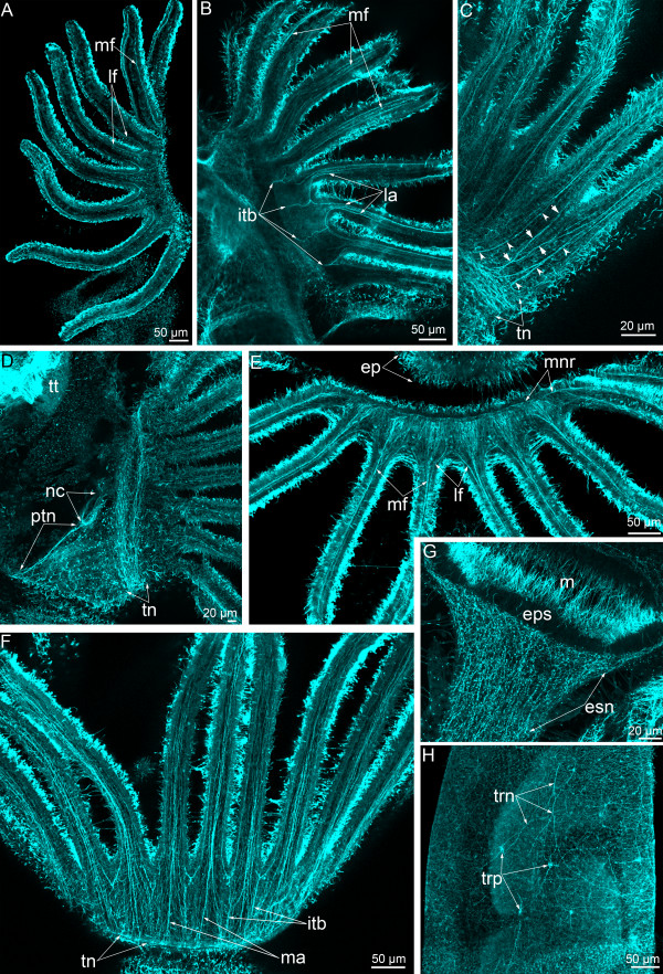
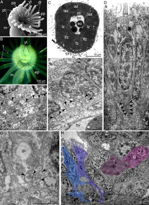
Results: During metamorphosis, the juvenile consumes the nerve elements of the larval hood. Two dorsolateral groups of larval perikarya remain and give rise to the dorsal ganglion, which appears as the "commissural brain". The juvenile inherits the main and minor tentacular nerve rings from the larva. Although the larval tentacles are directly inherited by the juvenile in P. harmeri, the ultrastructure and location of the definitive tentacular neurite bundles change greatly. Innervation of the juvenile lophophore exhibits a regular alternation of the intertentacular and abfrontal neurite bundles. The giant nerve fiber appears at early stage of metamorphosis and passes from the right group of dorsolateral perikarya to the left side of the body.

Discussion: THE METAMORPHIC REMODELING OF THE PHORONID NERVOUS SYSTEM OCCURS IN TWO DIFFERENT WAYS: with complete or incomplete destruction of organ systems. The morphology of the lophophore seems similar to those of the former members of "Lophophorata", but its innervation differs greatly. These findings support the separation of bryozoans from Lophophorata and establish a need for new data on the organization of the brachiopod nervous system. The nervous system of the phoronid juvenile is organized as an epidermal nerve plexus but exhibits a nerve center in the anterior portion of the body. The simultaneous presence of both the apical organ and anlage of the cerebral ganglion in phoronids at the larval stage, and the reduction of the apical organ during metamorphosis support the Trochea theory and allow to suggest the presence of two nervous centers in the last common ancestor of the Bilateria. Phoronids retained some plesiomorphic traits and can be regarded as one of the most primitive groups of lophotrochozoans.

Keywords: Deuterostomia; Evolution; Lophophorata; Metamorphosis; Nervous system; Phylogeny; Protostomia; The last common bilaterian ancestor.


Figures

Figure 1
Figure 1
Schemes of organization of the nerve system in juvenile (A, B, D) and competent larvae (C) of Phoronopsis harmeri. Nerve elements are shown by different colors. Although the homology between larval and definitive nerve elements is still uncertain, some nerve elements, which juvenile certainly inherits from larva, are indicated by the same colors in juvenile and in larva. (A) The scheme of cross section of tentacle. (B) The scheme of innervation of definitive tentacles, which are viewed from the abfrontal side. (C) The nervous system of competent larva, which is ready for metamorphosis. All nerve elements, which were observed by TEM and immunocytochemical staining, are shown. Larva is viewed from the left; the apical is at the top, the ventral side is to the left. Picture was changed from Temereva and Tsitrin [8]; abbreviation are shown in the Table 1 and in Figure 4[8]. (D) Scheme of whole nervous system in 10-day-old juvenile. The lophophore is simplified; tentacles of the left side are removed. The oral side is to the left. The number of nerve fibers along the body reflects the density of the nerve plexus in different body parts. Abbreviations: a – anus; amp – ampulla; apt – anterior portion of the body; az – abfrontal zone; c – commissure; c2 – mesocoel; e – erythrocyte; fz – frontal zone; gf – giant fiber; gp – group of perikarya; itb – intertentacular branch; la – lateroabfrontal neurites; laz – lateroabfrontal zone; lf – laterofrontal neurites; lfz – laterofrontal zone; lz – lateral zone; m – mouth; ma – medioabfrontal neurites; mdn – most dorsal neurites, which probably correspond to second group of larval neurites (sg); mf – mediofrontal neurites; mnr – minor nerve ring; nc – nephridial channel; pt – posterior portion of the body; tn – tentacular (main) nerve ring; trn – trunk neurites; trp – trunk perikarya.
Figure 2
Figure 2
Serotonin-like immunoreactive nervous system in Phoronopsis harmeri during the first stages of metamorphosis. (A-G) Animals at one of the first stages of metamorphosis (the “hood-eating stage”). (H-J) Animals at the stage when the postoral ciliated band is ingested. In all images, the apical is at the top, and the oral side is to the right. Z-projections (B, D, E, G, H) of animals after mono- and double staining for serotonin (yellow) and phalloidin (blue). (A) Whole animal viewed from the top; SEM. (B) Anterior portion of the body. The intertentacular branches are indicated by opened arrowheads. (C) Sagittal semithin section of the anterior portion of the body. (D) Optical sagittal section of the anterior part of the body. The serotonin-like immunoreactive cells in the esophagus are indicated by closed arrowheads. The nerve ring around the anus is indicated by arrows. (E) The portion of tentacular (main) nerve ring with intertentacular branches (arrowheads). (F) Middle part of the body with nerve plexus and nonsensory perikarya (double close arrowheads). (G) Whole anterior part of the body. (H) Anterior part of the body; SEM. (I) Whole anterior part of the animal. The main nerve ring is indicated by double arrowheads. (J) Part of the main nerve ring and lateroabfrontal neurites. The intertentacular branches are indicated by open arrowheads. Abbreviations: ao – apical organ; bc – blastocoel; c3 – trunk coelom; es – esophagus; gp – groups of perikarya; m – mouth; la – lateroabfrontal neurite bundles; of – oral field; p – proctodaeum; pl – preoral lobe; po – postoral ciliated band; pp – remnant of the hood; pt – posterior part of the larval body with the telotroch; sg – neurites of the second group; st – stomach; t – tentacle; tn – tentacular (main) nerve ring; tt – telotroch; yt – youngest tentacles.
Figure 3
Figure 3
Serotonin-like immunoreactive nervous system in 3-day-old Phoronopsis harmeri. In all images, the apical is at the top, and the oral side is to the right. Z-projections (B-E) of animals after mono- and double staining for serotonin (yellow) and phalloidin (blue). (A) Live whole animal. (B) Anterior portion of the body viewed from the right side. (C) A part of the main nerve ring with multipolar perikarya, which give rise to the tentacular neurites. (D) Neurites and immunoreactive perikarya in tentacles. (E) The most dorsal portion of the main nerve ring with group of dorsolateral perikarya and the most dorsal neurites around the nephridial channel. Abbreviations: ep – epistome; gp – dorsolateral group of perikarya; mdn – most dorsal neurites; nc – nephridial channel; st – stomach; t – tentacle; ten – tentacular neurites; tep – perikarya in the tentacles; tn – tentacular (main) nerve ring; trn – neurites innervating the trunk; trp – perikarya innervating the trunk; tt – telotroch.
Figure 4
Figure 4
Serotonin-like immunoreactive nervous system in 10-day-old Phoronopsis harmeri. In all images, the apical is at the top. Z-projections (B-D) of animals after mono- and double staining for serotonin (yellow) and phalloidin (blue). (A) Live animal; anterior part of the body. The anal side is to the right. (B) Anterior portion of the body viewed from the anal side. (C) Optical sections of the several tentacles with tentacular neurites and solitary perikarya (arrowheads). (D) The right half of the lophophore with the main nerve ring and tentacular neurites. (E) Optical frontal section of the anterior body part viewed from the oral side. Abbreviations: ah – area of the anal hill; cnb – central neurite bundles; ep – epistome; gp – dorsolateral group of perikarya; nc – nephridial channel; t – tentacle; ten – tentacular neurites; tn – tentacular (main) nerve ring; trp – perikarya innerveting the trunk.
Figure 5
Figure 5
FMRFamide-like immunoreactive nervous system in Phoronopsis harmeri at consecutive stages of metamorphosis. In all images, the apical is at the top. Z-projections (B-I) of animals after mono- and double staining for FMRFamide (green), phalloidin (blue), and alpha-tubulin (cyan). (A) Live whole animal at first stage of metamorphosis. The oral side is to the right. (B) The same stage; anterior portion of the body and the preoral lobe. The oral side is to the right. (C) The same stage; anterior part of the body with esophageal and anal nerve elements. The esophageal immunoreactive neurites and perikarya are indicated by open arrowheads. The tentacular nerve ring is indicated by double arrowheads. (D) The stage when the postoral ciliated band is ingested. Whole animal viewed from the right side. (E) A 3-day-old juvenile viewed from the oral side; anterior part of the body. (F) The same stage; a part of the tentacular nerve ring. (G) The same stage; the part of the midgut with neurites and perikarya (closed arrowheads). (H) A 3-day-old juvenile viewed from the anal side. (I) The same stage; the upper part of the esophagus. Abbreviations: ar – anal nerve ring; ao – apical organ;dn – dorsal neurites; bm – blood masses; ep – epistome; eps – epithelium of the esophagus; es – esophagus; esn – neurites innervating the esophagus; fo – frontal organ; m – mouth; mg – midgut; mgn – neurites in the midgut; mn – medial neurite bundle; pl – preoral lobe; pm – posterior marginal neurite bundle; pt – posterior part of the larval body with the telotroch; sd – stomach diverticulum; t – tentacle; ten – tentacular neurites; tn – tentacular (main) nerve ring; trn – neurites innervating the trunk; trp – perikarya innervating the trunk; tt – telotroch.
Figure 6
Figure 6
Innervation of the lophophore and alpha-tubulin immunoreactive elements in Phoronopsis harmeri at different stages of metamorphosis. In all images, the apical is at the top. (A) First stage of metamorphosis; several tentacles viewed from the frontal side. The oral side is to the right. (B) The same stage; a part of the lophophore with tentacles viewed from the abfrontal side. The oral side is to the right. (C) The stage when the postoral ciliated band is ingested, several tentacles viewed from the abfrontal side. Medioabfrontal neurite bundles are indicated by arrowheads. Intertentacular branches are indicated by arrows. The oral side is to the right. (D) The same stage; anterior part of the body viewed from the right. (E) A 3-day-old juvenile; a part of the lophophore with tentacles viewed from the frontal side. (F) The same stage; a part of the lophophore with tentacles viewed from the abfrontal side. (G) A 3-day-old juvenile; the upper part of the esophagus. (H) A 3-day-old juvenile; a part of the trunk body wall. Abbreviations: ep – epistome; eps – epithelium of the esophagus; itb – intertentacular branch; la – lateroabfrontal neurites in tentacle; lf – laterofrontal neurites in tentacle; m – mouth; ma – medioabfrontal neurites in tentacle; mf – mediofrontal neurites in tentacle; mnr – minor nerve ring; nc – nephridial channel; ptn – prominent trunk neurite bundle; tn – tentacular (main) nerve ring; trn – trunk neurites; trp – trunk perikarya; tt – telotroch.
Figure 7
Figure 7
Morphological and ultrasrtuctural changes of the larval hood and tentacles at the firsts stages of metamorphosis of Phoronopsis harmeri. (A-C) The anterior portion of the body of live animals. (D, F, G) Semithin sections. (E, H-K) Thin section. (A) First stage of metamorphosis: the hood (pl) remains its integrity. (B) The next (second) stage of metamorphosis: the hood (pl) turns into cellular debris and engulfed. (C) The third stage of metamorphosis: larval tentacles (t) form a “cup” and surround the hood. (D) The second stage of metamorphosis, sagittal section; the oral side is to the right, the anal side is to the left, the apical is at the top. (E) Thick basal lamina (bl) and spacious blastocoel (bc) under degenerated cells of the preoral lobe and the apical organ, which still remains synaptic vesicles (sv). (F) Sagittal section of the protocoel (c1), degenerated hood (pl), and the apical organ (ao). (G) The cross section of the tentacle, which starts to acquire the definitive style via the peeling of the postoral ciliated band (po). (H) The mediofrontal neurite bundle (arrowheads). (I) The medioabfrontal neurite bundles (arrowheads). (J) The lateroabfrontal neurite bundle (arrowheads), which is associated with gland cell (gc). (K) Laterofrontal neurite bundle (arrowheads) is associated with laterofrontal sensory cell (lfc), which undergoes the cell death. Abbreviations: az – abfrontal zone; bm – blood masses; bv – blood vessel; c2 – tentacular coelom; c3 – trunk coelom; es – esophagus; fz – frontal zone; lfz – laterofrontal zone; m – mouth; mc – muscle cell; mi – microvilli; mv – median blood vessel; n – nucleus; nf – nerve fiber; pt – posterior part of the larval body with the telotroch; sd – stomach diverticulum; st – stomach; tn – main nerve ring; tt – telotroch.
Figure 8
Figure 8
Organization of main nerve elements at first stages of metamorphosis of Phoronopsis harmeri. Sagittal semithin (A, C) and thin (B, D-E) sections. (A) One of dorsolateral groups of perikarya. (B) Perikarya (pink) of dorsolateral group, which is associated with the main nerve ring. (C) The main nerve ring. (D) Perikaryon (pink) in the main nerve ring. (E) The neuropil of the main nerve ring. Abbreviations: bc – blastocoel; c2 – tentacular coelom; c3 – trunk coelom; d – diaphragm; dcv – dense-core vesicle; gp – groups of perikarya; m – mitochondrion; mc – muscle cell; mt – microtubule; n – nucleus; nc – nephridial channel; nf – nerve fiber; pl – preoral lobe; sv – synaptic vesicle; tn – main nerve ring.
Figure 9
Figure 9
The latest stages of metamorphosis of Phoronopsis harmeri. (A) Metamorphic animal, which underwent all catastrophic changes in 30 minutes after metamorphosis start. (B-H) A 3-day-old juvenile. (A) The head region of metamorphic animal viewed from the oral side; SEM. (B) The head region of live juvenile viewed from the oral side. (C) The cross section of the tentacle: new postoral ciliated band is formed (the long microvilly of laterofrontal cell are shown by double arrowheads). (D) The laterofrontal cell, which is associated with laterofrontal neurite bundle (arrowheads). (E) The mediofrontal neurite bundle (arrowheads). (F) The medioabrfontal neurite bundles (arrowheads). (G) The lateroabfrontal neurite bundle (arrowheads), which is associated with gland cell. (H) The main nerve ring: sensory (blue) and nonsensory (pink) perikarya. Abbreviations: az – abfrontal zone; bl – basal lamina; bv – blood vessel; cc – coelomic lining; dcv – dense-core vesicle; dt – definitive tentacle; ec – erythrocyte; ep – epistome; fz – frontal zone; G – Golgi apparatus; gc – gland cell; laz – lateroabfrontal zone; lfc – laterofrontal cell; lfz – laterofrontal zone; m – mouth; mc – muscle cell; mi – microvilli; mt – microtubule; n – nucleus; nf – nerve fiber; pp – remnants of the hood; pt – posterior part of the larval body with the telotroch; yt – youngest tentacle.
Figure 10
Figure 10
Organization of the dorsal ganglion in 3-day-old juvenile of Phoronopsis harmeri. Cross semithin (A) and thin (B-D) sections of the head region. (A) Two dorsolateral groups of perikarya (pink boxes) connect via thick commissure. (B) A portion of one of the dorsolateral group of perikarya: sensory (blue) and nonsensory (pink) perikarya are visible. Perikarya of both types bear centrioles (arrowheads). (C) A portion of the commissure, which consists of nerve fibers of different types. (D) A proximal portion of the giant nerve fiber associated with epidermal cell (blue), which contacts the epidermis surface and bears the centriole (arrowhead). Abbreviations: bl – basal lamina; c – commissure; c2 – mesocoel; cc – coelomic lining; cv – clear (electron-lucent) synaptic vesicle; dcv – dense-core vesicle; er – erythrocyte; es – esophagus; G – Golgi apparatus; gf – giant nerve fiber; m – mouth; mc – mitochondria; mi – microvilli; mnr – minor nerve ring; mt – microtubule; n – nucleus; nf – nerve fiber; tt – telotroch.
Figure 11
Figure 11
Some nerve elements of the trunk in 3-day-old juvenile of Phoronopsis harmeri. Cross semithin (A) and thin (B-F) sections of the anterior body part. (A) Whole section with formed definitive digestive tract and blood vessels. (B) The giant nerve fiber, which is completely enveloped by two cells and accompanied by several nerve fibers of common diameter. (C) Group of nerve fibers (arrowheads) in the epithelium of descending branch of the digestive tract. (D) Large projection of nerve cell contains synaptic vesicles and located in the epithelium of descending branch of the digestive tract. (E) Projection of neurosecretory cell (arrowheads) in the epithelium of ascending branch of the digestive tract. (F) Neuron (pink) with dense-core synaptic vesicles and nerve fibers, some of which contain clear (electron-lucent) vesicles in the epithelium of the esophagus. Abbreviations: ab – ascending branch of digestive tract; am – anal mesentery; bc – blastocoel; bl – basal lamina; c3 – trunk coelom; cc – coelomic lining; cv – clear (electron-lucent) synaptic vesicle; db – descending branch of digestive tract; dcv – dense-core vesicle; ec – enveloping cell; ep – epidermis; llv – left lateral blood vessel; lm – longitudinal muscles; m – mitochondria; mc – muscle cells; mv – median blood vessel; n – nucleus; nf – nerve fiber; sv – synaptic vesicles.
Figure 12
Figure 12
Scenario of phoronid body plan evolution. Different elements of the nervous system are indicated by different color: orange – apical organ, yellow – cerebral ganglion; cyan – the commissure, blue – nerve tract, which innervates the tentacles, pink – neurites and perikarya of the nerve plexus. (A) Hypothetical pelagic ancestor with tentacles and ciliated epidermis. (B) Creeping ancestor. (C) The formation of ventral pouch, which is exploited as a tool to dig into soft substrata to avoid danger. The apical organ undergoes the reduction. (D) Appearance of actinotroch larva. Abbreviations: ao – apical organ; cg – cerebral ganglion; dao – degenerating apical organ; ep – epistome; gp – dorsolateral groups of perikarya; m – mouth; t – tentacle.

References

    1. Temereva EN. New data on distribution, morphology and taxonomy of phoronid larvae (Phoronida, Lophophorata) Invert Zool. 2009;6(1):47–64.
    1. Temereva EN, Neretina TV. A distinct phoronid larva: morphological and molecular evidence. Invert Syst. 2013;27(6):622–633.
    1. Temereva EN, Malakhov VV. The evidence of metamery in adult brachiopods and phoronids. Invert Zool. 2011;8:87–101.
    1. Altenburger A, Wanninger A, Holmer LE. Metamorphosis in Craniiformea revisited: Novocrania anomala shows delayed development of the ventral valve. Zoomorphology. 2013;132(4):379–387. doi: 10.1007/s00435-013-0194-3. - DOI
    1. Halanych KM, Bacheller JD, Aguinaldo AMA, Liva SM, Hillis DM, Lake JA. Evidence from 18S ribosomal DNA that lophophorates are protostome animals. Science. 1995;267:1641–1643. doi: 10.1126/science.7886451. - DOI - PubMed