

Cytoskeletal self-organization in neuromorphogenesis
- PMID: 24847718
- PMCID: PMC4199815
- DOI: 10.4161/bioa.29070
Cytoskeletal self-organization in neuromorphogenesis
Abstract
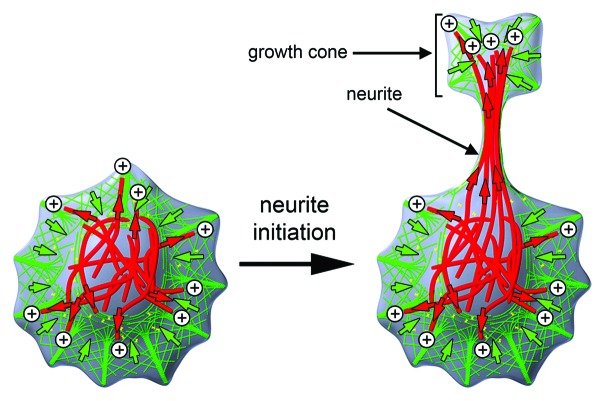
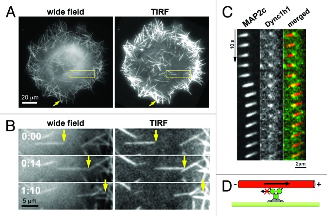
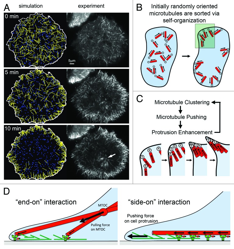
Self-organization of dynamic microtubules via interactions with associated motors plays a critical role in spindle formation. The microtubule-based mechanisms underlying other aspects of cellular morphogenesis, such as the formation and development of protrusions from neuronal cells is less well understood. In a recent study, we investigated the molecular mechanism that underlies the massive reorganization of microtubules induced in non-neuronal cells by expression of the neuronal microtubule stabilizer MAP2c. In that study we directly observed cortical dynein complexes and how they affect the dynamic behavior of motile microtubules in living cells. We found that stationary dynein complexes transiently associate with motile microtubules near the cell cortex and that their rapid turnover facilitates efficient microtubule transport. Here, we discuss our findings in the larger context of cellular morphogenesis with specific focus on self-organizing principles from which cellular shape patterns such as the thin protrusions of neurons can emerge.
Keywords: cellular morphogenesis; cortical dynein; cytoplasmic dynein; microtubules; neurite; neuromorphogenesis; self-organization.
Figures
Comment on
-
Direct observation of microtubule pushing by cortical dynein in living cells.Mol Biol Cell. 2014 Jan;25(1):95-106. doi: 10.1091/mbc.E13-07-0376. Epub 2013 Oct 30. Mol Biol Cell. 2014. PMID: 24173713 Free PMC article.
Similar articles
-
A microtubule-based, dynein-dependent force induces local cell protrusions: Implications for neurite initiation.Brain Cell Biol. 2006 Feb;35(1):39-56. doi: 10.1007/s11068-006-9001-0. Epub 2007 Mar 13. Brain Cell Biol. 2006. PMID: 17940912
-
Direct observation of microtubule pushing by cortical dynein in living cells.Mol Biol Cell. 2014 Jan;25(1):95-106. doi: 10.1091/mbc.E13-07-0376. Epub 2013 Oct 30. Mol Biol Cell. 2014. PMID: 24173713 Free PMC article.
-
She1-mediated inhibition of dynein motility along astral microtubules promotes polarized spindle movements.Curr Biol. 2012 Dec 4;22(23):2221-30. doi: 10.1016/j.cub.2012.10.017. Epub 2012 Nov 8. Curr Biol. 2012. PMID: 23142046 Free PMC article.
-
[Dynein and dynactin as organizers of the system of cell microtubules].Ontogenez. 2006 Sep-Oct;37(5):323-39. Ontogenez. 2006. PMID: 17066975 Review. Russian.
-
How Dynein Moves Along Microtubules.Trends Biochem Sci. 2016 Jan;41(1):94-105. doi: 10.1016/j.tibs.2015.11.004. Epub 2015 Dec 9. Trends Biochem Sci. 2016. PMID: 26678005 Free PMC article. Review.
Cited by
-
Polarity Sorting of Microtubules in the Axon.Trends Neurosci. 2018 Feb;41(2):77-88. doi: 10.1016/j.tins.2017.11.002. Epub 2017 Nov 30. Trends Neurosci. 2018. PMID: 29198454 Free PMC article. Review.
-
Robustness of the microtubule network self-organization in epithelia.Elife. 2021 Feb 1;10:e59529. doi: 10.7554/eLife.59529. Elife. 2021. PMID: 33522481 Free PMC article.
-
Force Generation by Molecular-Motor-Powered Microtubule Bundles; Implications for Neuronal Polarization and Growth.Front Cell Neurosci. 2015 Nov 10;9:441. doi: 10.3389/fncel.2015.00441. eCollection 2015. Front Cell Neurosci. 2015. PMID: 26617489 Free PMC article.
References
Publication types
MeSH terms
Substances
LinkOut - more resources
Full Text Sources
Other Literature Sources