. 2014 Oct;34(7):973-85.
doi: 10.1007/s10571-014-0073-6. Epub 2014 May 22.

Neuroprotective effects of bikaverin on H2O2-induced oxidative stress mediated neuronal damage in SH-SY5Y cell line

Affiliations

Neuroprotective effects of bikaverin on H2O2-induced oxidative stress mediated neuronal damage in SH-SY5Y cell line

D Nirmaladevi et al. Cell Mol Neurobiol. 2014 Oct.

Abstract

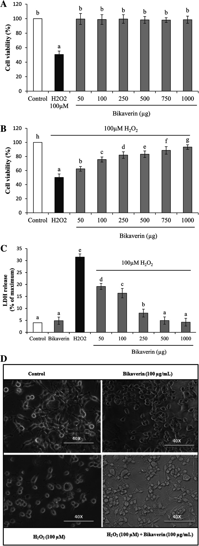
The generation of free radicals and oxidative stress has been linked to several neurodegenerative diseases including Parkinson's disease, Alzheimer's disease, Huntington's disease, and Amyotrophic lateral sclerosis. The use of free radical scavenging molecules for the reduction of intracellular reactive oxygen species is one of the strategies used in the clinical management of neurodegeneration. Fungal secondary metabolism is a rich source of novel molecules with potential bioactivity. In the current study, bikaverin was extracted from Fusarium oxysporum f. sp. lycopersici and its structural characterization was carried out. Further, we explored the protective effects of bikaverin on oxidative stress and its anti-apoptotic mechanism to attenuate H2O2-induced neurotoxicity using human neuroblastoma SH-SY5Y cells. Our results elucidate that pretreatment of neurons with bikaverin attenuates the mitochondrial and plasma membrane damage induced by 100 µM H2O2 to 82 and 26% as evidenced by MTT and LDH assays. H2O2 induced depletion of antioxidant enzyme status was also replenished by bikaverin which was confirmed by Realtime Quantitative PCR analysis of SOD and CAT genes. Bikaverin pretreatment efficiently potentiated the H2O2-induced neuronal markers, such as BDNF, TH, and AADC expression, which orchestrate the neuronal damage of the cell. The H2O2-induced damage to cells, nuclear, and mitochondrial integrity was also restored by bikaverin. Bikaverin could be developed as a preventive agent against neurodegeneration and as an alternative to some of the toxic synthetic antioxidants.


Conflict of interest statement

Authors declare that there is no conflict of interest.

Figures

Fig. 1
Fig. 1
Effect of bikaverin and H2O2 on SH-SY5Y cell viability (a), Dose-dependent protective effect of bikaverin on H2O2-induced cytotoxicity in SH-SY5Y cells. (b), Effect of bikaverin on LDH leakage (c) and The effects of bikaverin in H2O2-induced morphological alterations in SH-SY5Y cells by light microscopic observation of cell morphology (d). A: SH-SY5Y cells were exposed to various concentrations of Bikaverin and 100 µM H2O2. B: SH-SY5Y cells treated with various concentrations of Bikaverin were exposed to 100 µM H2O2 for 2 h. Cell proliferation was determined by MTT assay. C: SH-SY5Y cells treated with various concentrations of Bikaverin were exposed to 100 µM H2O2 for 2 h. LDH levels in the cells supernatant was determined using ELISA. D: Morphological changes in SH-SY5Y cells in presence of bikaverin, toxicant and cells pretreated with bikaverin and exposed to toxicant, observations were made under bright filed microscopy at 40× magnification. Results represent mean ± SD (n = 3) for each concentration. In each series, mean values labeled with the same lower case alphabets are not significantly different (at p < 0.05) according to the DMRT
Fig. 2
Fig. 2
Effect of bikaverin on the intracellular ROS formation in SH-SY5Y cells. a The ROS production in SH-SY5Y cells was monitored by fluorescence microscopy. Control cells without any treatment, 100 µg/mL bikaverin, 100 µM H2O2, and cells pretreated with 100 µg/mL bikaverin then treated with 100 µM H2O2, fluorescent images were taken using ZEISS fluorescent microscope under 40× magnification. b Estimation of ROS production by 2′,7′,DCFH-DA using spectrofluorimeter. The fluorescence intensity was expressed as relative value of control (% of control). Results represent mean ± SD (n = 3) for each concentration. In each series, mean values labeled with the same lower case alphabets are not significantly different (at p < 0.05) according to the DMRT
Fig. 3
Fig. 3
Attenuation effect of bikaverin on H2O2 induced decrease of mitochondrial membrane potential. Results represent mean ± SD (n = 3) for each concentration. In each series, mean values labeled with the same lower case alphabets are not significantly different (at p < 0.05) according to the DMRT
Fig. 4
Fig. 4
Effect of bikaverin on DNA damage induced by H2O2 in SH-SY5Y cells determined using comet assay. The photomicrographs represent the levels of DNA damage in SH-SY5Y cells following treatment with bikaverin and 100 µM H2O2, fluorescent images were taken using ZEISS fluorescent microscope under 40× magnification. b The tail lengths of the comet was measured in each cell using Image pro® plus software and  % OTM is represented as mean ± SD for each concentration (n = 3). In each series, mean values labeled with the same lower case alphabets are not significantly different (at p < 0.05) according to the DMRT
Fig. 5
Fig. 5
a Real-time PCR for quantification of antioxidant enzymes expression levels following exposure to bikaverin, bikaverin ± H2O2, b Real-time PCR for quantification of neuronal marker expression levels following exposure to bikaverin, bikaverin ± H2O2. The fold change was calculated based on normalization with β-2 myoglobulin gene expression. The analysis was performed with Light Cycler and relative quantification software. Each experiment was performed in triplicates. Results represent mean ± SD (n = 3) for each treatment. In each series, mean values labeled with the same lower case alphabets are not significantly different (at p < 0.05) according to the DMRT. CAT catalase, SOD superoxide dismutase, BDNF brain-derived neurotrophic factor, TH tyrosin hydroxylase, AADC amino acid decarboxylase

References

    1. Andersen JK (2004) Oxidative stress in neurodegeneration: cause or consequence? Nat Med 5:S18–S25 - PubMed
    1. Balan J, Fuska J, Kuhr I, Kuhrová V (1970) Bikaverin, an antibiotic from Gibberella fujikuroi, effective against Leishmania brasiliensis. Folia Microbiol 15:479–484 - PubMed
    1. Baraccaa A, Gianluca S, Giancarlo S, Giorgio L (2003) Rhodamine 123 as a probe of mitochondrial membrane potential: evaluation of proton flux through F0 during ATP synthesis. Biochim Biophys Acta 1606:137–146 - PubMed
    1. Bell AA, Wheeler MH, Liu J, Stipanovic RD, Puckhaber LS, Orta H (2003) United States Department of Agriculture-Agricultural Research Service. Studies on polyketide toxins of Fusarium oxysporum f. sp. vasinfectum: potential targets for disease control. Pest Manag Sci 59:736–747 - PubMed
    1. Brewer D, Arsenault GP, Wright C, Vining LC (1973) Production of bikaverin by Fusarium oxysporum and its identity with lycopersin. J Antibiot 26:778–781

Publication types

MeSH terms

LinkOut - more resources