. 2014 May 21;9(5):e97434.
doi: 10.1371/journal.pone.0097434. eCollection 2014.

Cytoplasmic localization of p21 protects trophoblast giant cells from DNA damage induced apoptosis

Affiliations

Cytoplasmic localization of p21 protects trophoblast giant cells from DNA damage induced apoptosis

Christelle de Renty et al. PLoS One. .

Abstract

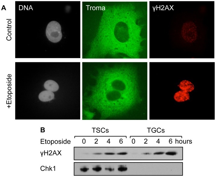
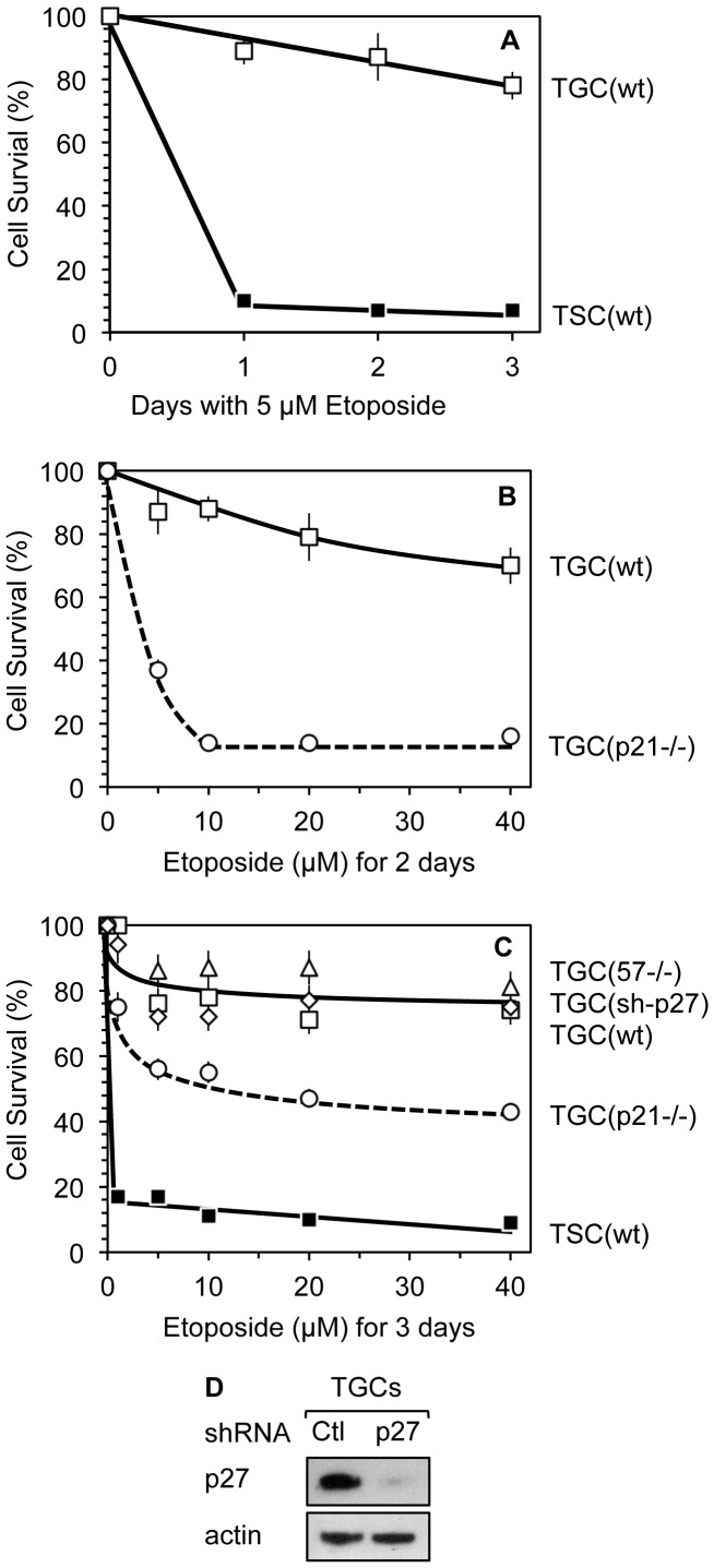
Proliferating trophoblast stem cells (TSCs) can differentiate into nonproliferating but viable trophoblast giant cells (TGCs) that are resistant to DNA damage induced apoptosis. Differentiation is associated with selective up-regulation of the Cip/Kip cyclin-dependent kinase inhibitors p57 and p21; expression of p27 remains constant. Previous studies showed that p57 localizes to the nucleus in TGCs where it is essential for endoreplication. Here we show that p27 also remains localized to the nucleus during TSC differentiation where it complements the role of p57. Unexpectedly, p21 localized to the cytoplasm where it was maintained throughout both the G- and S-phases of endocycles, and where it prevented DNA damage induced apoptosis. This unusual status for a Cip/Kip protein was dependent on site-specific phosphorylation of p21 by the Akt1 kinase that is also up-regulated in TGCs. Although cytoplasmic p21 is widespread among cancer cells, among normal cells it has been observed only in monocytes. The fact that it also occurs in TGCs reveals that p57 and p21 serve nonredundant functions, and suggests that the role of p21 in suppressing apoptosis is restricted to terminally differentiated cells.


Conflict of interest statement

Competing Interests: The authors have declared that no competing interests exist.

Figures

Figure 1
Figure 1. Only the Cip/Kip p27 protein is expressed in TSCs.
Proliferating TSCs were fixed, stained for nuclear DNA (Hoechst 33342), and stained with antibodies against p21, p27 or p57 proteins. Images were acquired with a confocal microscope (63x objective). Scale bars represent 50 µm.
Figure 2
Figure 2. The p27 and p57 proteins are localized to the nucleus in G-phase TGCs.
After three days of FGF4 deprivation, wild-type TGCs were cultured for 20 min in the presence of EdU (10 µM) to label S-phase cells. After fixation, EdU was detected using Click-iT chemistry (green), and the cells were stained with either anti-p57 (cyan) (A, C) or anti-p27 (red) (B, C) antibodies, and with Hoechst 33342 to visualize nuclear DNA (gray). Images were acquired with a confocal microscope (63x objective). The bottom panels are higher magnifications of merged images in order to visualize differential staining of EdU, p27 and p57. Scale bars represent 50 µm.
Figure 3
Figure 3. The p21 protein localized to the cytoplasm in TGCs.
After three days of FGF4 deprivation, wild-type and p21−/− TGCs were stained for nuclear DNA (gray), p21 (green) and p57 (red), as in figure 1. The p21−/− TGCs expressed nuclear p57 protein, but not cytoplasmic p21 protein. Images were acquired with an epifluorescence microscope (60x objective). The same exposure time was used for wild-type and p21−/− TGCs acquisitions.
Figure 4
Figure 4. The p21 protein localized to the cytoplasm of both S-phase and G-phase TGCs.
(A) Wild-type TGCs were prepared as in figure 2 and stained for nuclear DNA (gray), p57 (red), and p21 (green). Images were acquired with an epifluorescence microscope using a 40x objective. (B) The same TGCs were also cultured for 30 min with BrdU (10 µM). Following cell fixation and DNA denaturation, the cells were stained with anti-BrdU (green) and anti-p21 (red) antibodies. Images were acquired with an epifluorescence microscope with a 60x objective.
Figure 5
Figure 5. Akt1 is required for p21 stabilization in TGCs, and Akt1 phosphorylates p21 at T140.
(A) C-terminal sequence of mouse p21 protein with the putative Akt1 phosphorylation site (RKRRQTS) and the essential RKR nuclear localization signal in bold face. (B) Extracts of TSCs and TGCs were assayed by Western immuno-blotting using antibodies specific for Akt1, p21, p27 and p57 proteins. The phosphorylated form of p21 was recognized as a p21 protein that migrated slower than unphosphorylated p21, and by its reaction with an anti-p21 antibody specific for Thr-145 phosphorylation in human p21. (C) After three days of FGF4 deprivation, TGCs were stained with 4,6-diamidino-2-phenylindole (DAPI) to visualize nuclear DNA (gray) and with anti-Akt1 antibody (green). (D) Wild-type p21 and two p21 mutant forms with a T140V or a S141A substitution were tested as substrates for phosphorylation by Akt1 in vitro. (E) NIH3T3 fibroblasts were co-transfected with a plasmid expressing the tetracycline repressor and a plasmid encoding the indicated p21 protein whose expression was regulated by a tetracycline inducible promoter. Each protein carried a [His]6-cMyc-epitope tag fused to its C-terminus. After 24 hours of transfection, the cells were cultured for 18 hours in the presence of tetracycline (1 µg/ml) in order to induce expression of the indicated recombinant p21 protein. Cells were then harvested at 0, 6, 12 and 24 hours after release, and extracts were analyzed for the indicated protein by Western immuno-blotting using a Myc-Tag specific antibody. Wild-type (wt) p21 and the T140V (T/V) and S141A (S/A) p21 mutants, as well as a double mutant (T/V+S/A) were tested. Actin served as a loading control in each case.
Figure 6
Figure 6. Phosphomimetic mutations in the Akt1 phosphorylation site induced cytoplasmic localization of p21.
(A) Representative immuno-fluorescence images (60x objective) of ectopically expressed p21 protein 24 hours post-transfection of NIH3T3 cells using anti-Myc-Tag antibody illustrate nuclear, cytoplasmic, and nuclear plus cytoplasmic p21 localization. (B) Recombinant mouse p21 proteins were expressed in NIH3T3. Localization of the indicated p21 protein was detected by immuno-staining with an anti-Myc-Tag antibody, 24 hours after transfection. In addition to wild-type (wt) p21 and the T140V (T/V) and S141A (S/A) p21 mutants, T140E (T/E) and S141D (S/D) p21 phosphomimetic mutants, and a double phosphomimetic mutant (T/E+S/D) were also tested. More than 100 cells were scored for each transfection.
Figure 7
Figure 7. DNA damage can be induced in TGCs in the absence of the Chk1 kinase.
(A) Wild-type TGCs at three days post-FGF4 deprivation (control) were treated with 5 µM etoposide (+ Etoposide) for 3 days. Cells were stained with DAPI to visualize nuclear DNA (gray), Troma-1 antibody to visualize the cytoplasm (green), and anti-γH2AX to visualize areas where double stranded DNA breaks occurred (red). (B) Wild-type TSCs and TGCs three days after FGF4 deprivation were treated with 5 µM etoposide for the indicated times. Chk1 protein and histone H2AX (γH2AX) phosphorylation were assayed by western immuno-blotting.
Figure 8
Figure 8. Cytoplasmic p21 provided TGCs with resistance to DNA damage induced apoptosis.
(A) Wild-type TSCs (▪, 6×10⁵ cells), cultured in standard TSC medium, were treated with 5 µM etoposide for up to three days. Wild-type TGCs (□, 6×10⁵ cells) were produced by FGF4 deprivation of TSCs for three days, 5 µM etoposide was then added to the medium for up to three days. The number of cells remaining attached to the dish was recorded and the results calculated as the percentage of cells that survived. (B) Same as in A except that wild-type TGCs (□) and p21−/− TGCs (○, broken line) had been deprived of FGF4 for only two days before treating them with the indicated concentration of etoposide for two days. Wild-type and p21−/− TSCs were indistinguishable during their proliferation and their differentiation into TGCs. (C) Wild-type TSCs (▪) and TGCs (□), cultured as in panels A and B, were exposed to indicated concentration of etoposide for three days and cell survival was assessed as in B. Wild-type TGCs infected with lentivirus containing shRNA against p27 (⋄), p57−/− TGCs (▵) and p21−/− TGCs (○, broken line) were treated as well. (D) Depletion of p27 protein in TGCs treated either with shRNA against p27 RNA or scramble control shRNA (Ctl) was assayed by Western immuno-blotting. Errors bars indicate standard error of the mean for triplicate samples of each datum point.
Figure 9
Figure 9. Inhibition of Akt1 activity induced apoptosis.
(A) After three days of FGF4 deprivation, TGCs were transduced with lentiviruses expressing either control ‘scramble shRNA’ (Ctl) or shRNA targeted against Akt1. Two days after transduction, total cell lysates of surviving cells were analyzed by Western immuno-blotting for the indicated proteins. (B) DIC pictures of the cells three days after transduction (10x objective). (C) The fraction of cells remaining attached to the dish 2 days after lentivirus transduction are indicated for wild-type TSCs and TGCs, and for p21−/− TGCs. (D) TGCs 3 days post-FGF4 deprivation were treated with the indicated concentration of ‘Akt1 inhibitor-X’ (Santa Cruz) for the indicated period of time and cell survival calculated as in (C). Results are shown for wild-type TGCs (black bars), p57−/− TGCs (dark gray bars) and p21−/− TGCs (light gray bars). Error bars represent standard deviation.

Similar articles

Cited by

References

    1. Dillon RL, White DE, Muller WJ (2007) The phosphatidyl inositol 3-kinase signaling network: implications for human breast cancer. Oncogene 26: 1338–1345. - PubMed
    1. Dillon RL, Muller WJ (2010) Distinct biological roles for the akt family in mammary tumor progression. Cancer Res 70: 4260–4264. - PMC - PubMed
    1. Song G, Ouyang G, Bao S (2005) The activation of Akt/PKB signaling pathway and cell survival. J Cell Mol Med 9: 59–71. - PMC - PubMed
    1. Heron-Milhavet L, Franckhauser C, Rana V, Berthenet C, Fisher D, et al. (2006) Only Akt1 is required for proliferation, while Akt2 promotes cell cycle exit through p21 binding. Mol Cell Biol 26: 8267–8280. - PMC - PubMed
    1. Zhou BP, Liao Y, Xia W, Spohn B, Lee MH, et al. (2001) Cytoplasmic localization of p21Cip1/WAF1 by Akt-induced phosphorylation in HER-2/neu-overexpressing cells. Nat Cell Biol 3: 245–252. - PubMed

Publication types

MeSH terms

Substances