. 2014 Jun;6(6):504-10.
doi: 10.1038/nchem.1944. Epub 2014 May 11.

The cytotoxicity of (-)-lomaiviticin A arises from induction of double-strand breaks in DNA

Affiliations

The cytotoxicity of (-)-lomaiviticin A arises from induction of double-strand breaks in DNA

Laureen C Colis et al. Nat Chem. 2014 Jun.

Abstract

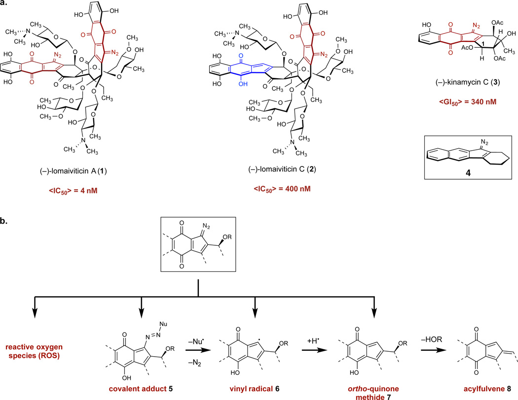
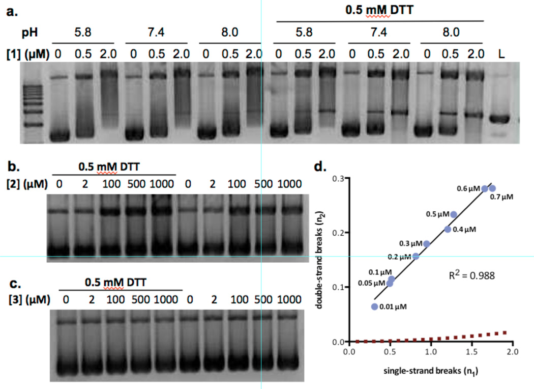
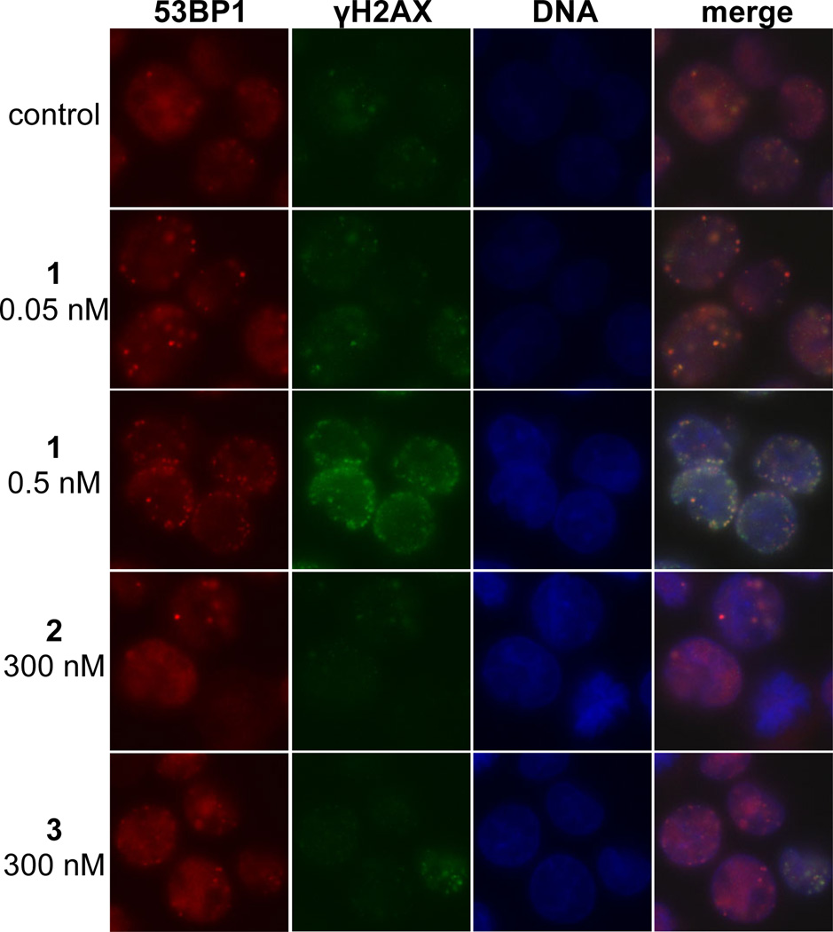
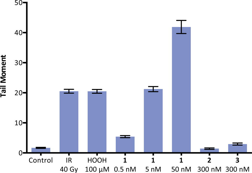
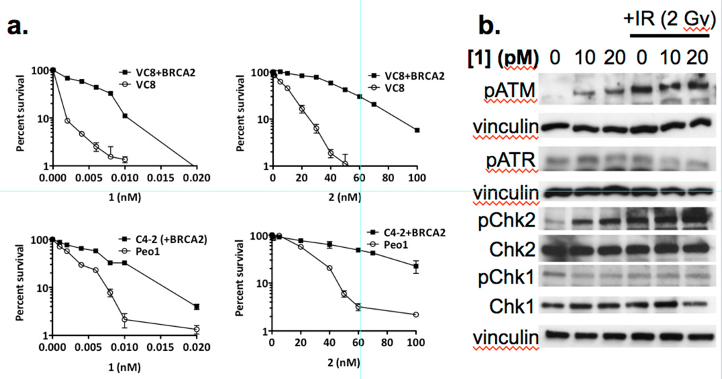
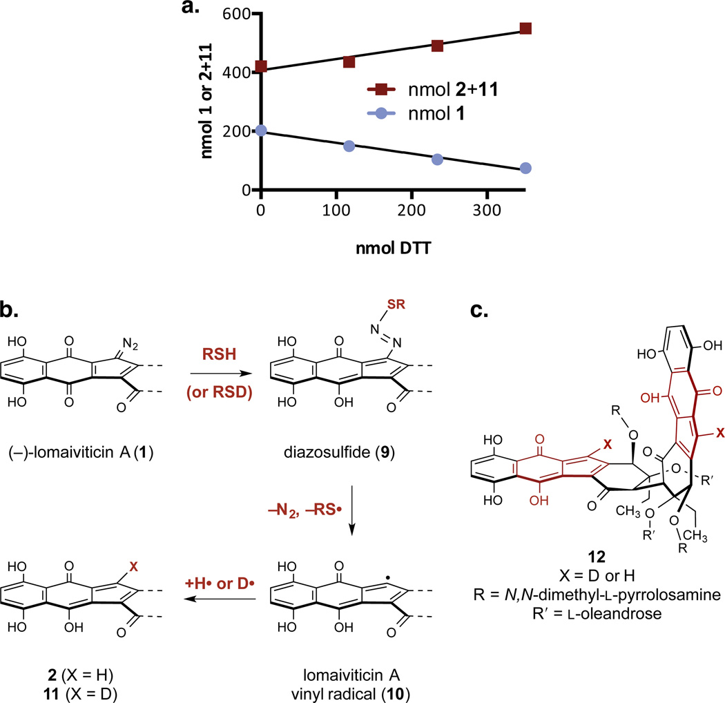
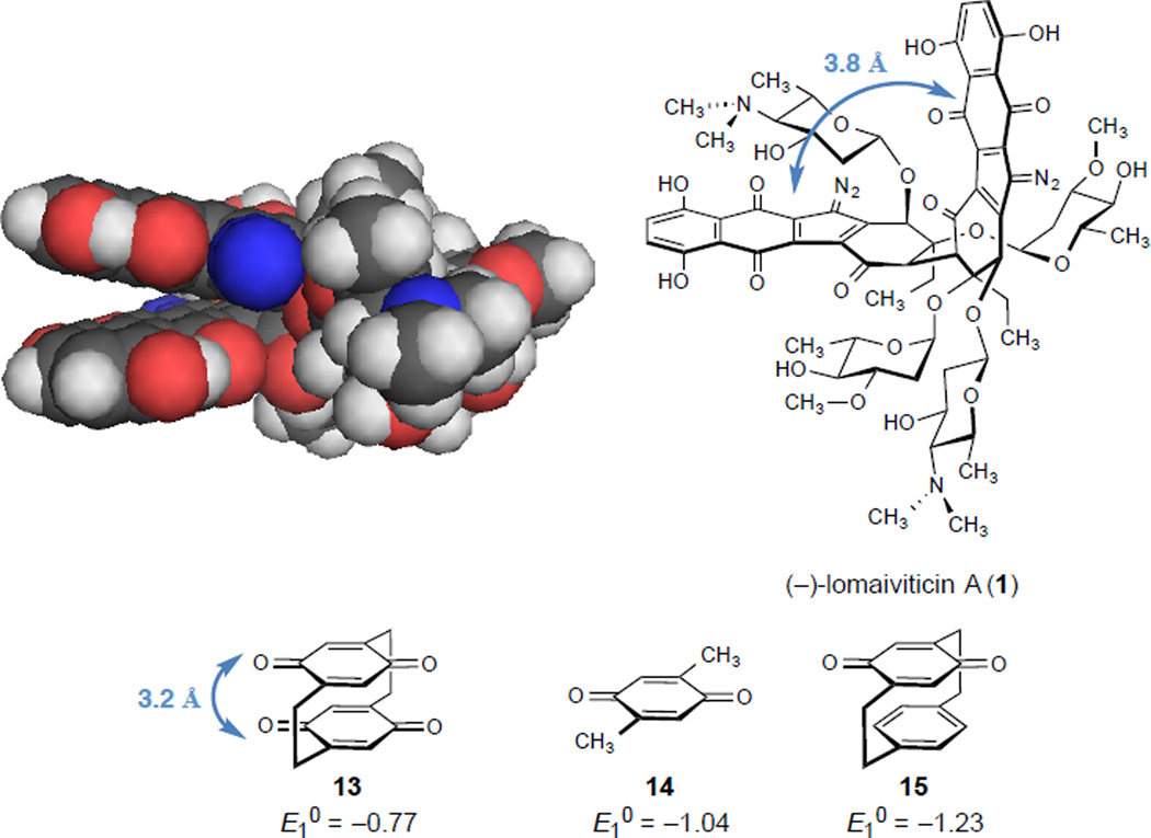
The metabolite (-)-lomaiviticin A, which contains two diazotetrahydrobenzo[b]fluorene (diazofluorene) functional groups, inhibits the growth of cultured human cancer cells at nanomolar-picomolar concentrations; however, the mechanism responsible for the potent cytotoxicity of this natural product is not known. Here we report that (-)-lomaiviticin A nicks and cleaves plasmid DNA by a pathway that is independent of reactive oxygen species and iron, and that the potent cytotoxicity of (-)-lomaiviticin A arises from the induction of DNA double-strand breaks (dsbs). In a plasmid cleavage assay, the ratio of single-strand breaks (ssbs) to dsbs is 5.3 ± 0.6:1. Labelling studies suggest that this cleavage occurs via a radical pathway. The structurally related isolates (-)-lomaiviticin C and (-)-kinamycin C, which contain one diazofluorene, are demonstrated to be much less effective DNA cleavage agents, thereby providing an explanation for the enhanced cytotoxicity of (-)-lomaiviticin A compared to that of other members of this family.


Figures

Figure 1
Figure 1
Structures of the metabolites employed in this study and their proposed reaction pathways. a. Structures of (–)-lomaiviticin A (1), (–)-lomaiviticin C (2), (–)-kinamycin C (3), and the diazotetrahydrobenzo[b]fluorene (diazofluorene, 4) functional group. b. Cytotoxic species proposed to form from the 1-diazo-1H-indene-4,7-dione. Red denotes the 1-diazo-1H-indene-4,7-dione functional group of 1, 2, and 3. Blue denotes the hydrofulvene functional group of 2.
Figure 2
Figure 2
Analysis of nicked and cleaved plasmid pBR322 DNA by (–)-lomaiviticin A (1), (–)-lomaiviticin C (2), and (–)-kinamycin C (3). a. Plasmid damage induced by 1, [1] = 0.5 or 2.0 µM, pH = 5.8, 7.4, or 8.0, [DTT] = 0 or 0.5 mM, 37 °C, 16 h. b. Plasmid damage induced by 2, [2] = 2–1000 µM, pH 7.4, [DTT] = 0 or 0.5 mM, 37 °C, 16 h. c. Plasmid damage induced by 3, [3] = 2–1000 µM, pH 7.4, [DTT] = 0 or 0.5 mM, 37 °C, 16 h. Top band: Form II (nicked) DNA, middle band: Form III (linearized) DNA, bottom band: Form I (supercoiled) DNA. d. Ratio of DNA dsbs () to ssbs () per DNA molecule in a plasmid cleavage assay using 1. Data points (left to right) correspond to 0.01, 0.05, 0.1, 0.2, 0.3, 0.4, 0.5, 0.6, 0.7 µM of 1. The dashed line represents the Freifelder–Trumbo relationship, defined as n2=n12(2h+1)/4L where h = 23.8 and L = 4361. Quantitative analyses and statistical treatments are presented in the Supporting Information.
Figure 3
Figure 3
Immunofluorescence imaging of γH2AX and 53BP1 foci in K562 cells treated with (–)-lomaiviticin A (1), (–)-lomaiviticin C (2), or (–)-kinamycin C (3). γH2AX and 53BP1 are commonly used markers (refs. 31–33) for DNA dsbs. Immunofluorescence imaging shows that these foci are induced and colocalize in K562 cells treated with 1 (0.05–0.5 nM), but are sparse or undetectable in cells treated with 300 nM 2 or 3. Columns (left to right), 53BP1 (red), γH2AX (green), nucleus (blue), merge. Rows (top to bottom): control, 0.05 nM 1, 0.5 nM 1, 300 nM (–)-lomaiviticin C (2), 300 nM (–)-kinamycin C (3). K562 cells in exponential growth phase were incubated with 0.05 nM 1, 0.5 nM 1, 300 nM 2, or 300 nM 3 for 4 h. Immunological detection was performed using a primary antibody [rabbit polyclonal anti-53BP1 antibody (Novus Biologicals) and mouse monoclonal anti-phospho-histone H2AX (SER139) antibody (Upstate)] and visualized with Alexa 488 (goat-anti-mouse IgG) and Alexa 594 (goat-anti-rabbit IgG). Mounting medium contained DAPI to visualize nuclear DNA.
Figure 4
Figure 4
Neutral comet unwinding assay of K562 cells treated with (–)-lomaiviticin A (1), (–)-lomaiviticin C (2), or (–)-kinamycin C (3). (–)-Lomaiviticin A (1) induces DNA dsb formation in K562 cells at 0.5–50 nM concentrations, while 2 and 3 induce minimal production of DNA double-strand breaks (dsbs) at 300 nM concentrations. Tail moment obtained in a neutral comet unwinding assay employing 1 (0.5, 5.0, or 50 nM), 2 (300 nM), or 3 (300 nM) and K562 cells. Drug exposure was 30 min. Bars represent mean tail moment (60–140 cells), error bars represent standard error of the mean. IR = ionizing radiation. Tail moment represents the extent of DNA cleavage and is defined as the product of the tail length and the fraction of DNA in the tail. The neutral comet assay is a method for the selective detection of DNA dsbs in tissue culture (see ref. 34).
Figure 5
Figure 5
Clonogenic survival curves and western blot analysis of cells treated with (–)-lomaiviticin A (1) or (–)-lomaiviticin C (2). a. Clonogenic survival curves for BRCA2-deficient VC8 and Peo1 cells and the corresponding isogenic cell lines transfected with and expressing a functional BRCA2 gene (VC8+BRCA2 and C4-2, respectively). Solid data points represent the BRCA2-proficient cell lines (VC8+BRCA2 and C4-2), unfilled data points represent the BRCA2-deficient cell lines (VC8 and Peo1). Left graphs: cells treated with 1. Right graphs: cells treated with 2. These data support DNA as the target of 1 and 2 as BRCA2 is involved in DNA repair and BRCA2-deficient cell lines are hypersensitive to DNA damaging agents (ref. 36). b. (–)-Lomaiviticin A (1) upregulates pATM and pChk2, but not pATR and pChk1, in MCF-7 cells. Western blot analysis of pATM, pATR, pChk2, Chk2, pChk1, and Chk1 in MCF-7 cells treated with 1 (10 or 20 pM). These data support production of DNA dsbs by 1 as pATM and pChk2 are involved in DNA dsb repair, while pATR and pChk1 respond primarily to replication fork stalling (ref. 37).
Figure 6
Figure 6
Relative reactivity studies and mechanistic pathways for reduction of the diazofluorene. a. Competition hydrodediazotization experiment between (–)-lomaiviticin A (1) and (–)-lomaiviticin C (2). 1/m = –0.01163, 0.01132 for 1, 2, respectively. Conditions: 1 (202 nmol), 2 (421 nmol), DTT (3 × 117 nmol), methanol-d4 (400 µL), 21 °C. b. Postulated pathway for the transformation of 1 to 2 and 11. c. Structure of the double hydrodediazotization product 12.
Figure 7
Figure 7

Comment in

References

    1. He H, et al. Lomaiviticins A and B, Potent Antitumor Antibiotics from Micromonospora lomaivitiensis. J. Am. Chem. Soc. 2001;123:5362–5363. - PubMed
    1. Woo CM, Beizer NE, Janso JE, Herzon SB. Isolation of Lomaiviticins C–E. Transformation of Lomaiviticin C to Lomaiviticin A, Complete Structure Elucidation of Lomaiviticin A, and Structure–Activity Analyses. J. Am. Chem. Soc. 2012;134:15285–15288. - PubMed
    1. Kersten RD, et al. Bioactivity-Guided Genome Mining Reveals the Lomaiviticin Biosynthetic Gene Cluster in Salinispora tropica. Chembiochem. 2013;14:955–962. - PMC - PubMed
    1. Ito S, Matsuya T, Ōmura S, Otani M, Nakagawa A. A New Antibiotic, Kinamycin. J. Antibiot. 1970;23:315–317. - PubMed
    1. Hata T, Ōmura S, Iwai Y, Nakagawa A, Otani M. A New Antibiotic, Kinamycin: Fermentation, Isolation, Purification and Properties. J. Antibiot. 1971;24:353–359. - PubMed

Publication types