. 2014 May 21;9(5):e97448.
doi: 10.1371/journal.pone.0097448. eCollection 2014.

Modulated expression of genes encoding estrogen metabolizing enzymes by G1-phase cyclin-dependent kinases 6 and 4 in human breast cancer cells

Affiliations

Modulated expression of genes encoding estrogen metabolizing enzymes by G1-phase cyclin-dependent kinases 6 and 4 in human breast cancer cells

Yi Jia et al. PLoS One. .

Abstract

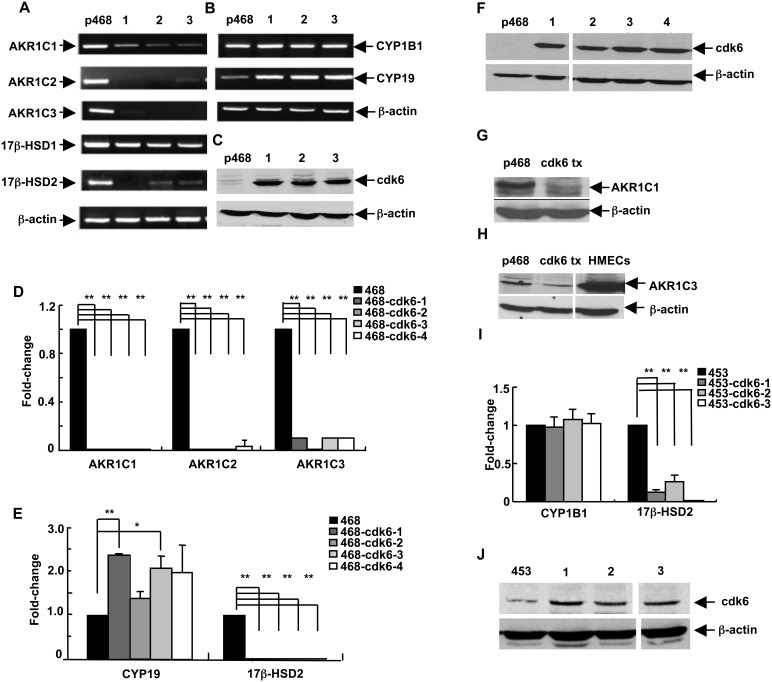
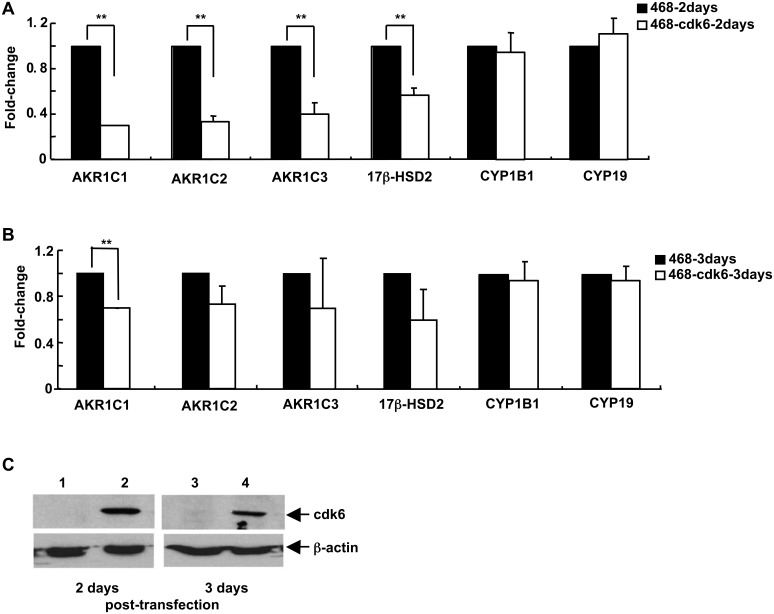
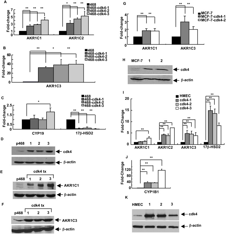
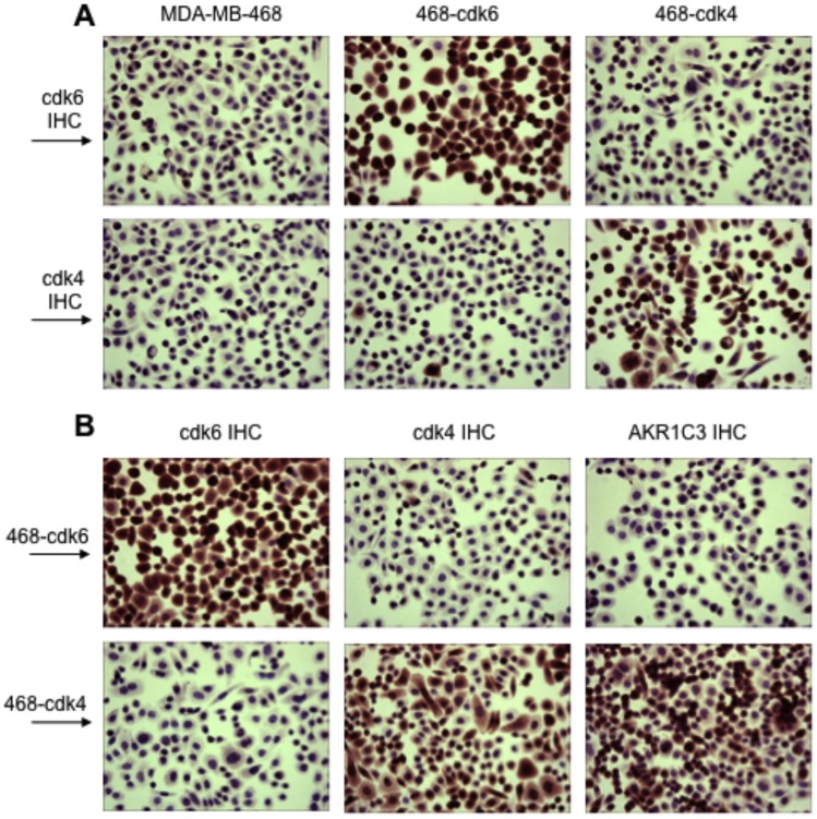
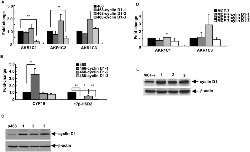
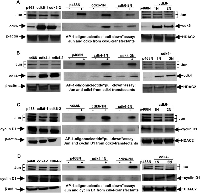
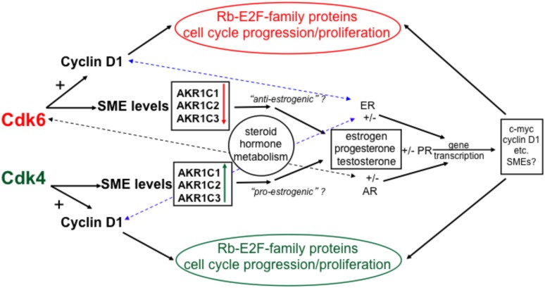
G1-phase cell cycle defects, such as alterations in cyclin D1 or cyclin-dependent kinase (cdk) levels, are seen in most tumors. For example, increased cyclin D1 and decreased cdk6 levels are seen in many human breast tumors. Overexpression of cdk6 in breast tumor cells in culture has been shown to suppress proliferation, unlike the growth stimulating effects of its close homolog, cdk4. In addition to directly affecting proliferation, alterations in cdk6 or cdk4 levels in breast tumor cells also differentially influence levels of numerous steroid metabolic enzymes (SMEs), including those involved in estrogen metabolism. Overexpression of cdk6 in tumor cell lines having low cdk6 resulted in decreased levels of mRNAs encoding aldo-keto reductase (AKR)1C1, AKR1C2 and AKR1C3, which are hydroxysteroid dehydrogenases (HSDs) involved in steroid hormone metabolism. In contrast, increasing cdk4 dramatically increased these transcript levels, especially those encoding AKR1C3, an enzyme that converts estrone to 17β-estradiol, a change that could result in a pro-estrogenic state favoring tumor growth. Effects on other estrogen metabolizing enzymes, including cytochrome P450 (CYP) 19 aromatase, 17β-HSD2, and CYP1B1 transcripts, were also observed. Interactions of cdk6 and cdk4, but not cyclin D1, with the promoter region of a cdk-regulated gene, 17β-HSD2, were detected. The results uncover a previously unsuspected link between the cell cycle and hormone metabolism and differential roles for cdk6 and cdk4 in a novel mechanism for pre-receptor control of steroid hormone action, with important implications for the origin and treatment of steroid hormone-dependent cancers.


Conflict of interest statement

Competing Interests: The authors have declared that no competing interests exist.

Figures

Figure 1
Figure 1. The pattern of SME gene transcripts in breast cancer cells is altered in stably-transfected cell lines with increased cdk6 protein levels.
For panels (A) through (H), MDA-MB-468 cells were stably-transfected with a sequence encoding cdk6. (A) Levels of the 6 indicated transcripts were detected by RT-PCR in samples from parental MDA-MB-468 cells (“p468”) and in 3 stably-transfected clonal cell lines (labeled 1, 2, and 3). (B) Levels of the 3 indicated transcripts were detected by RT-PCR in the same cell lines. (C) Cdk6 protein levels were detected by immunoblot analysis, with β-actin levels as the loading control. (D) AKR1C1, AKR1C2, and AKR1C3 transcript levels in parental MDA-MB-468 cells (468) and in 4 stably-transfected cell lines (468-cdk6-1 through 468-cdk6-4) were detected and quantitated by qRT-PCR. (E) CYP19 and 17β-HSD2 transcript levels were quantitated in the 5 cell lines. (F) Cdk6 protein levels in parental MDA-MB-468 and the 4 cdk6-transfectant cell lines analyzed in (D) and (E) were detected by immunoblot analysis, with β-actin levels as the loading control. (G) AKR1C1 protein levels in parental MDA-MB-468 (p468) cells and a cdk6-transfectant cell line (cdk6 tx) were detected by immunoblot analysis, with β-actin as the loading control. (H) AKR1C3 protein levels in parental MDA-MB-468 cells (p468), in a cdk6 transfectant cell line (cdk6 tx) and in normal HMECs were detected by immunoblot analysis, with β-actin as the loading control. For panels (I) and (J), MDA-MB-453 breast epithelial cells were stably-transfected with sequences encoding cdk6. (I) CYP1B1 and 17β-HSD transcript levels in parental MDA-MB-453 cells (453) and in 3 stably-transfected cell lines (453-cdk6-1 through-4) were detected and quantitated by qRT-PCR. (J) Cdk6 protein levels in parental MDA-MB-453 and the 3 cdk6-transfectant cell lines analyzed in I were detected by immunoblot analysis, with β-actin levels as loading control. For panels (D), (E), and (I), the data are expressed as the mean±SEM, n = 3 times/group; *p<0.05 and **p<0.01. Note the differences in Y-axis scales on the graphs for panels (D), (E), and (I).
Figure 2
Figure 2. The pattern of SME gene transcripts in MDA-MB-468 breast cancer cells is altered by transient increased expression of cdk6.
MDA-MB-468 cells were transiently transfected with a sequence encoding cdk6. RNA was prepared from cells transfected with the cdk6 sequence at (A) 2 days and (B) 3 days after transfection and assayed by qRT-PCR for transcript levels of AKR1C1, AKR1C2, AKR1C3, 17β-HSD2, CYP1B1, and CYP19. (C) The levels of cdk6 protein were determined in mock-transfected (lanes 1 and 3) and cdk6-transfected (lanes 2 and 4) cells at 2 days (lanes 1 and 2) and 3 days (lanes 3 and 4) after transfection, with β-actin as the loading control. For panels (A) and (B), the data are expressed as the mean ± SEM, n = 3 times/group and **p<0.01.
Figure 3
Figure 3. The pattern of SME gene transcripts in breast epithelial cells is altered by expression of cdk4.
For panels (A) through (F), MDA-MB-468 breast epithelial cells were stably-transfected with a sequence encoding cdk4. (A) AKR1C1 and AKR1C2 transcript levels in parental MDA-MB-468 cells (468) and in 3 stably-transfected cell lines (468-cdk4-1 through 468-cdk4-3) were detected and quantitated by qRT-PCR. (B) AKR1C3 transcript levels were similarly quantitated in the 4 cell lines. (C) CYP19 and 17β-HSD2 levels were similarly quantitated in the 4 cell lines. (D) The cdk4 protein levels in parental cells (lane labeled p468) and the cdk4-transfectant cell lines (lanes labeled 1, 2, and 3) were detected by immunoblot analysis, with β-actin as loading control. Increased expression of both (E) AKR1C1 protein and (F) AKR1C3 protein in cells stably-transfected with the cdk4 sequence was demonstrated by immunoblot analysis, with β-actin as the loading control. For panels (G) and (H), MCF-7 breast epithelial cells were stably-transfected with a sequence encoding cdk4. (G) AKR1C1 and AKR1C3 transcript levels in parental MCF-7 cells and in 2 stably-transfected cell lines (MCF-7-cdk4-1 and MCF-7-cdk4-2) were detected and quantitated by qRT-PCR. (H) The cdk4 protein levels in parental cells (lane labeled MCF-7) and the cdk4-transfectant cell lines (lanes labeled 1 and 2) were detected by immunoblot analysis, with β-actin as the loading control. For panels (I) through (K), normal HMECs were transfected with a sequence encoding cdk4. (I) AKR1C1, AKR1C2, AKR1C3, and 17β-HSD2 transcript levels were quantitated in cells that had been transfected with cdk4, incubated for 48 hrs in regular medium, for 2 weeks in medium containing G418 and an additional week in medium without the selective agent. Results are shown for mock-transfected HMECs and 3 transfected cultures (cdk4-1 through cdk4-3). (J) CYP1B1 transcript levels were also determined in the cells. (K) Levels of cdk4 protein in the cultures were determined by immunoblot analysis, with β-actin as loading control. For panels (A), (B), (C), (G), (I), and (J), the data are expressed as the mean ± SEM, n = 3 times/group; *p<0.05 and **p<0.01. Note the differences in Y-axis scales on the graphs for panels (A), (B), (C), (G), (I), and (J).
Figure 4
Figure 4. Immunohistochemical detection of cdk6, cdk4, and AKR1C3 in MDA-MB-468 cell lines transfected to express increased levels of cdk6 or cdk4.
In the top row of panel (A), cdk6 was detected by immunohistochemistry (cdk6 IHC) in parental MDA-MB-468 cells and in cell lines transfected to express increased levels of either cdk6 (“468-cdk6” cells) or cdk4 (“468-cdk4” cells). In the second row, the 3 cell types were assessed for cdk4 levels. In the top row of panel (B), cells expressing increased amounts of cdk6 (“468-cdk6” cells) were analyzed by IHC for cdk6, cdk4, or AKR1C3. In the second row, cells expressing increased amounts of cdk4 (“468-cdk4” cells) were analyzed for the 3 molecules by IHC. Magnification: x200. The results indicate that nearly all of the cdk4-transfected cells have greatly increased levels of AKR1C3 protein.
Figure 5
Figure 5. The pattern of SME gene transcripts in breast cancer cells is largely independent of cyclin D1 levels.
For panels (A) through (C), MDA-MB-468 breast epithelial cells were stably-transfected with a sequence encoding cyclin D1. (A) AKR1C1, AKR1C2, and AKR1C3 transcript levels in parental MDA-MB-468 cells (468) and in 3 stably-transfected cell lines (468-cyclin D1-1 through 468-cyclin D1-3) were detected and quantitated by qRT-PCR. (B) CYP19 and 17β-HSD2 transcript levels were similarly quantitated in the 4 cell lines. (C) The cyclin D1 protein levels in parental MDA-MB-468 cells and the 3 cyclin D1-transfectant cell lines analyzed in panels (A) and (B) were detected by immunoblot analysis, with β-actin levels as the loading control. For panels (D) and (E), MCF-7 breast epithelial cells were stably-transfected with a sequence encoding cyclin D1. (D) AKR1C1 and AKR1C3 transcript levels in parental MCF-7 cells and in 3 stably-transfected cell lines (MCF-7-cyclin D1-1 through MCF-7-cyclin D1–3) were detected and quantitated by qRT-PCR. (E) The cyclin D1 protein levels in parental MCF-7 and the 3 cyclin D1-transfectant cell lines analyzed in panel (D) were detected by immunoblot analysis, with β-actin levels as the loading control. For panels (A), (B), and (D), the data are expressed as the mean ± SEM, n = 3 times/group; *p<0.05 and **p<0.01. Note the differences in Y-axis scales on the graphs for panels (A), (B), and (D).
Figure 6
Figure 6. Association of cdk6 and cdk4, but not cyclin D1, with the AP-1 promoter sequence of the 17β-HSD2 gene.
A biotin-labeled oligonucleotide representing the sequence was used in oligonucleotide “pull-down” assays with nuclear extracts from MDA-MB-468 parental cells and from 2 cell lines stably-transfected with cdk6 (panels (A) and (C)) and 2 cell lines transfected with cdk4 (panels (B) and (D)). The oligonucleotide and associated nuclear proteins were collected by binding to streptavidin-conjugated beads. The proteins associated with the oligonucleotide (shown in the lanes marked “+” in the 2 center immunoblots in each panel ((A) through (D)) were probed for Jun (top immunoblot in (A) through (D)) and either cdk6 (A), cdk4 (B), or cyclin D1 ((C) and (D)), respectively (bottom immunoblots). Lanes designated as “−” are negative control samples in which the nuclear extracts were incubated without the oligonucleotide but then subjected to collection using streptavidin-conjugated beads. Immunoblots to the left of the center blots show the levels of Jun, β-actin, and either (A) cdk6, (B) cdk4, or (C) and (D) cyclin D1 in whole cell extracts from the parental MDA-MB-468 cells (p468) and the cdk6 (cdk6-1 and cdk6-2) and cdk4 (cdk4-1 and cdk4-2) transfectants. Immunoblots to the right of the center blots show the levels of Jun, HDAC2, and either (A) cdk6, (B) cdk4, or (C) and (D) cyclin D1 in nuclear extracts from the parental MDA-MB-468 cells (p468N) and from the cdk6 (cdk6-1N and cdk6-2N) and cdk4 (cdk4-1N and cdk4-2N) transfectants.
Figure 7
Figure 7. Regulatory functions of cdk6 and cdk4 in breast tumor epithelial cells: interactions of the cell cycle and steroid hormone metabolism and function.
Cdk4 and cdk6 interact with cyclin D1 to regulate cell cycle progression and proliferation, through Rb and E2F-family proteins. The cdks can also regulate levels of SMEs, including the AKR1C-family of enzymes. Induction or suppression of AKR1C-family enzymes by cdk4 or cdk6, respectively, could induce either a pro- or anti-estrognic state in a breast tumor. The dashed blue lines indicate that the ER can interact with cyclin D1 to affect gene transcription, even in the absence of estrogen , . The dashed black line indicates the interaction of the androgen receptor with cdk6, which can stimulate androgen receptor-directed gene transcription, independently of cyclin D1 . Steroid hormones bound to cognate steroid receptors regulate transcription of many genes that can affect tumor cell growth and function, including those encoding proteins that directly affect the cell cycle, such as c-myc and cyclin D1 , and perhaps SME genes themselves .

Similar articles

Cited by

References

    1. Ho A, Dowdy SF (2002) Regulation of (G)1 cell-cycle progression by oncogenes and tumor suppressor genes. Curr Opin Genet Dev 12: 47–52. - PubMed
    1. Fu M, Wang C, Li Z, Sakamaki T, Pestell RG (2004) Cyclin D1: normal and abnormal functions. Endocrinol 145: 5439–5447. - PubMed
    1. Yu Q, Sicinska E, Geng Y, Ahnstrom M, Zagozdzon A, et al. (2006) Requirement for cdk4 kinase function in breast cancer. Cancer Cell 9: 23–32. - PubMed
    1. Grossel MJ, Hinds PW (2006) Beyond the cell cycle: A new role for cdk6 in differentiation. J Cell Biochem 97: 485–493. - PubMed
    1. Anders L, Ke N, Hydbring P, Choi YJ, Widlund HR, et al. (2011) A systematic screen for CDK4/6 substrates links FOXM1 phosphorylation to senescence suppression in cancer cells. Cancer Cell 20: 620–634. - PMC - PubMed

Publication types

MeSH terms

Substances