. 2014 Aug 15;74(16):4515-25.
doi: 10.1158/0008-5472.CAN-13-3275. Epub 2014 May 21.

β-catenin activation in a novel liver progenitor cell type is sufficient to cause hepatocellular carcinoma and hepatoblastoma

Affiliations

β-catenin activation in a novel liver progenitor cell type is sufficient to cause hepatocellular carcinoma and hepatoblastoma

Sharada Mokkapati et al. Cancer Res. .

Abstract

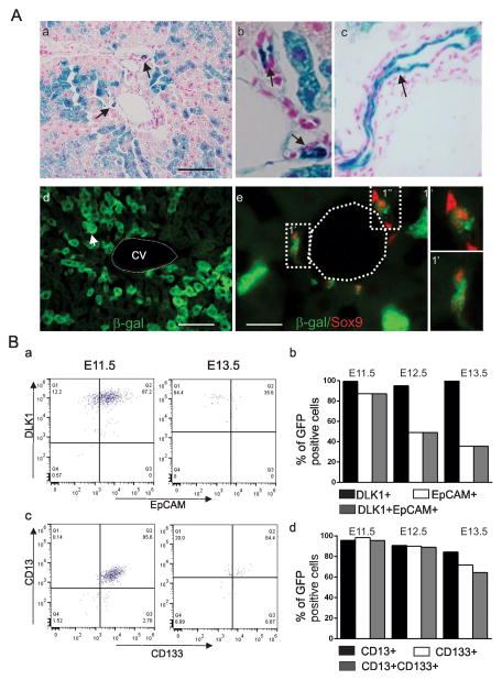
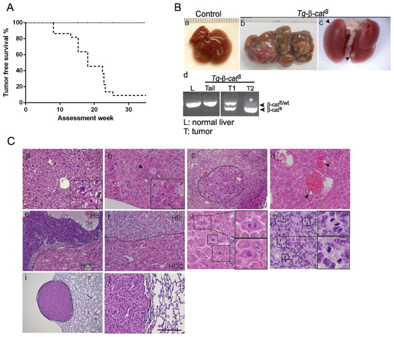
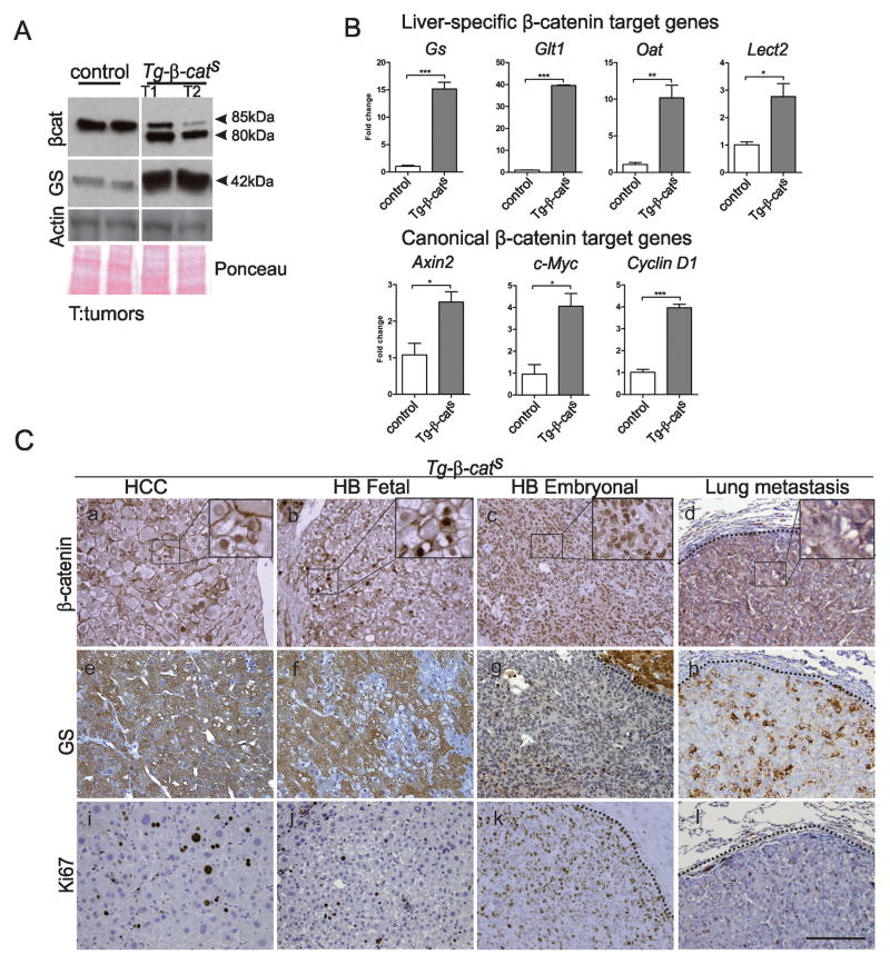
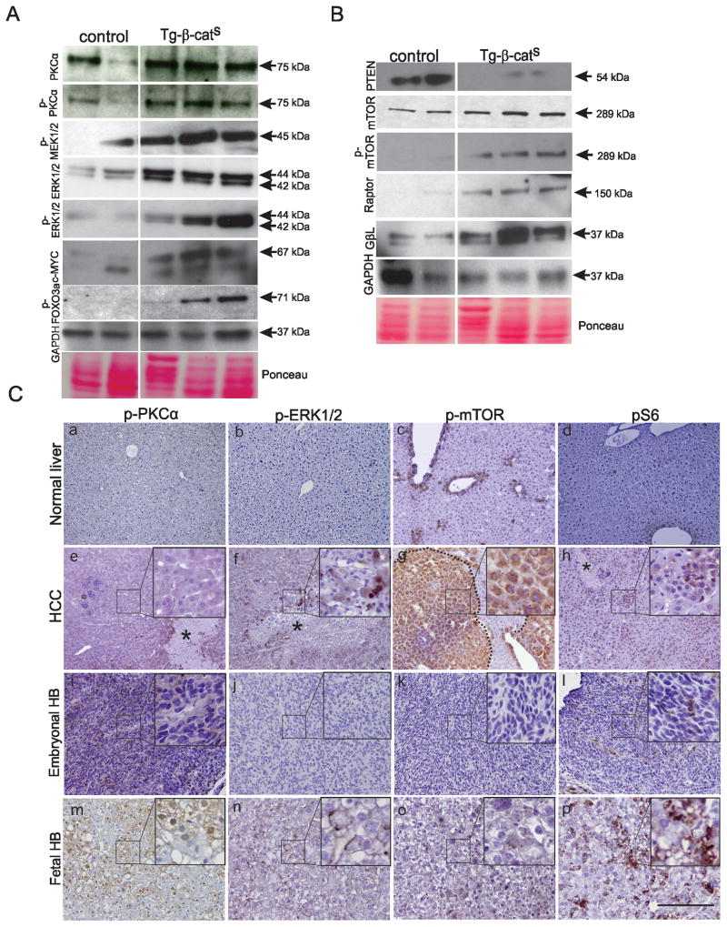
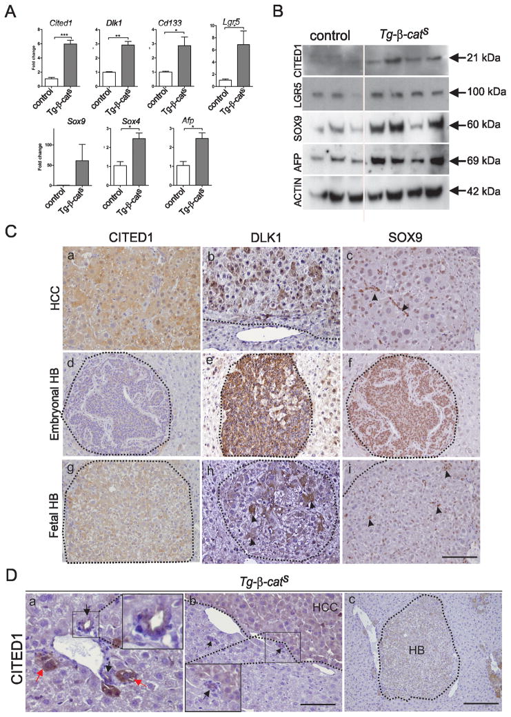
Hepatocellular carcinoma (HCC) was thought historically to arise from hepatocytes, but gene expression studies have suggested that it can also arise from fetal progenitor cells or their adult progenitor progeny. Here, we report the identification of a unique population of fetal liver progenitor cells in mice that can serve as a cell of origin in HCC development. In the transgenic model used, mice carry the Cited1-CreER(TM)-GFP BAC transgene in which a tamoxifen-inducible Cre (CreER(TM)) and GFP are controlled by a 190-kb 5' genomic region of Cited1, a transcriptional coactivator protein for CBP/p300. Wnt signaling is critical for regulating self-renewal of progenitor/stem cells and has been implicated in the etiology of cancers of rapidly self-renewing tissues, so we hypothesized that Wnt pathway activation in CreER(TM)-GFP(+) progenitors would result in HCC. In livers from the mouse model, transgene-expressing cells represented 4% of liver cells at E11.5 when other markers were expressed, characteristic of the hepatic stem/progenitor cells that give rise to adult hepatocytes, cholangiocytes, and SOX9(+) periductal cells. By 26 weeks of age, more than 90% of Cited1-CreER(TM)-GFP;Ctnnb1(ex3(fl)) mice with Wnt pathway activation developed HCC and, in some cases, hepatoblastomas and lung metastases. HCC and hepatoblastomas resembled their human counterparts histologically, showing activation of Wnt, Ras/Raf/MAPK, and PI3K/AKT/mTOR pathways and expressing relevant stem/progenitor cell markers. Our results show that Wnt pathway activation is sufficient for malignant transformation of these unique liver progenitor cells, offering functional support for a fetal/adult progenitor origin of some human HCC. We believe this model may offer a valuable new tool to improve understanding of the cellular etiology and biology of HCC and hepatoblastomas and the development of improved therapeutics for these diseases.


Conflict of interest statement

Conflict of interest

No conflicts to disclose.

Figures

Figure 1
Figure 1. Characterization of CreER-GFP+ cells in fetal liver
A. P60 liver sections showing X-gal+ liver cells, hepatocytes and bile ducts (arrows, a), higher magnification showing the small (arrows, b) and large bile ducts (c). Antibody staining of cryosections (P60) for β-galactosidase in hepatocytes (arrow, d) and in SOX9 positive (red nuclei) bile duct cells (e). 1′ and 1″ show higher magnification. Scale bar, 160 μm (a, d) and 32 μm (e). B. FACS analysis for DLK1 and EPCAM (a,b), CD13 and CD133 (c,d) in the CreER-GFP+ cells at E11.5 – E13.5.
Figure 2
Figure 2. Development of liver tumors in Tg-β-catS mice
A. Kaplan-Meier analysis of tumor incidence in mutant animals (dotted line) and littermate controls (solid line). B. Gross appearance of normal liver (a), liver with multifocal tumors in Tg-β-catS mice (b), and lung metastases (arrowheads, c). Representative PCR from Tg-β-catS mice showing wildtype/floxed bands in tail and liver DNA of control mice and mutant tumors (d). Asterisk (*) indicates loss of the wildtype allele in T2. C. Histological analysis of liver tumors (b–h) and associated lung metastasis (i,j). H&E staining of normal liver (a), altered hepatocytes (denoted by arrow, b) adenoma (demarcated by dotted line, c) HCC (d–f), HB embryonal (e) and HB pure fetal (f). Insets in (a) and (b) provide higher magnification (3X). Mitotic figures of HCC (g) and embryonal undifferentiated HB (h). Lung metastasis (dotted lines, i), and higher magnification (j). Scale bar, 80 μm (a,b,d,j), 500 μm (c,e,f), and 1280 μm (i).
Figure 3
Figure 3. Activation of Wnt pathway in Tg-β-catS tumors
A. Western blot analysis of Tg-β-catS tumors and normal control livers for β-catenin and glutamine synthetase (GS). β-Actin and Ponceau stained membrane (P) were used as loading controls. B. Q-PCR analysis of liver-specific β-catenin target genes (Gs), glutamate transporter 1 (Glt1), ornithine aminotransferase (Oat) and Lect2 in Tg-β-catS tumors (n=3) and canonical target genes Axin2, c-Myc, and CyclinD, compared to littermate normal livers (n=3). C. β-catenin IHC in HCC (a), HB pure fetal (b), embryonal undifferentiated HB (c), and lung metastasis (d). GS staining in HCC, fetal HB, embryonal HB, and lung metastasis (e–h). Ki67 staining in HCC, HB, and metastasis (i–l). Scale bar, 80 μm.
Figure 4
Figure 4. Activation of the MAPK and mTOR pathways in Tg-β-catS tumors
A, B. Western blot analysis of MAPK and mTOR pathway components in HCC from Tg-β-catS HCCs compared to control livers. GAPDH and Ponceau stained membranes were used as loading controls. C. IHC analysis of phospho-PKCα (a,e,I,m), phospho-ERK1/2 (b,f,j,n), phospho-mTOR (c,g,k,o), and pS6 (d,h,l,p) in the normal liver (a–d), HCCs (eh), embryonal HB (i–l) and fetal HB (m–p) of Tg-β-catS mice. Insets show higher magnification (e–p). Necrotic areas denoted by *. Scale bar, 80μm.
Figure 5
Figure 5. Expression of hepatic stem/progenitor markers in Tg-β-catS tumors
A. RT-PCR analysis of HCCs (n=3) and control livers (n=3) for expression of stem/progenitor markers. B. Western blot analysis of stem/progenitor markers in Tg-β-catS HCCs. C. IHC showing expression of CITED1 (a,d,g), DLK1 (b,e,h), and SOX9 (arrowheads, c, i and f) in HCC (a–c) embryonal HB (d–f) and fetal HB (g–i). Scale bar, 70 μm. D. IHC showing CITED1 expression in bile duct epithelium (black arrows, a) and in hepatocytes emerging around the bile ducts (red arrows, a). Tumors (HCC, b and HB, c) originating around the bile ducts. Insets in (a) and (b) show CITED1 expression in bile duct epithelium. Scale bar; a,b, 70 μm; c, 160 μm.

Similar articles

Cited by

References

    1. El-Serag HB. CURRENT CONCEPTS Hepatocellular Carcinoma. New England Journal of Medicine. 2011;365:1118–27. - PubMed
    1. Thorgeirsson SS, Lee JS, Heo J, Libbrecht L, Chu IS, Kaposi-Novak P, et al. A novel prognostic subtype of human hepatocellular carcinoma derived from hepatic progenitor cells. Nature Medicine. 2006;12:410–6. - PubMed
    1. Whittaker S, Marais R, Zhu AX. The role of signaling pathways in the development and treatment of hepatocellular carcinoma. Oncogene. 2010;29:4989–5005. - PubMed
    1. Guichard C, Imbeaud S, Amaddeo G, Ben Maad I, Letouze E, Pelletier L, et al. Landscape of Somatic Mutation in Hepatocellular Carcinoma. Journal of Hepatology. 2012;56:S8-S.
    1. Monga SPS. Role of Wnt/beta-catenin signaling in liver metabolism and cancer. International Journal of Biochemistry & Cell Biology. 2011;43:1021–9. - PMC - PubMed

Publication types

MeSH terms