. 2014 Oct;131(2):219-28.
doi: 10.1111/jnc.12766. Epub 2014 Jun 13.

A vesicular sequestration to oxidative deamination shift in myocardial sympathetic nerves in Parkinson's disease

Affiliations

A vesicular sequestration to oxidative deamination shift in myocardial sympathetic nerves in Parkinson's disease

David S Goldstein et al. J Neurochem. 2014 Oct.

Abstract

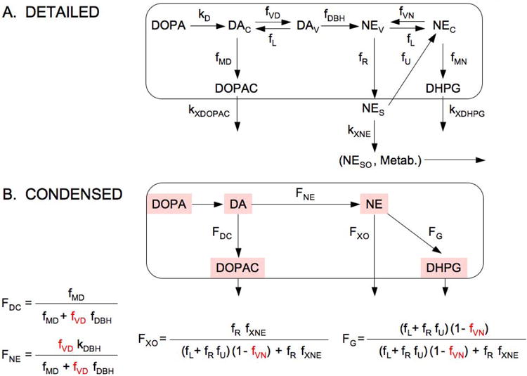
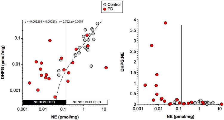
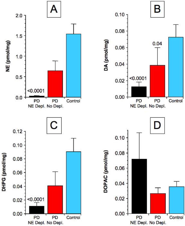
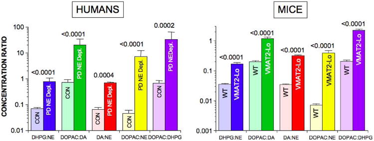
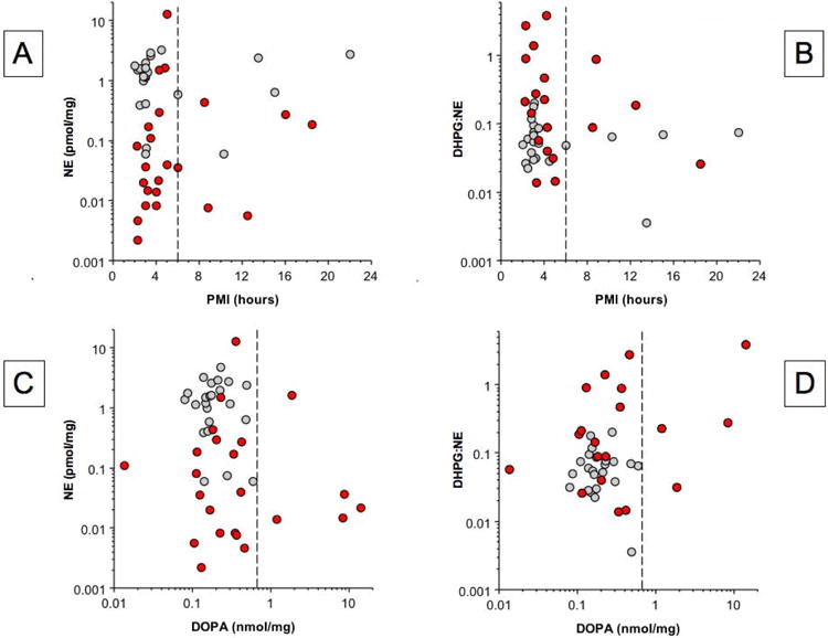
In Parkinson's disease (PD), profound putamen dopamine (DA) depletion reflects denervation and a shift from vesicular sequestration to oxidative deamination of cytoplasmic DA in residual terminals. PD also involves cardiac sympathetic denervation. Whether PD entails myocardial norepinephrine (NE) depletion and a sequestration-deamination shift have been unknown. We measured apical myocardial tissue concentrations of NE, DA, and their neuronal metabolites 3,4-dihydroxyphenylglycol (DHPG), and 3,4-dihydroxyphenylacetic acid (DOPAC) from 23 PD patients and 23 controls and ascertained the extent of myocardial NE depletion in PD. We devised, validated in VMAT2-Lo mice, and applied 5 neurochemical indices of the sequestration-deamination shift-concentration ratios of DOPAC:DA, DA:NE, DHPG:NE, DOPAC:NE, and DHPG:DOPAC-and used a kinetic model to estimate the extent of the vesicular storage defect. The PD group had decreased myocardial NE content (p < 0.0001). The majority of patients (70%) had severe NE depletion (mean 2% of control), and in this subgroup all five indices of a sequestration-deamination shift were increased compared to controls (p < 0.001 for each). Vesicular storage in residual nerves was estimated to be decreased by 84-91% in this subgroup. We conclude that most PD patients have severe myocardial NE depletion, because of both sympathetic denervation and decreased vesicular storage in residual nerves. We found that the majority (70%) of Parkinson's disease (PD) patients have profound (98%) myocardial norepinephrine depletion, because of both cardiac sympathetic denervation and a shift from vesicular sequestration to oxidative deamination of cytoplasmic catecholamines in the residual nerves. This shift may be part of a final common pathogenetic pathway in the loss of catecholaminergic neurons that characterizes PD.

Keywords: Parkinson's disease; catecholamines; neurochemistry; neurodegenerative mechanisms; sympathetic nervous system.


Conflict of interest statement

Disclosures: The Authors have no conflicts of interest to disclose.

Conflicts of interest: none

⇒ if ‘none’, insert “The authors have no conflict of interest to declare.”

⇒ otherwise insert info unless it is already included

Figures

Figure 1
Figure 1. Concept diagrams about sources and metabolic fates of catecholamines in sympathetic nerves
Under resting conditions, loss of norepinephrine (NE) from the neurons is due mainly to passive leakage from the vesicles (NEv) into the cytosol (NEc), followed by enzymatic deamination catalyzed by monoamine oxidase (MAO). Cytosolic NE is taken up into the vesicles via the type 2 vesicular monoamine transporter (VMAT). Release by exocytosis from the vesicles, with escape of reuptake via the cell membrane NE transporter (NET), is a minor determinant of NE turnover. NE loss is balanced by catecholamine biosynthesis from the action of cytosolic L-aromatic-amino-acid decarboxylase (LAAAD) on 3,4-dihydroxyphenylalanine (DOPA) produced from tyrosine (TYR) by tyrosine hydroxylase (TH) and of dopamine-beta-hydroxylase (DBH), which is localized in the vesicles. The action of MAO on cytosolic DA produces 3,4-dihydroxyphenylacetaldehyde (DOPAL) and on NE produces 3,4-dihydroxyphenylglycolaldehyde (DOPEGAL). DOPEGAL is mainly reduced by aldehyde/aldose reductase (AR), to form 3,4-dihydroxyphenylglycol (DHPG), and DOPAL is mainly oxidized by aldehyde dehydrogenase (ALDH) to form 3,4-dihydroxyphenylacetic acid (DOPAC). Red X marks indicate decreased vesicular uptake. When vesicular uptake is attenuated, as in VMAT2-Lo mice, myocardial NE depletion reflects decreased NE synthesis, because less DA is taken up into the vesicles and more is deaminated to form DOPAC. Furthermore, decreased reuptake of NE that leaks from vesicles into the cytoplasm, where the NE is deaminated and is converted to DHPG, accelerates the turnover of NE.
Figure 2
Figure 2. Detailed and condensed kinetic models for the fate of catecholamines in sympathetic nerves
Abbreviations: FDC = fraction of DA that is metabolized to DOPAC; FNE = fraction of DA that is converted to NE; FG = fraction of vesicular NE that is lost by oxidative deamination; FXO = fraction of NE that is lost by release; kMD = rate constant for cytosolic DA being deaminated to form DOPAC; kVD = rate constant for uptake of cytosolic DA into the vesicles; kL = rate constant for leakage of vesicular DA or NE into the cytosol; kR = rate constant for NE release into the interstitial fluid; kU = rate constant for uptake of NE from the interstitial fluid into the cytosol via the cell membrane NE transporter; kXNE = rate constant for loss of NE from the interstitial fluid by metabolism or spillover (NESO) into the bloodstream; kXDOPAC = rate constant for DOPAC entry into the interstitial fluid; and kXDHPG = rate constant for DHPG entry into the interstitial fluid.
Figure 3
Figure 3. Individual values for myocardial DHPG and DHPG:NE as a function of NE in patients with PD (red circles) and controls (gray circles)
Vertical line separates PD subgroups with or without NE depletion. Dashed line shows line of best fit for the relationship between DHPG and NE in controls. Equation is for the line of best fit in controls.
Figure 4
Figure 4. Mean (± SEM) values for catechols in PD subgroups with NE depletion (PD NE Depl.) or no NE depletion (PD No Depl.) and in controls (Control)
(A) NE, (B) DA, (C) DHPG, (D) DOPAC. P values indicate significant differences from Control.
Figure 5
Figure 5. Myocardial mean (± SEM) ratios of DHPG:NE, DOPAC:DA, DA:NE, DOPAC:NE in control subjects (CON, light colors) and patients with Parkinson disease (PD, dark colors) and in wild-type (WT) mice (light colors) and mice with very low activity of the type 2 vesicular monoamine transporter (VMAT2-Lo, dark colors)
P values are for PD vs. CON and WT vs. VMAT2-Lo groups for each ratio.
Figure 6
Figure 6. Individual values for myocardial NE content and DHPG:NE ratios expressed as functions of post-mortem intervals (PMIs, A, B) and DOPA content (C, D) in patients with PD (red circles) and controls (gray circles)
Vertical dashed lines in A and B indicate PMI of 6 hours and in C and D indicate the upper range of control values.

References

    1. Amino T, Orimo S, Takahashi A, Uchihara T, Mizusawa H. Profound cardiac sympathetic denervation occurs in Parkinson disease. Brain Path. 2005;15:29–34. - PMC - PubMed
    1. Braak H, Ghebremedhin E, Rub U, Bratzke H, Del Tredici K. Stages in the development of Parkinson's disease-related pathology. Cell Tissue Res. 2004;318:121–134. - PubMed
    1. Burke WJ, Kumar VB, Pandey N, et al. Aggregation of alpha-synuclein by DOPAL, the monoamine oxidase metabolite of dopamine. Acta Neuropathol. 2008;115:193–203. - PubMed
    1. Caudle WM, Richardson JR, Wang MZ, et al. Reduced vesicular storage of dopamine causes progressive nigrostriatal neurodegeneration. J Neurosci. 2007;27:8138–8148. - PMC - PubMed
    1. Cotzias GC. Levodopa in the treatment of Parkinsonism. JAMA. 1971;218:1903–1908. - PubMed

Publication types

MeSH terms