. 2014 Aug;99(8):2995-3004.
doi: 10.1210/jc.2014-1139. Epub 2014 May 21.

IGF1R signaling is necessary for FSH-induced activation of AKT and differentiation of human Cumulus granulosa cells

Affiliations

IGF1R signaling is necessary for FSH-induced activation of AKT and differentiation of human Cumulus granulosa cells

Sarah C Baumgarten et al. J Clin Endocrinol Metab. 2014 Aug.

Abstract

Context: FSH is routinely administered to in vitro fertilization patients to induce follicle maturation. During this process, granulosa cells differentiate and acquire specific functional characteristics that are required to coordinate ovulation and oocyte maturation.

Objective: The objective of the study was to gain insight into the molecular mechanisms regulating human granulosa cell differentiation. Design, Setting, Patients, and Interventions: Cumulus and mural granulosa cells were isolated from the follicular aspirates of in vitro fertilization patients and analyzed immediately or cultured in serum-free media in the presence of FSH, IGFs, or an inhibitor of type I IGF receptor (IGF1R) activity.

Main outcome: We quantified the mRNA and protein levels of steroidogenic enzymes, components of the IGF system, and gonadotropin receptors; measured 17β-estradiol levels; and examined the activation of intracellular signaling pathways to assess the granulosa cell differentiation as well as the FSH and IGF actions in both cumulus and mural cells.

Results: In freshly isolated cells, LH receptor (Lhr) and steroidogenic acute regulator (Star) were expressed at lower levels in cumulus than mural cells, whereas FSH receptor (Fshr) and anti-Müllerian hormone (Amh) were expressed at higher levels in cumulus than mural cells. In vitro, the expression of Igf2, the differentiation markers Lhr, Star, and Cyp19a1 (aromatase) as well as 17β-estradiol production remained low in untreated cumulus cells but increased significantly after FSH treatment. Strikingly, this stimulatory effect of FSH was abolished by the inhibition of IGF1R activity. FSH-induced activation of v-akt murine thymoma viral oncogene homolog 3 (AKT) required IGF1R activity, and overexpression of constitutively active AKT rescued the induction of differentiation markers and 17β-estradiol production by FSH in the presence of the IGF1R inhibitor.

Conclusions: The cumulus cell response to FSH resembles the differentiation of preantral to preovulatory granulosa cells. This differentiation program requires IGF1R activity and subsequent AKT activation.


Figures
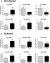
Figure 1.
Figure 1.
Cumulus and mural cells have distinct gene expression profiles. A, RNA was isolated from freshly purified, uncultured cumulus and mural cells collected from patients undergoing IVF. The expression of key gene markers of granulosa cell differentiation and luteinization, including Lhr, Cyp19a1, Fshr, Cyp11a1, Star, and Amh, were measured by qPCR. Columns represent the mean ± SEM for six independent experiments. B, RNA was isolated from cumulus and mural granulosa cells after culture in serum-free media for 72 hours, and the expression of key differentiation and luteinization genes was measured by qPCR. Columns represent the mean ± SEM for 14 independent experiments. *, P < .05; **, P < .01, t test.
Figure 2.
Figure 2.
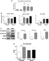
Cumulus cells differentiate in response to FSH. A, Fshr expression was measured in cumulus cells cultured for increasing periods of time (0–96 h) by qPCR. Columns represent the mean ± SEM for three independent experiments. B, Cumulus and mural cells were cultured in serum-free media for 24 hours and then treated with FSH (50 ng/mL) or left untreated (control) for 48 hours. RNA was isolated from the cells, and the expression of Cyp19a1, Cyp11a1, and Star was measured by qPCR. Columns represent the mean ± SEM for 10 independent experiments. C, Cumulus cells were cultured in serum-free media for 24 hours and then treated with FSH (50 ng/mL) or left untreated (Ctrl) for 48 hours. Total protein was isolated from these cells and CYP19A1, CYP11A1, StAR, and ACTB protein levels were assessed by Western blot. Columns represent the mean ± SEM for three independent experiments. D, 17β-Estradiol levels were measured in media collected from cultures of cumulus and mural cells that received FSH treatment (50 ng/mL) or no treatment (control) for 48 hours. Columns represent the mean ± SEM for three independent experiments. *, P < .05; **, P < .01, t test.
Figure 3.
Figure 3.
Cumulus granulosa cells express Igf2 but not Igf1. Cumulus cells were cultured in serum-free media for 72 hours before RNA isolation. A, Cumulus cell RNA from four different patients as well as human liver RNA was reverse transcribed, and PCR was used to detect expression of Igf1, Igf2, Igf1r, Igf2r, and the housekeeping gene Rpl19. PCR products were visualized on agarose gels. Igf1r and Igf2r expression levels in cumulus cells were quantified by qPCR. B, Cumulus cells were cultured in serum-free media for 24 hours and then treated with FSH (50 ng/mL) or left untreated for 48 hours. RNA was isolated from these cells, and the expression of Igf2, Igf1r, and Igf2r was measured by qPCR. For qPCR results, columns represent the mean ± SEM of at least seven independent experiments. **, P < .01, t test.
Figure 4.
Figure 4.
FSH action in cumulus granulosa cells requires IGF1R activity. Cumulus cells were cultured in serum-free media for 24 hours and then pretreated with AEW (0.5 μM) or vehicle (dimethyl sulfoxide, DMSO) for 1 hour before the addition of FSH (50 ng/mL) for 48 hours. RNA (A) and total protein (B) were isolated and the mRNA and protein levels of CYP19A1, CYP11A1, and StAR were measured by qPCR and Western blot. For mRNA quantification, columns represent the mean ± SEM for five independent experiments. For protein quantification, columns represent the mean ± SEM for the same three independent experiments presented in Figure 2. C, 17β-Estradiol levels were measured in media collected from cultures of cumulus cells by ELISA. Columns represent the mean ± SEM for three independent experiments. *, P < .05; **, P < .01; ***, P < .001, one-way ANOVA.
Figure 5.
Figure 5.
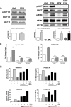
FSH-induced AKT phosphorylation requires IGF1R activity. A, left panel, Cumulus cells were cultured in serum-free media for 24 hours and then treated with FSH (50 ng/mL) for 1 hour. Total protein was isolated from these cells and phosphorylated and total IGF1R were detected by Western blot. Protein levels were normalized to ACTB, and the ratio of phosphorylated to total IGF1R is reported. Columns represent the mean ± SEM for three independent experiments. A, right panel, Cumulus cells were cultured in serum-free media for 24 hours and pretreated with AEW (0.5 μM) or vehicle (dimethylsulfoxide) for 1 hour before the addition of FSH (50 mg/mL) for 1 hour. Phosphorylated and total AKT and ERK were assessed by Western blot of whole-cell lysates. Protein levels were normalized to ACTB, and the ratio of phosphorylated to total AKT and ERK are reported. Columns represent the mean ± SEM for three independent experiments. B, Cumulus granulosa cells were cultured in serum-free media for 24 hours and then pretreated with AEW (0.5 μM), MK2206 (1 μM), or vehicle (dimethyl sulfoxide, DMSO) for 1 hour followed by treatment with FSH (50 ng/mL) for 48 hours. RNA was isolated from the cells, and the expression of Cyp19a1 was measured by qPCR. Columns represent the mean ± SEM for five independent experiments. 17β-Estradiol was measured in media collected from treated cells, and columns represent the mean ± SEM for two patients. C, Cumulus granulosa cells were cultured in serum-free media for 24 hours and then pretreated with AEW (0.5 μM) for 1 hour before the addition of FSH (50 ng/mL) for 48 hours in the presence of lentivirus-expressing green fluorescent protein or caAKT. The expression of Cyp19a1 as well as the production of 17β-estradiol is reported for two patients. *, P < .05; **, P < .01; ***, P < .001, one-way ANOVA. Ctrl, control; ns, not significant.

References

    1. Thoma ME, McLain AC, Louis JF, et al. Prevalence of infertility in the United States as estimated by the current duration approach and a traditional constructed approach. Fertil Steril. 2013;99:1324–1331-e1 - PMC - PubMed
    1. Li RH, Ng EH. Management of anovulatory infertility. Best practice, research. Clin Obstet Gynaecol. 2012;26:757–768 - PubMed
    1. Centers for Disease Control and Prevention, American Society for Reproductive Medicine, Society for Assisted Reproductive Technology. 2011 assisted reproductive technology fertility clinic success rates report. Atlanta (GA): US Department of Health and Human Services; 2013
    1. Sunderam S, Kissin DM, Flowers L, et al. Assisted reproductive technology surveillance—United States, 2009. MMWR Surveill Summ. 2012;61:1–23 - PubMed
    1. Fiedler K, Ezcurra D. Predicting and preventing ovarian hyperstimulation syndrome (OHSS): the need for individualized not standardized treatment. Reprod Biol Endocrinol. 2012;10:32. - PMC - PubMed

Publication types

Substances