. 2014 Aug;56(6):434-47.
doi: 10.1111/dgd.12140. Epub 2014 May 22.

MicroRNA expression profiling of the developing murine upper lip

Affiliations

MicroRNA expression profiling of the developing murine upper lip

Dennis R Warner et al. Dev Growth Differ. 2014 Aug.

Abstract

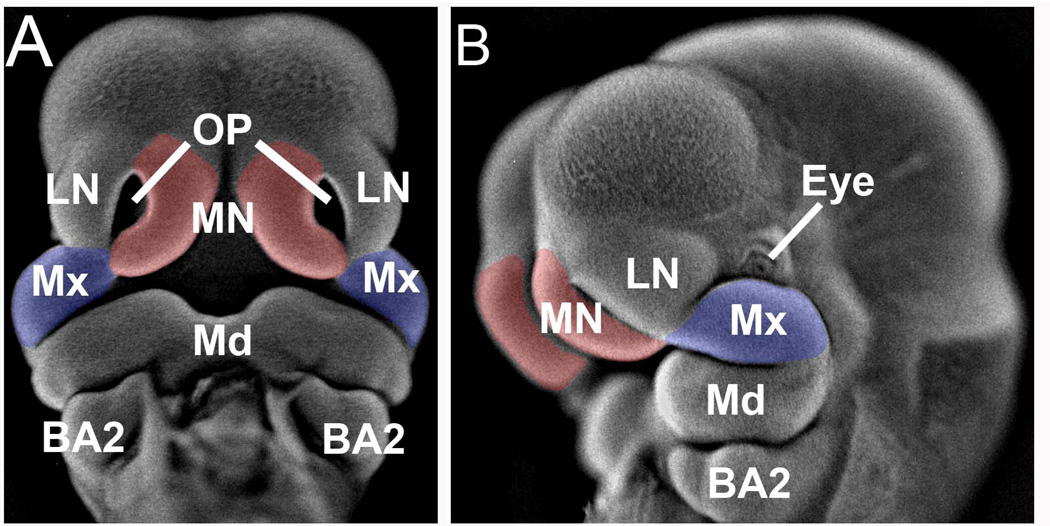
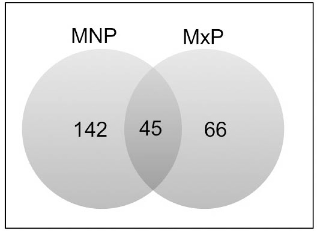
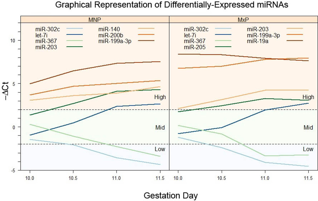
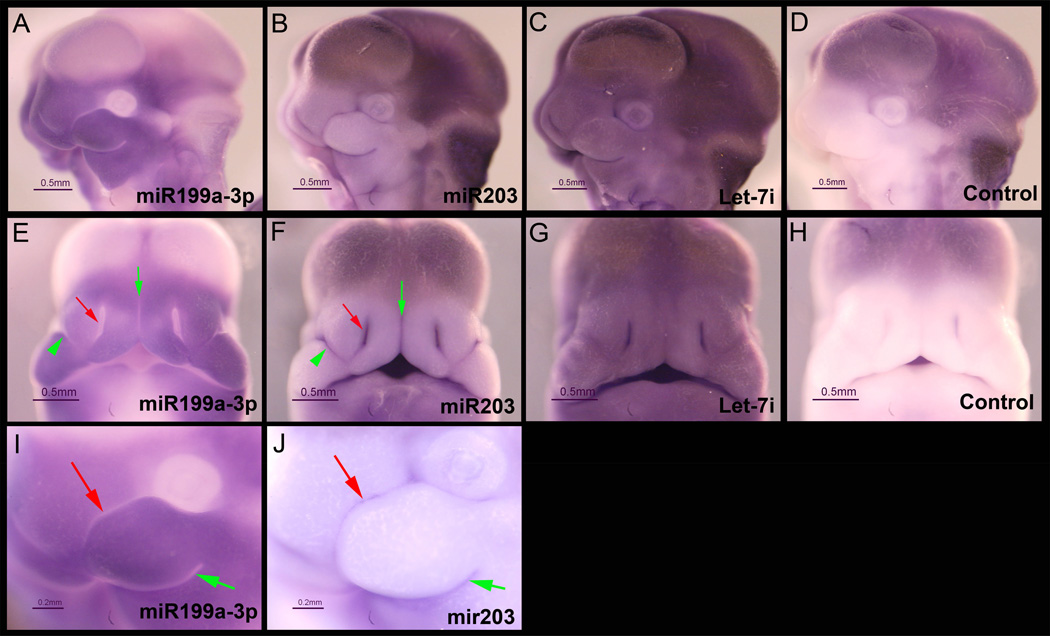
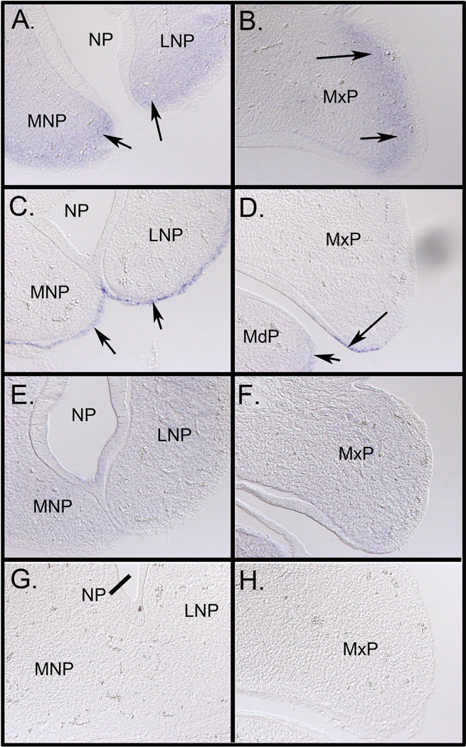
Clefts of the lip and palate are thought to be caused by genetic and environmental insults but the role of epigenetic mechanisms underlying this common birth defect are unknown. We analyzed the expression of over 600 microRNAs in the murine medial nasal and maxillary processes isolated on GD10.0-GD11.5 to identify those expressed during development of the upper lip and analyzed spatial expression of a subset. A total of 142 microRNAs were differentially expressed across gestation days 10.0-11.5 in the medial nasal processes, and 66 in the maxillary processes of the first branchial arch with 45 common to both. Of the microRNAs exhibiting the largest percent increase in both facial processes were five members of the Let-7 family. Among those with the greatest decrease in expression from GD10.0 to GD11.5 were members of the microRNA-302/367 family that have been implicated in cellular reprogramming. The distribution of expression of microRNA-199a-3p and Let-7i was determined by in situ hybridization and revealed widespread expression in both medial nasal and maxillary facial process, while that for microRNA-203 was much more limited. MicroRNAs are dynamically expressed in the tissues that form the upper lip and several were identified that target mRNAs known to be important for its development, including those that regulate the two main isoforms of p63 (microRNA-203 and microRNA-302/367 family). Integration of these data with corresponding proteomic datasets will lead to a greater appreciation of epigenetic regulation of lip development and provide a better understanding of potential causes of cleft lip.

Keywords: cleft lip; craniofacial development; microRNA; mouse; p63.


Figures

Figure 1
Figure 1. Facial prominences dissected for miRNA expression analysis
Shown is a GD11.5 mouse fetus that has been incubated with DAPI and photographed under epi-fluorescence according to the method detailed in (Sandell et al., 2012). Panel A, frontal view and panel B, lateral view. The medial nasal processes are indicated in red and the maxillary processes in blue. MN, medial nasal process; LN, lateral nasal process; Mx, maxillary process of the first branchial arch; OP, olfactory pit; Md, mandibular process of first branchial arch; BA2, second branchial arch.
Figure 2
Figure 2. Venn diagram illustrating the number of miRNAs differentially regulated
There were 143 and 67 miRNAs differentially regulated in the MNP (blue) and MxP (yellow), respectively. Of those, 46 miRNAs were common to both facial processes.
Figure 3
Figure 3. Longitudinal profiles of differentially-expressed miRNAs
Differentially-expressed miRNAs in MNP (left panel) or MxP (right panel) tissue were categorized by their initial ΔCt values (those on GD10) as low, mid, or high expressers, as indicated in each panel. Each line represents the ΔCt value for a specific miRNA on GD10.0-GD11.5. For clarity, only a selected few miRNAs are shown on each category.
Figure 4
Figure 4. Expression of miR-199a-3p, miR-203, and Let-7i in GD11.5 fetuses
Mouse fetuses were dissected on GD11.5 and fixed overnight in 4% paraformaldehyde and then processed for in situ hybridization as described in the Methods section. Fetuses were incubated with locked nucleic acid (LNA)-modified DNA probes (Exiqon) targeting miR-199a-3p (panels A, E, and I), miR-203 (panels B, F, and J), Let-7i (panels C and G), or a scrambled sequence control probe (Control, panels D and H). Images shown in panels A-H were taken under identical lighting conditions and exposure time. Panels A-D and I-J are lateral views and Panels E-F are the corresponding frontal views. Panels I and J are higher magnification images of the same fetuses shown in A and B, respectively. There was variable trapping of the LNA probes in the ventricles of the brain, however, there was no non-specific binding detected in the medial and lateral nasal processes or the maxillary and mandibular processes of the first branchial arch (Panels D and H). miR-199a-3p was expressed in both the medial and lateral nasal processes and in the maxillary process (Panels A and E). In contrast, the expression of miR-203 was more restricted (arrows in Panels B and F) and appeared to be mutually exclusive with miR-199a-3p. The expression of Let-7i was ubiquitous (Panels C and G). Scales bars are 0.5 mm.
Figure 5
Figure 5. Expression of miR-199a-3p, miR-203, and Let-7i in GD11.5 mouse facial processes
To determine the specific cellular expression pattern of each miR analyzed, the fetuses shown in Figure 3 were equilibrated in 30% sucrose/PBS, embedded and frozen in OCT medium, and sectioned at a thickness of 16 µm. Panel A shows the expression of miR-199a-3p in the mesenchyme of the MNP and LNP (arrows) and in the MxP (Panel B, arrows). Panels C and D reveal epithelium-specific expression of miR-203 in the MNP and LNP and MxP, respectively (arrows). Expression was also noted in the MdP (panel D, arrow). The expression of Let-7i was more uniform and widespread than that for miR-199a-3p and miR-203 (panels E and F). The negative control probe is shown in panels G and H. MNP, medial nasal process; LNP, lateral nasal process; MxP, maxillary and MdB, mandibular aspect of the first branchial arch; NP, nasal pit. All images were taken at 200X magnification.

References

    1. Ambros V. microRNAs: tiny regulators with great potential. Cell. 2001;107:823–826. - PubMed
    1. Barritt LC, Miller JM, Scheetz LR, et al. Conditional deletion of the human ortholog gene Dicer1 in Pax2-Cre expression domain impairs orofacial development. Indian J Hum Genet. 2012;18:310–319. - PMC - PubMed
    1. Bartel DP. MicroRNAs: target recognition and regulatory functions. Cell. 2009;136:215–233. - PMC - PubMed
    1. Bazzini AA, Lee MT, Giraldez AJ. Ribosome profiling shows that miR-430 reduces translation before causing mRNA decay in zebrafish. Science. 2012;336:233–237. - PMC - PubMed
    1. Benjamini Y, Hochberg Y. Controlling the false discovery rate: a practical and powerful approach to multiple testing. J R Stat Soc B. 1995;57:289–300.

Publication types

LinkOut - more resources