. 2014 May 21;34(21):7281-92.
doi: 10.1523/JNEUROSCI.0646-14.2014.

Anti-ApoE antibody given after plaque onset decreases Aβ accumulation and improves brain function in a mouse model of Aβ amyloidosis

Affiliations

Anti-ApoE antibody given after plaque onset decreases Aβ accumulation and improves brain function in a mouse model of Aβ amyloidosis

Fan Liao et al. J Neurosci. .

Abstract

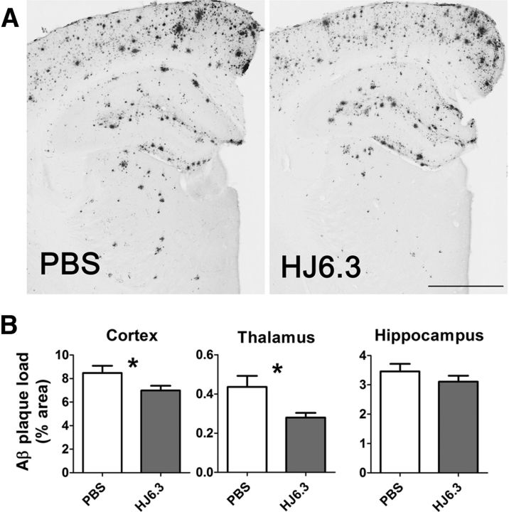
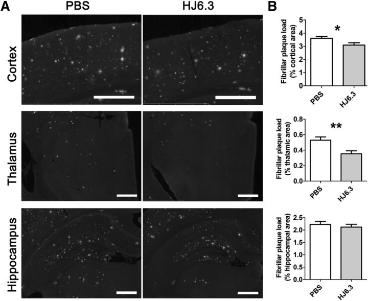
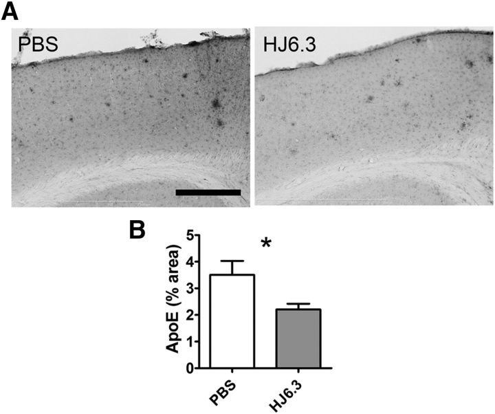
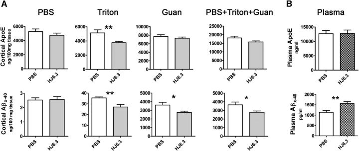
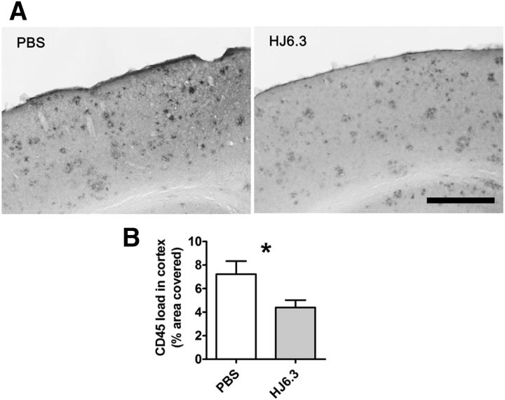
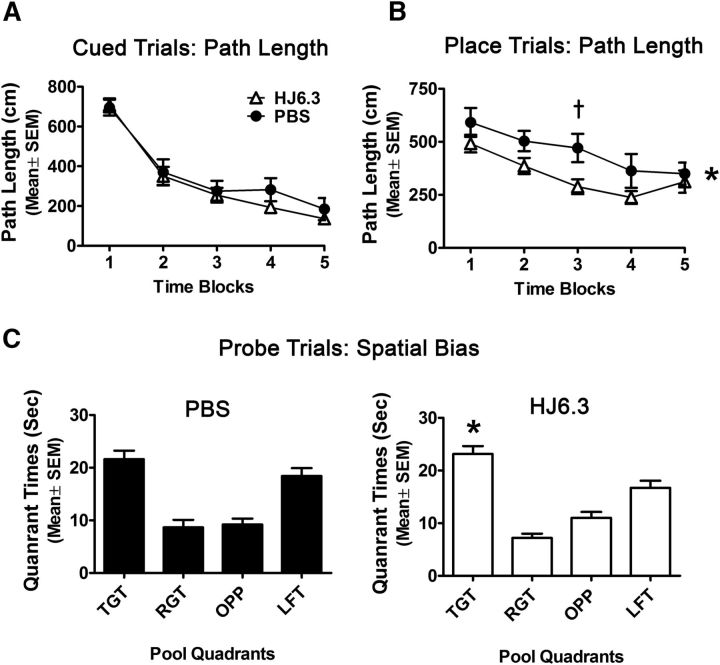
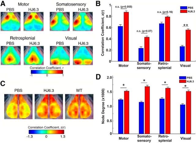
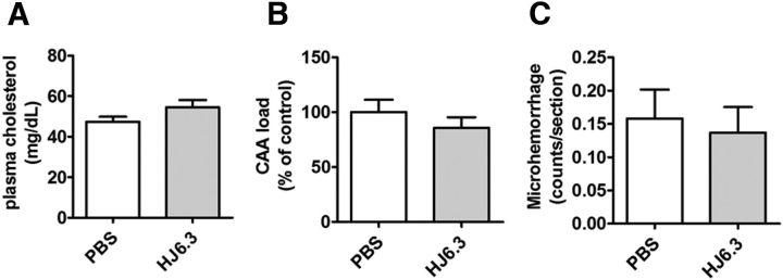
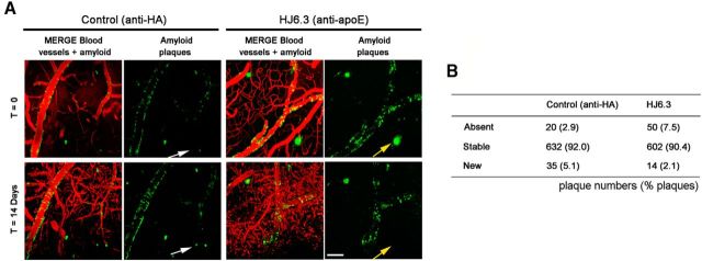
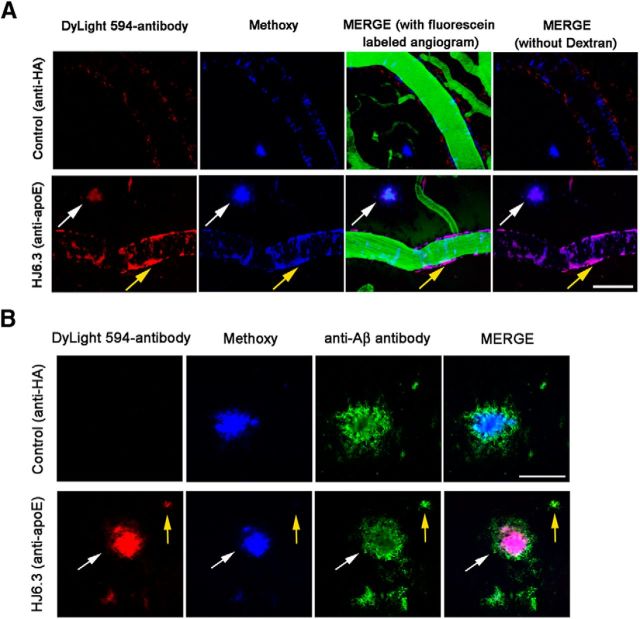
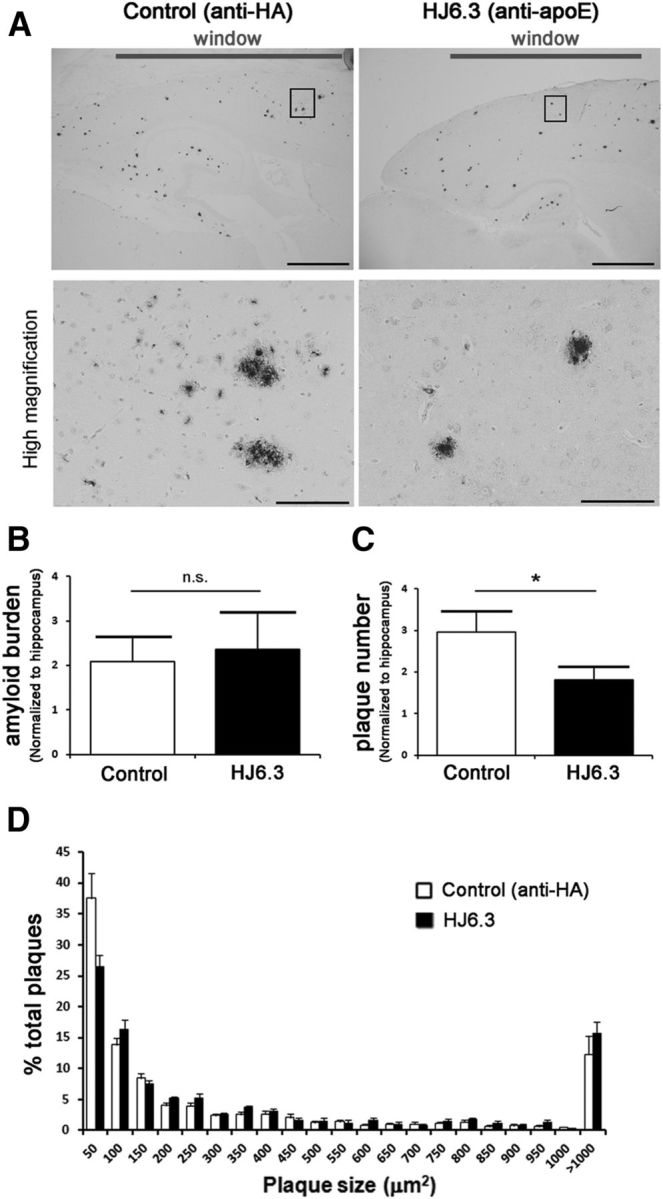
Apolipoprotein E (apoE) is the strongest known genetic risk factor for late onset Alzheimer's disease (AD). It influences amyloid-β (Aβ) clearance and aggregation, which likely contributes in large part to its role in AD pathogenesis. We recently found that HJ6.3, a monoclonal antibody against apoE, significantly reduced Aβ plaque load when given to APPswe/PS1ΔE9 (APP/PS1) mice starting before the onset of plaque deposition. To determine whether the anti-apoE antibody HJ6.3 affects Aβ plaques, neuronal network function, and behavior in APP/PS1 mice after plaque onset, we administered HJ6.3 (10 mg/kg/week) or PBS intraperitoneally to 7-month-old APP/PS1 mice for 21 weeks. HJ6.3 mildly improved spatial learning performance in the water maze, restored resting-state functional connectivity, and modestly reduced brain Aβ plaque load. There was no effect of HJ6.3 on total plasma cholesterol or cerebral amyloid angiopathy. To investigate the underlying mechanisms of anti-apoE immunotherapy, HJ6.3 was applied to the brain cortical surface and amyloid deposition was followed over 2 weeks using in vivo imaging. Acute exposure to HJ6.3 affected the course of amyloid deposition in that it prevented the formation of new amyloid deposits, limited their growth, and was associated with occasional clearance of plaques, a process likely associated with direct binding to amyloid aggregates. Topical application of HJ6.3 for only 14 d also decreased the density of amyloid plaques assessed postmortem. Collectively, these studies suggest that anti-apoE antibodies have therapeutic potential when given before or after the onset of Aβ pathology.

Keywords: Alzheimer's; amyloid; antibody; apolipoprotein E.


Figures

Figure 1.
Figure 1.
Reduction of Aβ plaque deposition by the anti-apoE antibody HJ6.3. Seven-month-old female APP/PS1 mice received 10 mg/kg weekly intraperitoneal injections of HJ6.3 or PBS for 21 weeks. A, Brain sections of the HJ6.3- or PBS-treated mice were stained with the anti-Aβ antibody HJ3.4B. Scale bar, 1 mm. B, Aβ plaque load in cortex, thalamus, and hippocampus were quantified (n = 13–17/group; *p < 0.05).
Figure 2.
Figure 2.
Attenuation of fibrillar amyloid deposition by the anti-apoE antibody HJ6.3. Seven-month-old female APP/PS1 mice received 10 mg/kg weekly intraperitoneal injections of HJ6.3 or PBS for 21 weeks until 12 months of age. A, Fibrillar amyloid plaques on brain sections of the HJ6.3- or PBS-treated mice were stained with ThioS. Scale bar, 500 μm. B, Quantification of fibrillar amyloid plaque load in cortex, thalamus, and hippocampus (n = 13–17/group; *p < 0.05; **p < 0.01).
Figure 3.
Figure 3.
Effects of the anti-apoE antibody HJ6.3 on brain apoE. Seven-month-old female APP/PS1 mice received 10 mg/kg weekly intraperitoneal injections of HJ6.3 or PBS for 21 weeks. ApoE in the cortex of the HJ6.3- or PBS-treated mice was immunostained with HJ6.8B (A) and the percentage area covered by the staining was quantified (B) (n = 15–18/group; *p < 0.05; **p < 0.01). Scale bar, 400 μm.
Figure 4.
Figure 4.
Effects of the anti-apoE antibody HJ6.3 on Aβ and apoE levels in the brain and blood. A, The cortices of the HJ6.3- or PBS-treated mice were homogenized in PBS, followed by 1% Triton X-100 and 5 m guanidine (Guan). ApoE and Aβx-40 levels in the brain tissue lysates in the PBS, Triton X-100, and Guan fractions were measured by ELISA. The level of apoE and Aβx-40 in all fractions combined is also shown (n = 10–18/group). B, Plasma apoE and Aβx-40 levels in HJ6.3- and PBS-treated animals were measured by ELISA (n = 15–18/group; *p < 0.05; **p < 0.01).
Figure 5.
Figure 5.
Effects of the anti-apoE antibody HJ6.3 on microglial activation. Seven-month-old female APP/PS1 mice received 10 mg/kg weekly intraperitoneal injections of HJ6.3 or PBS for 21 weeks until 12 months of age. Activated microglia in the cortex were immunostained with anti-CD45 antibody (A) and the area covered by CD45 staining was quantified (B) (n = 15–18/group; *p < 0.05). Scale bar, 400 μm.
Figure 6.
Figure 6.
HJ6.3 treatment improved spatial learning performance in the MWM. Seven month-old female APP/PS1 mice received 10 mg/kg weekly intraperitoneal injections of HJ6.3 or PBS for 21 weeks until 12 months of age. During the last 4 weeks of treatment, the performance of HJ6.3-treated (n = 19) and PBS-treated (n = 18) mice in the MWM task was tested. A, The HJ6.3- and PBS-treated mice did not differ in performance with regard to path length during the cued trials. B, However, the HJ6.3-treated mice exhibited significantly improved performance in terms of showing decreased path length during the place (spatial learning) trials compared with the PBS-treated group (treatment effect: *p = 0.035), with differences being greatest during block 3 (†p = 0.019). C, PBS-treated mice did not show a spatial bias for the target quadrant during the probe trial because they did not spend significantly more time in that quadrant compared with the times in each of the other quadrants. In contrast, the HJ6.3-treated mice did show a spatial bias for the target quadrant because they spent significantly more time in the target quadrant compared with the times spent in each of the other pool quadrants (*p < 0.014).
Figure 7.
Figure 7.
HJ6.3 treatment improved brain functional connectivity assessed by fcOIS. Seven-month-old female APP/PS1 mice received 10 mg/kg weekly intraperitoneal injections of HJ6.3 or PBS for 21 weeks. At the age of 12 months, fcOIS was performed to assess the resting state functional connectivity of the brain before euthanizing the mice. A, Composite, group-averaged, functional correlation maps of motor, somatosensory, retrosplenial, and visual cortices in HJ6.3-treated (n = 15) and PBS-treated (n = 14) APP/PS1 mice. Black circles denote seed position. B, Regional bilateral functional correlation of PBS-treated (blue) and HJ6.3-treated (red) APP/PS1 mice. C, Consensus bilateral functional connectivity maps generated for HJ6.3 and PBS treated APP/PS1 mice and age-matched untreated wild-type B6C3 mice. The maps for age-matched wild-type B6C3 mice were regenerated from the data reported in Bero et al. (2012). D, Node degree in motor, somatosensory, retrosplenial, and visual cortices (*p < 0.05; **p < 0.01).
Figure 8.
Figure 8.
No effects of HJ6.3 on plasma cholesterol, CAA, or CAA-associated microhemorrhage. Seven-month-old APP/PS1 mice received 10 mg/kg weekly intraperitoneal injections of HJ6.3 or PBS for 21 weeks until 12 months of age. A, Total plasma cholesterol levels. B, CAA on the surface of the cortex was identified by anti-Aβ antibody HJ3.4B immunostaining and the area covered by CAA was quantified. C, CAA-associated microhemorrhage was detected by Prussian blue and ThioS costaining (n = 13–18/group).
Figure 9.
Figure 9.
In vivo imaging of Aβ plaques after topical application of the anti-apoE antibody HJ6.3. Fifty microliters of the anti-apoE antibody HJ6.3 or anti-HA control antibody (1.0 mg/ml) were topically applied to the brain surface of 5- to 7-month-old APP/PS1 mice. Plaques were monitored through the cranial window using two-photon microscopy the day of surgery and 14 d after treatment, following the exact same fields of view over time. A, Representative in vivo images of amyloid plaques the day of surgery (t = 0) and 14 d (t = 14 d) after topical application of control (anti-HA) or the anti-apoE antibody HJ6.3. Yellow and white arrows respectively show “absent” and “new” plaques. Scale bar, 100 μm. B, Contingency table summarizing the number of “absent,” “stable,” and “new” plaques 2 weeks after topical application of the anti-apoE antibody HJ6.3 and anti-HA antibodies (n = 7/group; p < 0.0001, χ2 test).
Figure 10.
Figure 10.
Affinity of fluorescent-labeled DyLight 594-apoE antibody HJ6.3 to CAA and Aβ plaques. Fifty micrograms of fluorescently labeled DyLight 594-apoE antibody HJ6.3 or DyLight 594-anti-HA control antibody (1.0 mg/ml) were topically applied to the brain surface of 5- to 7-month-old APP/PS1 mice. Plaques and CAA were imaged by in vivo two-photon microscopy and immunohistochemistry with the rabbit anti-human Aβ (N) antibody (Immuno-Biological Laboratories). A, Representative photographs of in vivo imaging after 30 min of incubation with antibody, showing that only the DyLight 594-apoE antibody HJ6.3 can bind apoE in CAA (yellow arrow) and amyloid plaques (white arrow) after a single topical application to the brain. Scale bar, 100 μm. B, Representative photographs of immunohistochemistry after 2 h of incubation with antibody, showing that the DyLight 594-apoE antibody HJ6.3 can bind apoE in small, methoxy-XO4-negative amyloid plaque (yellow arrow) and in a methoxy-XO4-positive amyloid plaque (white arrow) after a single topical application to the brain. Scale bar, 50 μm.
Figure 11.
Figure 11.
In vivo dynamic measurement of Aβ plaques after treatment with HJ6.3. Fifty microliters of fluorescently labeled DyLight 594-apoE antibody HJ6.3 or DyLight 594-anti-HA control antibody (1.0 mg/ml) were topically applied to the brain surface of 5- to 7-month-old APP/PS1 mice. Plaques were monitored by two-photon microscopy. A, Representative in vivo images of the fluorescently labeled DyLight 594-HJ6.3 and methoxy-XO4 the day of application (t = 0) or 14 d after (t = 14 d), showing that the plaque that got cleared (yellow arrow) was previously labeled by DyLight 594-HJ6.3, as opposed to nonlabeled plaque (white arrow). Scale bar, 100 μm. B, Contingency table summarizing the number of “absent,” “stable,” and “new” plaques according to their initial binding with anti-apoE HJ6.3 and anti-HA antibodies. C, Representative in vivo images of the fluorescently labeled DyLight 594-HJ6.3 and methoxy-XO4 the day of application (t = 0) or 14 d after (t = 14 d), showing that the size of HJ6.3-labeled plaque decreased after topical application of labeled HJ6.3 (yellow arrow), as opposed to nonlabeled plaque (indicated by white arrow). Scale bar, 100 μm. D, Graph bar indicating the size ratio between day 0 and day 14 after topical application of the control antibody or the anti-apoE antibody HJ6.3 (n = 3/group; *p < 0.05, ***p < 0.001 for one-way ANOVA followed by post hoc Tukey test and p < 0.0001 for χ2 test).
Figure 12.
Figure 12.
Reduction of the number of amyloid plaques after acute exposure with the anti-apoE antibody HJ6.3. Fifty microliters of the anti-apoE antibody HJ6.3 or anti-HA control antibody (1.0 mg/ml) were topically applied to the brain surface of 5- to 7-month-old APP/PS1 mice. A, Fifteen days after application, amyloid plaques were stained with the rabbit anti-human Aβ (N) antibody (Immuno-Biological Laboratories). Scale bars: top, 2 mm; bottom, 100 μm. B, C, Bar graphs summarizing the amyloid load (B) and plaque number (C) quantified in the cortical region under the cranial window, normalized to the amount of plaques within the hippocampus. D, Graph bar representing the plaque size distribution between the application of the control antibody and the anti-apoE antibody HJ6.3 (n = 9/group; *p < 0.05, nonparametric Mann–Whitney test).

Similar articles

Cited by

References

    1. Ashford JW. ApoE4: is it the absence of good or the presence of bad? J Alzheimers Dis. 2002;4:141–143. - PubMed
    1. Bacskai BJ, Klunk WE, Mathis CA, Hyman BT. Imaging amyloid-beta deposits in vivo. J Cereb Blood Flow Metab. 2002;22:1035–1041. doi: 10.1097/00004647-200209000-00001. - DOI - PubMed
    1. Bales KR, Verina T, Cummins DJ, Du Y, Dodel RC, Saura J, Fishman CE, DeLong CA, Piccardo P, Petegnief V, Ghetti B, Paul SM. Apolipoprotein E is essential for amyloid deposition in the APP(V717F) transgenic mouse model of Alzheimer's disease. Proc Natl Acad Sci U S A. 1999;96:15233–15238. doi: 10.1073/pnas.96.26.15233. - DOI - PMC - PubMed
    1. Bard F, Cannon C, Barbour R, Burke RL, Games D, Grajeda H, Guido T, Hu K, Huang J, Johnson-Wood K, Khan K, Kholodenko D, Lee M, Lieberburg I, Motter R, Nguyen M, Soriano F, Vasquez N, Weiss K, Welch B, et al. Peripherally administered antibodies against amyloid beta-peptide enter the central nervous system and reduce pathology in a mouse model of Alzheimer disease. Nat Med. 2000;6:916–919. doi: 10.1038/78682. - DOI - PubMed
    1. Belinson H, Michaelson DM. Pathological synergism between amyloid-beta and apolipoprotein E4–the most prevalent yet understudied genetic risk factor for Alzheimer's disease. J Alzheimers Dis. 2009;17:469–481. doi: 10.3233/JAD-2009-1065. - DOI - PubMed

Publication types

MeSH terms