. 2014 Jul 4;289(27):18928-42.
doi: 10.1074/jbc.M114.551929. Epub 2014 May 21.

SCUBE3 (signal peptide-CUB-EGF domain-containing protein 3) modulates fibroblast growth factor signaling during fast muscle development

Affiliations

SCUBE3 (signal peptide-CUB-EGF domain-containing protein 3) modulates fibroblast growth factor signaling during fast muscle development

Cheng-Fen Tu et al. J Biol Chem. .

Abstract

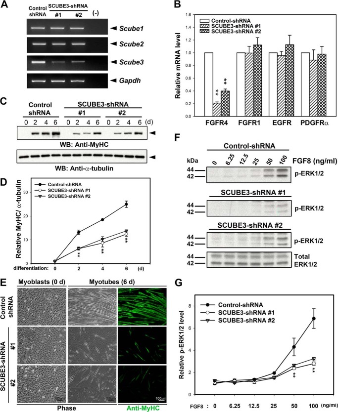
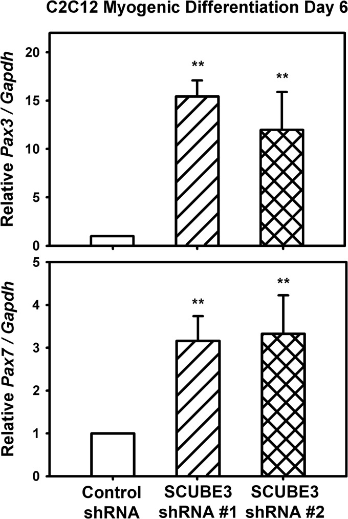
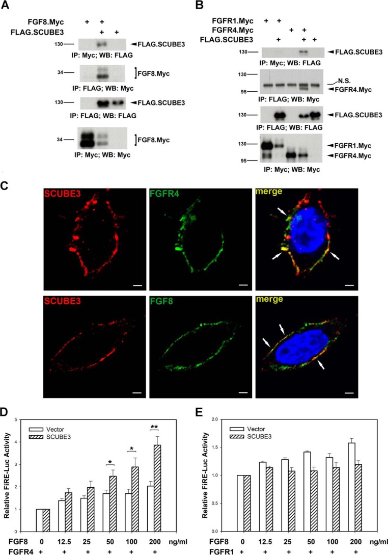
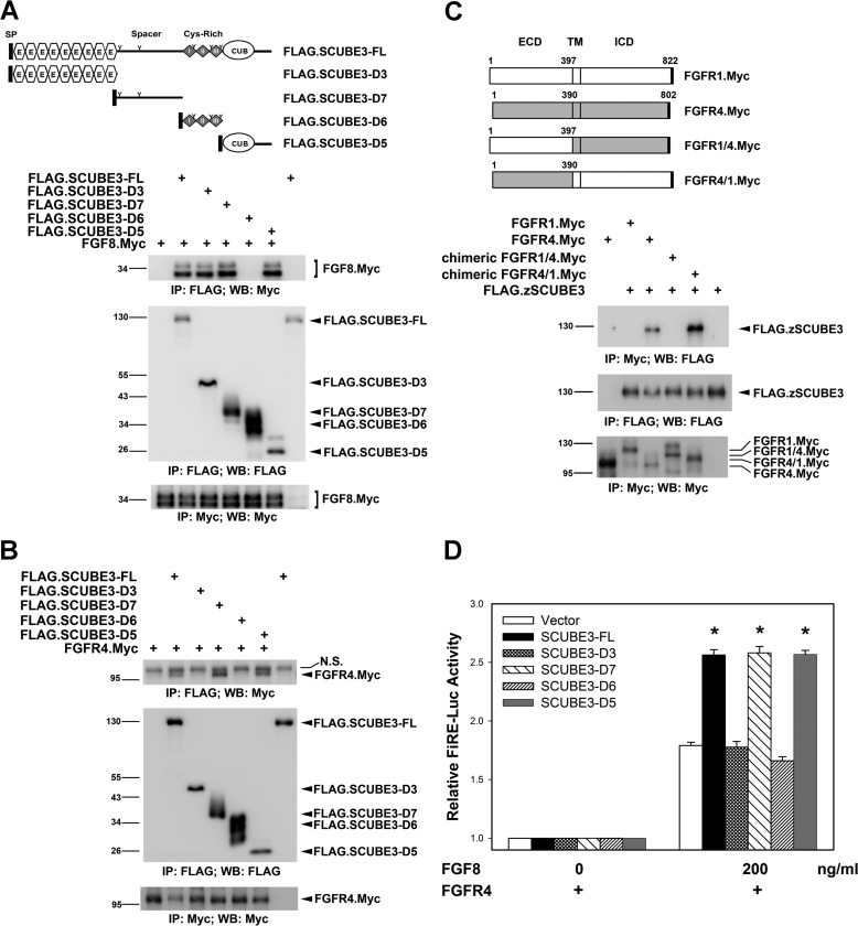
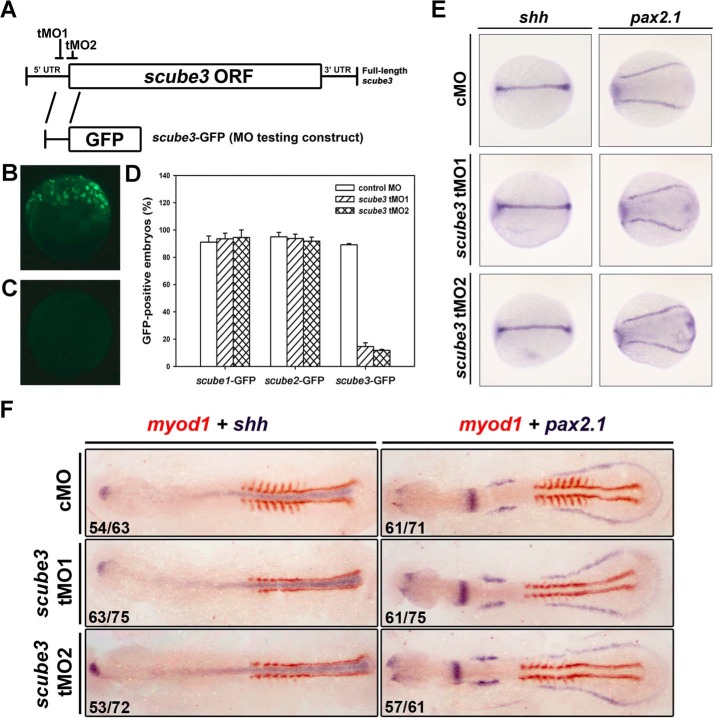
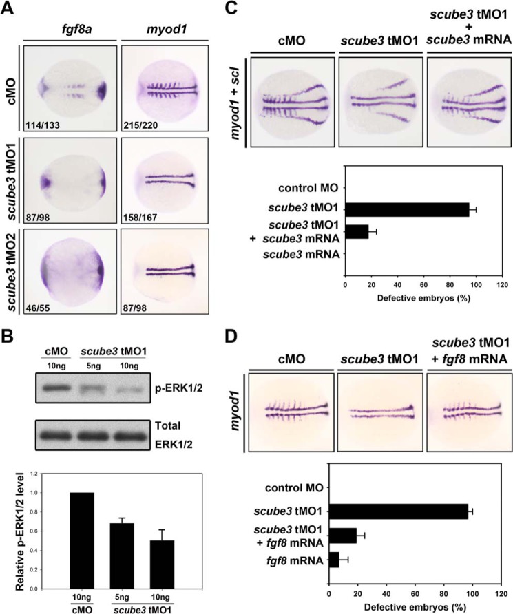
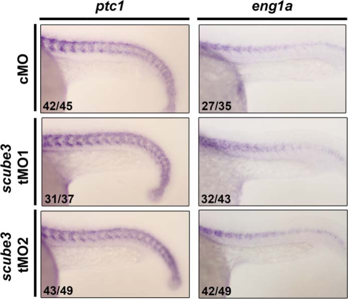
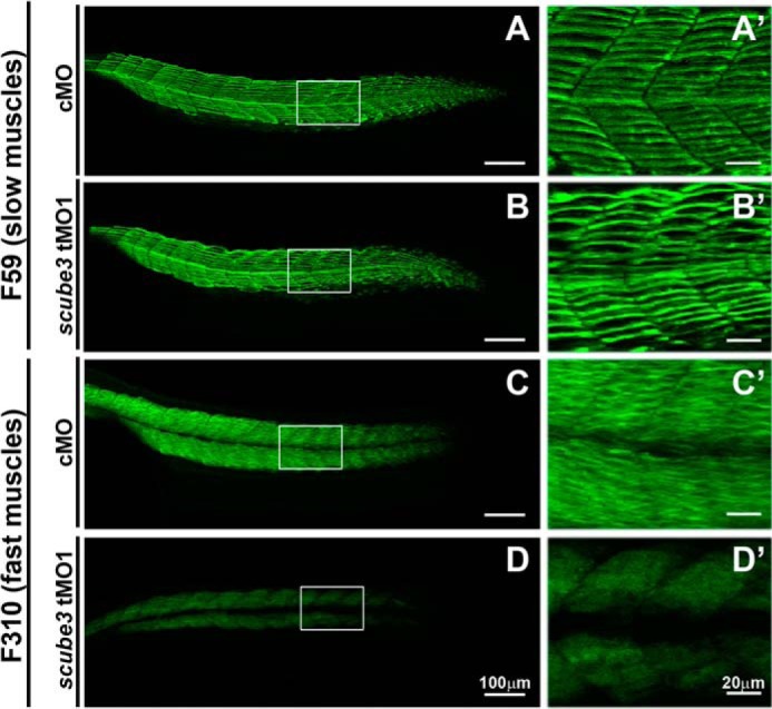
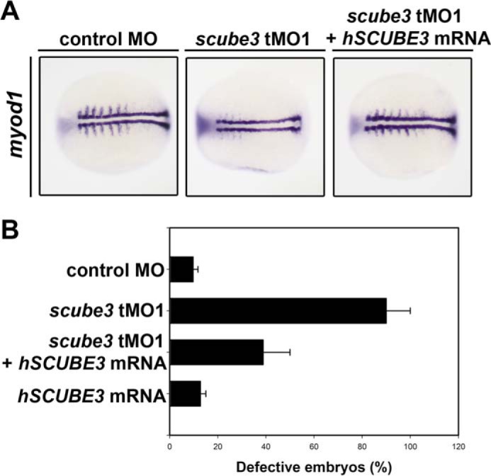
SCUBE3 (signal peptide CUB-EGF-like domain-containing protein 3) belongs to a newly identified secreted and cell membrane-associated SCUBE family, which is evolutionarily conserved in vertebrates. Scube3 is predominantly expressed in a variety of developing tissues in mice such as somites, neural tubes, and limb buds. However, its function during development remains unclear. In this study, we first showed that knockdown of SCUBE3 in C2C12 myoblasts inhibited FGF receptor 4 expression and FGF signaling, thus resulting in reduced myogenic differentiation. Furthermore, knockdown of zebrafish scube3 by antisense morpholino oligonucleotides specifically suppressed the expression of the myogenic marker myod1 within the lateral fast muscle precursors, whereas its expression in the adaxial slow muscle precursors was largely unaffected. Consistent with these findings, immunofluorescent staining of fast but not slow muscle myosin was markedly decreased in scube3 morphants. Further genetic studies identified fgf8 as a key regulator in scube3-mediated fast muscle differentiation in zebrafish. Biochemical and molecular analysis showed that SCUBE3 acts as a FGF co-receptor to augment FGF8 signaling. Scube3 may be a critical upstream regulator of fast fiber myogenesis by modulating fgf8 signaling during zebrafish embryogenesis.

Keywords: Cell Differentiation; Fast Muscle; Fibroblast Growth Factor (FGF); Fibroblast Growth Factor Receptor (FGFR); Myogenesis; Zebrafish; scube3.


Figures

FIGURE 1.
FIGURE 1.
Scube3 knockdown causes defective myogenic differentiation and reduced FGF8 signaling. A, knockdown of Scube3 mRNA expression in murine C2C12 myoblasts. C2C12 myoblasts were infected with a lentiviral vector to generate a stable clone expressing an shRNA control or shRNA specific for mouse Scube3 (SCUBE3-shRNAs 1 and 2), respectively. These cells were maintained in normal growth medium for 24 h after transfection and supplemented with puromycin (selection antibiotics). The efficiency and specificity of Scube3 knockdown but not Scube1 and Scube2 in the selected stable pool was confirmed by RT-PCR analysis. Gapdh mRNA level was used as an internal control. B, specific down-regulation of FGFR4 expression in Scube3-silenced C2C12 myoblasts. The expression of FGFR4, FGFR1, EGF receptor, and PDGFRα mRNA was assessed by quantitative real time PCR normalized to GAPDH expression with individual primer sets. The data are means ± S.E. **, p < 0.01 (compared with the control). C, suppression of myosin heavy chain (MyHC) expression by SCUBE3 knockdown. Control and SCUBE3-shRNA 1 and 2 myoblasts were induced to differentiate for 0, 2, 4, and 6 days in differentiation medium. Cell lysates were analyzed for the expression of MyHC by Western blot analysis. Tubulin levels were used as a loading control. D, quantification of MyHC protein levels during myogenic differentiation. Relative MyHC levels were quantified by densitometric scanning and normalized by α-tubulin. The data are means ± S.E. of four experiments. **, p < 0.01 (compared with control). E, effect of SCUBE3 knockdown on morphological transformation and anti-MyHC immunostaining after myogenic differentiation. Control and SCUBE3-shRNA 1 and 2 myoblasts were analyzed by phase contrast microscopy or immunofluorescence staining for MyHC after 6 days of differentiation. F, effect of SCUBE3 knockdown on FGF8 signaling. Control myoblasts and Scube3 knockdown myoblasts (SCUBE3-shRNA 1 or 2) were treated with the indicated concentrations of FGF8 for 10 min. Cell lysates (20 μg) underwent Western blot analysis with antibody specific for phosphorylated ERK1/2 (p-ERK1/2; representative blots are shown in the top three panels) or total ERK1/2 protein (representative blots are shown in the bottom panel). G, quantification of relative phosphorylated ERK1/2 levels by densitometric scanning and normalized by total amount of ERK1/2. The data are means ± S.E. of two experiments. **, p < 0.01 (compared with control). d, days; WB, Western blot.
FIGURE 2.
FIGURE 2.
Effects of Scube3 knockdown on the expression of Pax3 or Pax7. Control and SCUBE3-shRNA 1 and 2 myoblasts were induced to differentiate for 6 days in differentiation medium. Cells were collected to analyze the expression of Pax3 or Pax7 by quantitative RT-PCR. The data are means ± S.E. of three experiments. **, p < 0.01 (compared with control).
FIGURE 3.
FIGURE 3.
SCUBE3 forms a complex with FGF8 and FGFR4 and augments FGF signaling. A and B, SCUBE3 interacts with FGF8 and FGFR4. The expression plasmid encoding the Myc-tagged FGF8 (A) or Myc-tagged FGFR1 or FGFR4 (B) was transfected alone or with FLAG-tagged SCUBE3 in HEK-293T cells. After 2 days, cell lysates were prepared and underwent immunoprecipitation, followed by Western blot analysis with indicated antibodies to determine protein-protein interactions. Double bands seen for the FGF8.Myc were caused by differential glycosylation of this protein. N.S., nonspecific band; IP, immunoprecipitation; WB, Western blot. C, co-localization of SCUBE3 and FGF8 or FGFR4 was visualized by confocal immunofluorescence microscopy. SCUBE3 localization was detected by mouse anti-FLAG antibody and Alexa Fluor 594-conjugated goat anti-mouse IgG (red). FGF8 or FGFR4 was seen with rabbit anti-Myc antibody and Alexa Fluor 488-conjugated goat anti-rabbit IgG (green). The overlay demonstrates co-localization of SCUBE3 with FGF8 or FGFR4 on the plasma membrane (arrows). Scale bar, 2 μm. D and E, SCUBE3 augments FGF signaling through FGFR4. HEK-293T cells were transfected with FGF-responsive luciferase reporter (FiRE-Luc) and pRL-TK alone or with the expression plasmid encoding SCUBE3 combined with FGFR4 (D) or FGFR1 (E) expression plasmid. Transfected cells were incubated for 24 h with or without an increasing concentration of FGF8, and then luciferase activity was measured. Firefly luciferase values were normalized to that of Renilla activity for relative luciferase activity. The data are means ± S.E. *, p < 0.05; **, p < 0.01 (compared with the vector control).
FIGURE 4.
FIGURE 4.
Molecular mapping for the functional domain of SCUBE3. A and B, the spacer and CUB domain of SCUBE3 can interact with FGF8 and FGFR4. The expression plasmid encoding Myc-tagged FGF8 (A) and Myc-tagged FGFR4 (B) was transfected alone or together with a series of FLAG-tagged SCUBE3 deletion constructs in HEK-293T cells as depicted on top of A. After 2 days, cell lysates were prepared and underwent immunoprecipitation, followed by Western blot analysis with indicated antibodies to determine the protein-protein interactions. IP, immunoprecipitation; N.S., nonspecific band; WB, Western blot. C, FGFR4 interacts with SCUBE3 through its extracellular domain. Myc-tagged FGFR1, FGFR4, and chimeric FGFR1/4 or FGFR4/1 expression plasmids were transfected alone or with the FLAG-tagged SCUBE3 in HEK-293T cells. Co-immunoprecipitation experiments were performed as described above to examine the protein-protein interactions. ECD, extracellular domain; TM, transmembrane domain; ICD, intracellular domain. D, SCUBE3 augments FGF signaling through its spacer region or CUB domain. HEK-293T cells were transfected with FiRE-Luc and pRL-TK alone (blank bars) or with the expression plasmid encoding FL or various SCUBE3 domain deletion constructs combined with FGFR4 expression plasmid. Transfected cells were incubated with or without FGF8 (200 ng/ml) for 24 h, and then luciferase activity was measured. Firefly luciferase values were normalized to Renilla activity to obtain relative luciferase activity. The data are means ± S.E. *, p < 0.05.
FIGURE 5.
FIGURE 5.
Flow cytometry of cell surface expression of SCUBE3. A, domain structure of SCUBE3-FL and its deletion constructs. A FLAG tag was added immediately after the signal peptide sequences, thus at the NH2 terminus, for easy detection. FL, amino acids 1–993; D3, amino acids 1–401; D6, amino acid 398–629; D7, amino acids 632–803; D5, amino acids 804–993. FL, full-length; SP, signal peptide; E, EGF-like repeats; Cys-Rich, cysteine-rich motifs; CUB, CUB domain. B, the spacer region, cysteine-rich motifs, or CUB domain of SCUBE3 is capable of tethering on cell surface. At 24 h after transfection of empty vector or these expression plasmids, cells were detached and stained with anti-FLAG monoclonal antibody to determine the cell surface expression by flow cytometry. Empty vector-transfected cells are presented as the basal expression level (dotted line), and SCUBE3-transfected cells are shown as a thick line.
FIGURE 6.
FIGURE 6.
Identification, embryonic expression, and characterization of zebrafish scube3. A, graphic illustration of the domain organization of zebrafish Scube3 protein. SP, signal peptide sequence; E, EGF-like domain; Cys-rich, cysteine-rich repeats. B, phylogenetic tree of the SCUBE protein family. Similarity of human (h), mouse (m), and zebrafish (z) SCUBE protein sequences was analyzed by use of Lasergene MEGALIGN program (Clustal W algorithm). Branch order indicates structural relative, and branch length reflects sequence identity. C and D, embryonic expression patterns of zebrafish scube3. Scube3 transcripts are maternally deposited in zebrafish embryos and distributed widely in the embryos throughout development. RT-PCR analysis of scube3 mRNA expression in 1-cell to 3-dpf embryos. C, the Ef-1α gene was used as the internal control. D, the expression pattern of scube3 was examined by whole mount in situ hybridization at indicated stages. Stages are indicated at the upper right of each micrograph. c, cell; %, % of epiboly; s, somite; h, hours postfertilization. E, secretion of zebrafish Scube3 into the conditioned medium. HEK-293T cells were transfected with empty vector (Vector) or expression plasmid encoding FLAG-tagged zebrafish Scube3 (FLAG.zSCUBE3). At 48 h post-transfection, the serum-free conditioned medium was collected, and cells were detached. Samples from cell lysate (Cell) or the serum-free conditioned culture medium (medium) were separated by SDS-PAGE and transferred to polyvinylidene difluoride membranes. Recombinant FLAG.zSCUBE3 proteins were detected by Western blot (WB) analysis with anti-FLAG antibody. F, cell surface expression of zebrafish Scube3 protein. HEK-293T cells were transfected with empty vector (Vector) or expression plasmid encoding FLAG.zSCUBE3 protein. At 48 h post-transfection, cells were detached and probed with anti-FLAG antibody and underwent flow cytometry. G, N-linked glycosylation of zebrafish SCUBE1. HEK-293T cells were transfected with expression vector encoding FLAG.zSCUBE1 protein. Transfected cells were cultured without (−) or with (+) tunicamycin (an inhibitor of N-glycosylation) for 24 h (left panel). Alternatively, cell lysates were left untreated (−) or were treated (+) with peptide N-glycosidase F (PNGaseF) to remove the N-linked oligosaccharide chain (right panel). Cell lysates derived from each treatment were examined by Western blot analysis with anti-FLAG antibody. H, SCUBE3 protein was targeted into the membrane. HEK-293T cells were transfected with the expression plasmids encoding FLAG-tagged SCUBE protein: human (h) or zebrafish (z) orthologue, respectively. Two days after transfection, cells were collected and homogenized. Samples of supernatant were centrifuged at 100,000 × g for 30 min to yield the supernatant (cytosolic fraction) and the pellet (membrane fraction). Each subcellular fraction was subjected to SDS-PAGE and immunoblot analysis using anti-FLAG, anti-flotillin-1 (a lipid raft membrane protein), or anti-GAPDH (a cytosolic protein) antibody. I, cell surface localization of SCUBE3 visualized by confocal immunofluorescent microscopy. After transfection of expression plasmid encoding FLAG-tagged SCUBE3, in HeLa cells, SCUBE3 expression was detected by mouse anti-FLAG antibody and Alexa Fluor 594-conjugated goat anti-mouse IgG (red). Scale bar, 5 μm.
FIGURE 7.
FIGURE 7.
Specificity and effectiveness of scube3 tMOs and no effect of scube3 knockdown on the expression of other mesoderm genes. A, schematic presentation of the targeting sites of two scube3 tMOs and the scube3-GFP test construct. tMO1 targets the 5′-UTR, and tMO2 blocks at the AUG translation start site. B–D, efficiency of scube3 tMO. One-cell embryos were injected with 300 pg of tMO testing construct with 1 ng of control MO or scube3 tMO1 or tMO2, respectively. The expression of GFP was examined at 50–70% epiboly stage and photographed by epifluorescent microscopy. The images are representative of embryos injected with scube3-GFP plasmid in combination with control MO (B) or scube3-tMO1 (C). D, summary of translational blocking efficiency of scube3 tMOs. The lack of suppression effect on the scube1-GFP or scube2-GFP testing constructs confirms their specificity in blocking translation of scube3. The data are means ± S.E. E, whole mount in situ hybridization for shh and pax2.1 expression in the zebrafish embryos. Dorsal view of the eight-somite embryos. Anterior is to the left in all images. The expression of shh (the adaxial mesoderm marker) and pax2.1 (the intermediate mesoderm marker) was not affected in scube3 morphants. F, effect of scube3 knockdown on the expression of myod1. One-celled embryos were injected with control MO (cMO) or scube3 tMO1 or tMO2 (5 ng/embryo) and examined for expression of the indicated markers at the eight-somite stage by two-color RNA ISH. The embryos are shown in dorsal view with anterior to the left. Although the expression of shh (blue) and pax2.1 (blue) remained relatively unaltered, the expression level of myod1 (red) in the lateral somites were reduced in the scube3 morphants as compared with the control embryos. The numbers in the bottom left-hand corners of photographs are the numbers of affected embryos with phenotype similar to what is shown in the image (left) and the total number of observed embryos (right).
FIGURE 8.
FIGURE 8.
Knockdown of scube3 suppressed FGF target genes and reduced FGF signaling in zebrafish embryos. A, effect of scube3 knockdown on the expression of FGF target genes (fgf8 and myod1) evaluated by whole mount in situ hybridization. The images are the dorsal view of the eight-somite embryos with anterior to the left. In scube3 morphants, fgf8 and myod1 expression was markedly reduced in the somites. Of note, scube3 knockdown specifically suppressed myod1 expression in lateral fast muscle precursors but not adaxial cells that will differentiate into slow muscles. The numbers in the bottom left-hand corners of photographs are the numbers of affected embryos with phenotype similar to what is shown in the image (left column) and the total number of observed embryos (right column). B, ERK activation in control or scube3 knockdown embryos. After Scube3 tMO1 injection, protein lysates (20 μg) derived from embryos (70% epiboly) were blotted with antibody specific to the phosphorylated form (p-ERK) or total ERK protein and quantified by densitometric scanning (mean ± S.E.) from three independent experiments (bottom panel). Note that scube3 knockdown significantly decreased p-ERK expression in a dose-dependent manner, which supports the role of scube3 in FGF signaling during zebrafish development. C and D, forced expression of scube3 or fgf8 mRNA restored myod1 expression in fast muscle precursors of scube3 morphants. C, co-injection of scube3 morphants with scube3 mRNA rescued myod1 expression. Scube3 morphants showed rescued expression of lateral myod1expression in somites when injected with 100 pg of synthetic scube3 mRNA at the one- or two-cell stage. The images are shown as dorsal views with anterior to the left. Quantification data are shown in the bottom panel. D, forced expression of fgf8 mRNA restored myod1 expression in lateral somites. Scube3 morphants showed rescued expression of lateral myod1 expression in somites when injected with 0.0625 pg of synthetic fgf8 mRNA at the one- or two-cell stage. The images are shown as dorsal views with anterior to the left. Quantification data are shown in the bottom panel. The data are means ± S.E. cMO, control MO.
FIGURE 9.
FIGURE 9.
Knockdown of scube3 did not affect Hh target genes in zebrafish embryos. Effects of scube3 knockdown on the expression of Hh target genes (ptc1 and eng1a) were evaluated by whole mount in situ hybridization. The images are the lateral views of the 22-somite embryos with anterior to the left. No effect of scube3 knockdown on the expression of ptc1 and eng1a was observed. The numbers in the bottom left-hand corners of the photographs are the numbers of embryos with phenotype similar to what is shown in the image (left column) and the total number of observed embryos (right column). cMO, control MO.
FIGURE 10.
FIGURE 10.
Scube3 is required for fast muscle differentiation. A, A′, B, and B′, immunofluorescent staining of slow muscle with F59 antibody in control (A and A′) and scube3 morphants (B and B′) at the 26-somite stage. The images are presented as lateral views with anterior to the left. C, C′, D, and D′, immunostaining of fast muscle with F310 antibody in control (C and C′) and scube3 morphants (D and D′) at the 26-somite stage. Corresponding higher magnification views are shown in A′, B′, C′, and D′. cMO, control MO.
FIGURE 11.
FIGURE 11.
Human SCUBE3 mRNA restores myod1 expression in the lateral somites in the scube3 morphants. A, visualization of the rescue experiments using myod1 expression. The images are shown as dorsal views with anterior to the left. Scube3 morphants showed rescued expression of lateral myod1 expression in somites when injected with 5 pg of synthetic human SCUBE3 mRNA at the one- to two-cell stage. B, corresponding quantified data are shown. The data are means ± S.E.

Similar articles

Cited by

References

    1. Cheng C. J., Lin Y. C., Tsai M. T., Chen C. S., Hsieh M. C., Chen C. L., Yang R. B. (2009) SCUBE2 suppresses breast tumor cell proliferation and confers a favorable prognosis in invasive breast cancer. Cancer Res. 69, 3634–3641 - PubMed
    1. Grimmond S., Larder R., Van Hateren N., Siggers P., Hulsebos T. J., Arkell R., Greenfield A. (2000) Cloning, mapping, and expression analysis of a gene encoding a novel mammalian EGF-related protein (SCUBE1). Genomics 70, 74–81 - PubMed
    1. Haworth K., Smith F., Zoupa M., Seppala M., Sharpe P. T., Cobourne M. T. (2007) Expression of the Scube3 epidermal growth factor-related gene during early embryonic development in the mouse. Gene Expr. Patterns 7, 630–634 - PubMed
    1. Kawakami A., Nojima Y., Toyoda A., Takahoko M., Satoh M., Tanaka H., Wada H., Masai I., Terasaki H., Sakaki Y., Takeda H., Okamoto H. (2005) The zebrafish-secreted matrix protein you/scube2 is implicated in long-range regulation of hedgehog signaling. Curr. Biol. 15, 480–488 - PubMed
    1. Tsai M. T., Cheng C. J., Lin Y. C., Chen C. C., Wu A. R., Wu M. T., Hsu C. C., Yang R. B. (2009) Isolation and characterization of a secreted, cell-surface glycoprotein SCUBE2 from humans. Biochem. J. 422, 119–128 - PubMed

Publication types

MeSH terms