

Multicolor live-cell chemical imaging by isotopically edited alkyne vibrational palette
- PMID: 24849912
- PMCID: PMC4063185
- DOI: 10.1021/ja502706q
Multicolor live-cell chemical imaging by isotopically edited alkyne vibrational palette
Abstract
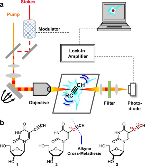
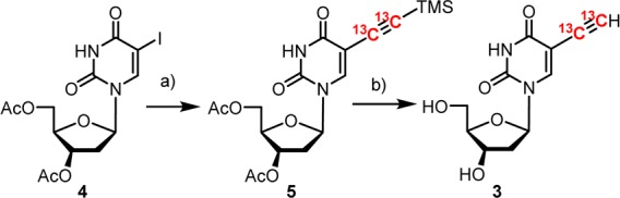
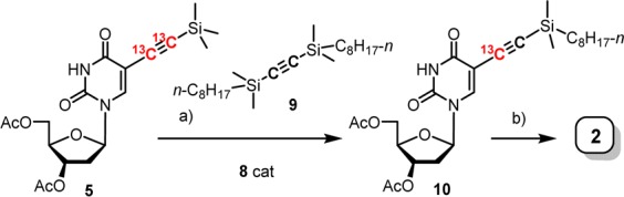
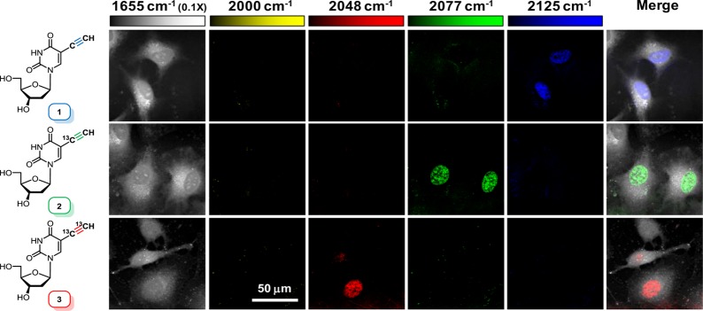
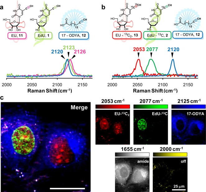
Vibrational imaging such as Raman microscopy is a powerful technique for visualizing a variety of molecules in live cells and tissues with chemical contrast. Going beyond the conventional label-free modality, recent advance of coupling alkyne vibrational tags with stimulated Raman scattering microscopy paves the way for imaging a wide spectrum of alkyne-labeled small biomolecules with superb sensitivity, specificity, resolution, biocompatibility, and minimal perturbation. Unfortunately, the currently available alkyne tag only processes a single vibrational "color", which prohibits multiplex chemical imaging of small molecules in a way that is being routinely practiced in fluorescence microscopy. Herein we develop a three-color vibrational palette of alkyne tags using a (13)C-based isotopic editing strategy. We first synthesized (13)C isotopologues of EdU, a DNA metabolic reporter, by using the newly developed alkyne cross-metathesis reaction. Consistent with theoretical predictions, the mono-(13)C ((13)C≡(12)C) and bis-(13)C ((13)C≡(13)C) labeled alkyne isotopologues display Raman peaks that are red-shifted and spectrally resolved from the originally unlabeled ((12)C≡(12)C) alkynyl probe. We further demonstrated three-color chemical imaging of nascent DNA, RNA, and newly uptaken fatty-acid in live mammalian cells with a simultaneous treatment of three different isotopically edited alkynyl metabolic reporters. The alkyne vibrational palette presented here thus opens up multicolor imaging of small biomolecules, enlightening a new dimension of chemical imaging.
Figures

Similar articles
-
Live-Cell Bioorthogonal Chemical Imaging: Stimulated Raman Scattering Microscopy of Vibrational Probes.Acc Chem Res. 2016 Aug 16;49(8):1494-502. doi: 10.1021/acs.accounts.6b00210. Epub 2016 Aug 3. Acc Chem Res. 2016. PMID: 27486796 Free PMC article. Review.
-
Live-cell stimulated Raman scattering imaging of alkyne-tagged biomolecules.Angew Chem Int Ed Engl. 2014 Jun 2;53(23):5827-31. doi: 10.1002/anie.201400328. Epub 2014 Apr 17. Angew Chem Int Ed Engl. 2014. PMID: 24753329
-
Deuteration of terminal alkynes realizes simultaneous live cell Raman imaging of similar alkyne-tagged biomolecules.Org Biomol Chem. 2021 Oct 6;19(38):8232-8236. doi: 10.1039/d1ob01479j. Org Biomol Chem. 2021. PMID: 34528645
-
Alkyne-tag Raman imaging for visualization of mobile small molecules in live cells.J Am Chem Soc. 2012 Dec 26;134(51):20681-9. doi: 10.1021/ja308529n. Epub 2012 Dec 14. J Am Chem Soc. 2012. PMID: 23198907
-
Applications of vibrational tags in biological imaging by Raman microscopy.Analyst. 2017 Oct 23;142(21):4018-4029. doi: 10.1039/c7an01001j. Analyst. 2017. PMID: 28875184 Free PMC article. Review.
Cited by
-
Anticancer, Antioxidant, and Catalytic Activities of Green Synthesized Gold Nanoparticles Using Avocado Seed Aqueous Extract.ACS Omega. 2023 Jul 11;8(29):26088-26101. doi: 10.1021/acsomega.3c02260. eCollection 2023 Jul 25. ACS Omega. 2023. PMID: 37521675 Free PMC article.
-
Biomedical applications, perspectives and tag design concepts in the cell - silent Raman window.RSC Chem Biol. 2024 Feb 12;5(4):273-292. doi: 10.1039/d3cb00217a. eCollection 2024 Apr 3. RSC Chem Biol. 2024. PMID: 38576725 Free PMC article. Review.
-
Toward the Next Frontiers of Vibrational Bioimaging.Chem Biomed Imaging. 2023 Mar 28;1(1):3-17. doi: 10.1021/cbmi.3c00004. eCollection 2023 Apr 24. Chem Biomed Imaging. 2023. PMID: 37122829 Free PMC article. Review.
-
Live Intracellular Biorthogonal Imaging by Surface Enhanced Raman Spectroscopy using Alkyne-Silver Nanoparticles Clusters.Sci Rep. 2018 Aug 23;8(1):12652. doi: 10.1038/s41598-018-31165-3. Sci Rep. 2018. PMID: 30140073 Free PMC article.
-
Far-Field Super-Resolution Vibrational Spectroscopy.Anal Chem. 2019 Jul 16;91(14):8723-8731. doi: 10.1021/acs.analchem.9b01731. Epub 2019 Jun 28. Anal Chem. 2019. PMID: 31251563 Free PMC article.
References
Publication types
MeSH terms
Substances
Grants and funding
LinkOut - more resources
Full Text Sources
Other Literature Sources
Miscellaneous
