Increase in hypotonic stress-induced endocytic activity in macrophages via ClC-3
- PMID: 24850147
- PMCID: PMC4044314
- DOI: 10.14348/molcells.2014.0031
Increase in hypotonic stress-induced endocytic activity in macrophages via ClC-3
Abstract
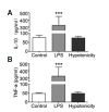
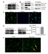
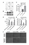
Extracellular hypotonic stress can affect cellular function. Whether and how hypotonicity affects immune cell function remains to be elucidated. Macrophages are immune cells that play key roles in adaptive and innate in immune reactions. The purpose of this study was to investigate the role and underlying mechanism of hypotonic stress in the function of bone marrow-derived macrophages (BMDMs). Hypotonic stress increased endocytic activity in BMDMs, but there was no significant change in the expression of CD80, CD86, and MHC class II molecules, nor in the secretion of TNF-α or IL-10 by BMDMs. Furthermore, the enhanced endocytic activity of BMDMs triggered by hypotonic stress was significantly inhibited by chloride channel-3 (ClC-3) siRNA. Our findings suggest that hypotonic stress can induce endocytosis in BMDMs and that ClC-3 plays a central role in the endocytic process.
Figures


Similar articles
-
IL-12 suppression, enhanced endocytosis and up-regulation of MHC-II and CD80 in dendritic cells during experimental endotoxin tolerance.Acta Pharmacol Sin. 2009 May;30(5):582-8. doi: 10.1038/aps.2009.34. Epub 2009 Apr 6. Acta Pharmacol Sin. 2009. PMID: 19349963 Free PMC article.
-
Oligosaccharide-dependent anti-inflammatory role of galectin-1 for macrophages in ulcerative colitis.J Gastroenterol Hepatol. 2020 Dec;35(12):2158-2169. doi: 10.1111/jgh.15097. Epub 2020 Jul 6. J Gastroenterol Hepatol. 2020. PMID: 32424849
-
Expression of the chloride channel ClC-2 in the murine small intestine epithelium.Am J Physiol Cell Physiol. 2000 Dec;279(6):C1787-94. doi: 10.1152/ajpcell.2000.279.6.C1787. Am J Physiol Cell Physiol. 2000. PMID: 11078693
-
A pseudopterane diterpene isolated from the octocoral Pseudopterogorgia acerosa inhibits the inflammatory response mediated by TLR-ligands and TNF-alpha in macrophages.PLoS One. 2013 Dec 16;8(12):e84107. doi: 10.1371/journal.pone.0084107. eCollection 2013. PLoS One. 2013. PMID: 24358331 Free PMC article.
-
Chloride channels and endocytosis: new insights from Dent's disease and ClC-5 knockout mice.Nephron Physiol. 2005;99(3):p69-73. doi: 10.1159/000083210. Nephron Physiol. 2005. PMID: 15637424 Review.
Cited by
-
Endocytosis in the adaptation to cellular stress.Cell Stress. 2020 Aug 18;4(10):230-247. doi: 10.15698/cst2020.10.232. Cell Stress. 2020. PMID: 33024932 Free PMC article. Review.
-
IL-33-induced alternatively activated macrophage attenuates the development of TNBS-induced colitis.Oncotarget. 2017 Apr 25;8(17):27704-27714. doi: 10.18632/oncotarget.15984. Oncotarget. 2017. PMID: 28423665 Free PMC article.
-
Interleukin-4 Inhibits Regulatory T Cell Differentiation through Regulating CD103+ Dendritic Cells.Front Immunol. 2017 Mar 3;8:214. doi: 10.3389/fimmu.2017.00214. eCollection 2017. Front Immunol. 2017. PMID: 28316599 Free PMC article.
-
Extracellular HMGB1 Contributes to the Chronic Cardiac Allograft Vasculopathy/Fibrosis by Modulating TGF-β1 Signaling.Front Immunol. 2021 Mar 10;12:641973. doi: 10.3389/fimmu.2021.641973. eCollection 2021. Front Immunol. 2021. PMID: 33777037 Free PMC article.
-
From Pinocytosis to Methuosis-Fluid Consumption as a Risk Factor for Cell Death.Front Cell Dev Biol. 2021 Jun 23;9:651982. doi: 10.3389/fcell.2021.651982. eCollection 2021. Front Cell Dev Biol. 2021. PMID: 34249909 Free PMC article. Review.
References
-
- Comes N., Gasull X., Gual A., Borras T. Differential expression of the human chloride channel genes in the trabecular meshwork under stress conditions. Exp. Eye Res. 2005;80:801–813. - PubMed
Publication types
MeSH terms
Substances
LinkOut - more resources
Full Text Sources
Other Literature Sources
Research Materials

