

Foxp3+ regulatory T cells promote lung epithelial proliferation
- PMID: 24850425
- PMCID: PMC4205163
- DOI: 10.1038/mi.2014.33
Foxp3+ regulatory T cells promote lung epithelial proliferation
Abstract
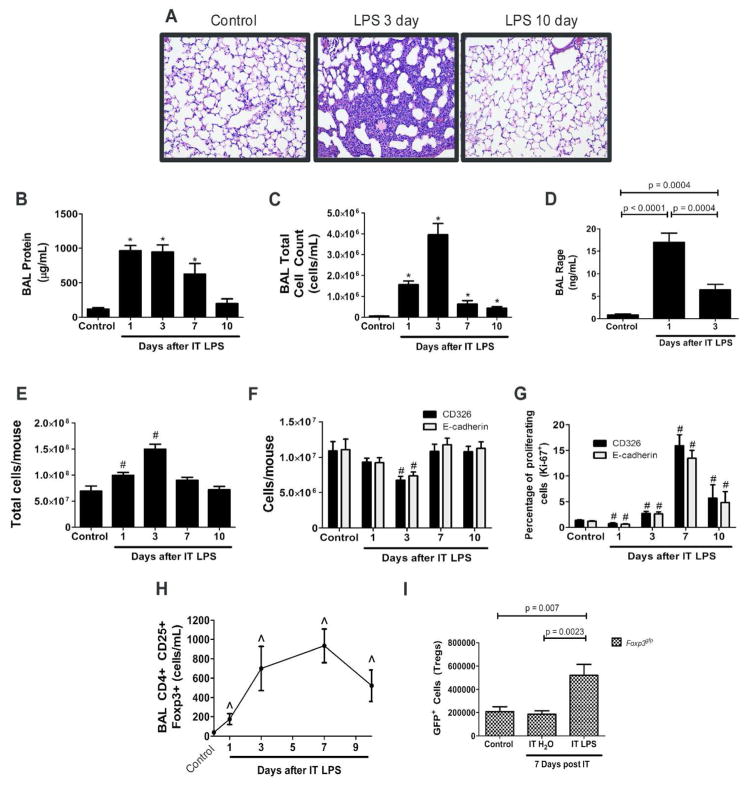
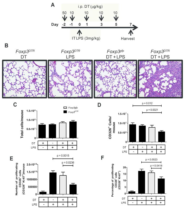
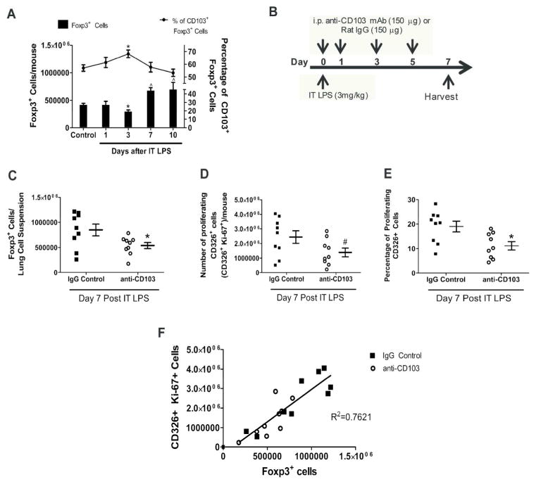
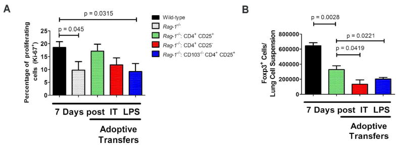
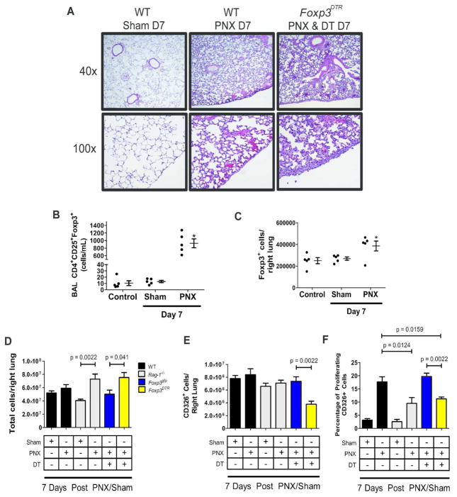
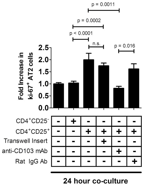
Acute respiratory distress syndrome (ARDS) causes significant morbidity and mortality each year. There is a paucity of information regarding the mechanisms necessary for ARDS resolution. Foxp3(+) regulatory T cells (Foxp3(+) T(reg) cells) have been shown to be an important determinant of resolution in an experimental model of lung injury. We demonstrate that intratracheal delivery of endotoxin (lipopolysaccharide) elicits alveolar epithelial damage from which the epithelium undergoes proliferation and repair. Epithelial proliferation coincided with an increase in Foxp3(+) T(reg) cells in the lung during the course of resolution. To dissect the role that Foxp3(+) T(reg) cells exert on epithelial proliferation, we depleted Foxp3(+) T(reg) cells, which led to decreased alveolar epithelial proliferation and delayed lung injury recovery. Furthermore, antibody-mediated blockade of CD103, an integrin, which binds to epithelial expressed E-cadherin decreased Foxp3(+) T(reg) numbers and decreased rates of epithelial proliferation after injury. In a non-inflammatory model of regenerative alveologenesis, left lung pneumonectomy, we found that Foxp3(+) T(reg) cells enhanced epithelial proliferation. Moreover, Foxp3(+) T(reg) cells co-cultured with primary type II alveolar cells (AT2) directly increased AT2 cell proliferation in a CD103-dependent manner. These studies provide evidence of a new and integral role for Foxp3(+) T(reg) cells in repair of the lung epithelium.
Figures
Similar articles
-
Foxp3+ Regulatory T Cell Expression of Keratinocyte Growth Factor Enhances Lung Epithelial Proliferation.Am J Respir Cell Mol Biol. 2017 Aug;57(2):162-173. doi: 10.1165/rcmb.2017-0019OC. Am J Respir Cell Mol Biol. 2017. PMID: 28296468 Free PMC article.
-
Cutting edge: CD47 controls the in vivo proliferation and homeostasis of peripheral CD4+ CD25+ Foxp3+ regulatory T cells that express CD103.J Immunol. 2008 Oct 15;181(8):5204-8. doi: 10.4049/jimmunol.181.8.5204. J Immunol. 2008. PMID: 18832672
-
The Ets-1 transcription factor controls the development and function of natural regulatory T cells.J Exp Med. 2010 Sep 27;207(10):2113-25. doi: 10.1084/jem.20092153. Epub 2010 Sep 20. J Exp Med. 2010. PMID: 20855499 Free PMC article.
-
CD103+ GALT DCs promote Foxp3+ regulatory T cells.Mucosal Immunol. 2008 Nov;1 Suppl 1:S34-8. doi: 10.1038/mi.2008.43. Mucosal Immunol. 2008. PMID: 19079226 Review.
-
Plasticity of T(reg) cells: is reprogramming of T(reg) cells possible in the presence of FOXP3?Int Immunopharmacol. 2011 May;11(5):555-60. doi: 10.1016/j.intimp.2010.11.024. Epub 2010 Nov 27. Int Immunopharmacol. 2011. PMID: 21115121 Review.
Cited by
-
The role of T cells in age-related diseases.Nat Rev Immunol. 2022 Feb;22(2):97-111. doi: 10.1038/s41577-021-00557-4. Epub 2021 Jun 7. Nat Rev Immunol. 2022. PMID: 34099898 Review.
-
Tissue regulatory T cells: regulatory chameleons.Nat Rev Immunol. 2021 Sep;21(9):597-611. doi: 10.1038/s41577-021-00519-w. Epub 2021 Mar 26. Nat Rev Immunol. 2021. PMID: 33772242 Free PMC article. Review.
-
Comprehensive Analysis of Potential ceRNA Network and Different Degrees of Immune Cell Infiltration in Acute Respiratory Distress Syndrome.Front Genet. 2022 Jun 1;13:895629. doi: 10.3389/fgene.2022.895629. eCollection 2022. Front Genet. 2022. PMID: 35719385 Free PMC article.
-
Interrelation Between Fibroblasts and T Cells in Fibrosing Interstitial Lung Diseases.Front Immunol. 2021 Nov 5;12:747335. doi: 10.3389/fimmu.2021.747335. eCollection 2021. Front Immunol. 2021. PMID: 34804029 Free PMC article. Review.
-
Regulatory T-cells promote pulmonary repair by modulating T helper cell immune responses in lipopolysaccharide-induced acute respiratory distress syndrome.Immunology. 2019 Jun;157(2):151-162. doi: 10.1111/imm.13060. Epub 2019 May 6. Immunology. 2019. PMID: 30919408 Free PMC article.
References
-
- Ware LB, Matthay MA. The acute respiratory distress syndrome. N Engl J Med. 2000;342:1334–1349. - PubMed
-
- Ranieri VM, et al. Acute respiratory distress syndrome: the Berlin Definition. JAMA. 2012;307:2526–2533. - PubMed
-
- Ventilation with lower tidal volumes as compared with traditional tidal volumes for acute lung injury and the acute respiratory distress syndrome. The Acute Respiratory Distress Syndrome Network. N Engl J Med. 2000;342:1301–1308. - PubMed
-
- Guerin C, et al. Prone positioning in severe acute respiratory distress syndrome. N Engl J Med. 2013;368:2159–2168. - PubMed
Publication types
MeSH terms
Substances
Grants and funding
LinkOut - more resources
Full Text Sources
Other Literature Sources
Medical
Research Materials
