. 2014 Nov;7(6):1480-91.
doi: 10.1038/mi.2014.36. Epub 2014 May 21.

Caspase-11 is expressed in the colonic mucosa and protects against dextran sodium sulfate-induced colitis

Affiliations

Caspase-11 is expressed in the colonic mucosa and protects against dextran sodium sulfate-induced colitis

D Demon et al. Mucosal Immunol. 2014 Nov.

Erratum in

  • Mucosal Immunol. 2014 Nov;7(6):1504

Abstract

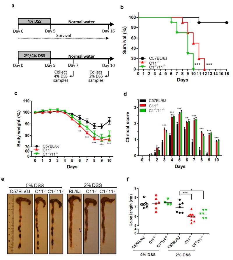
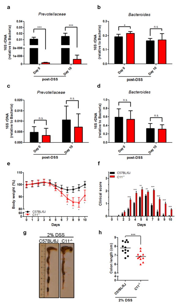
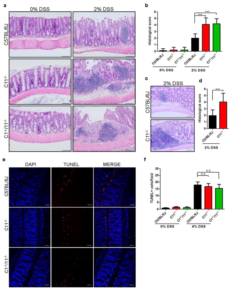
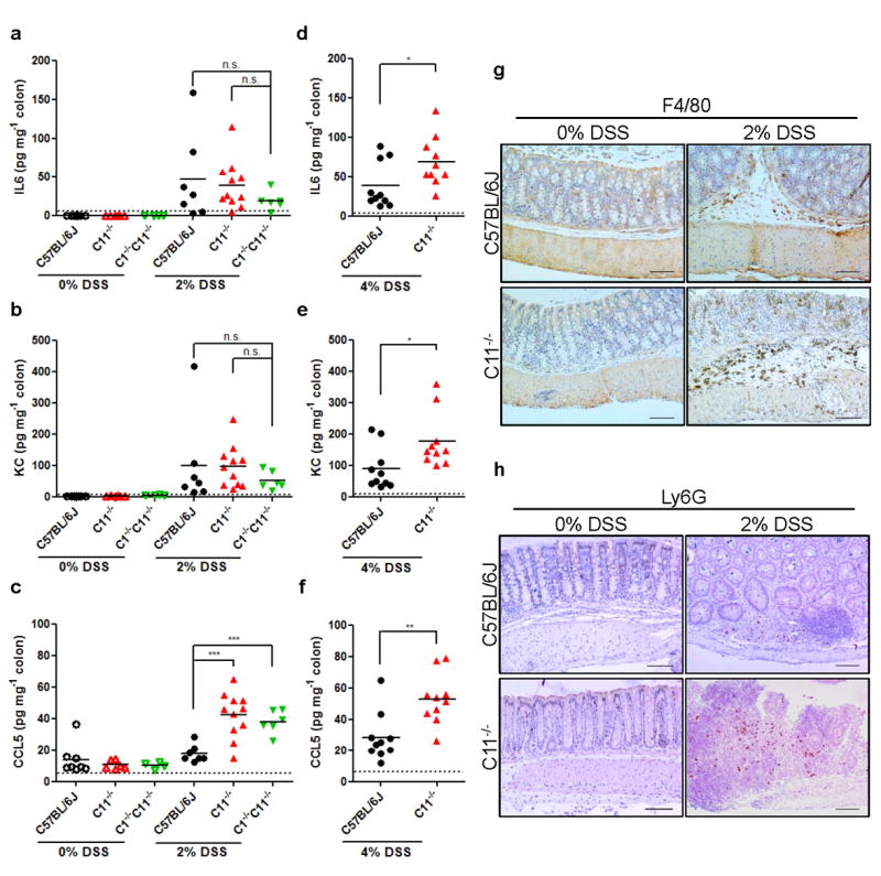
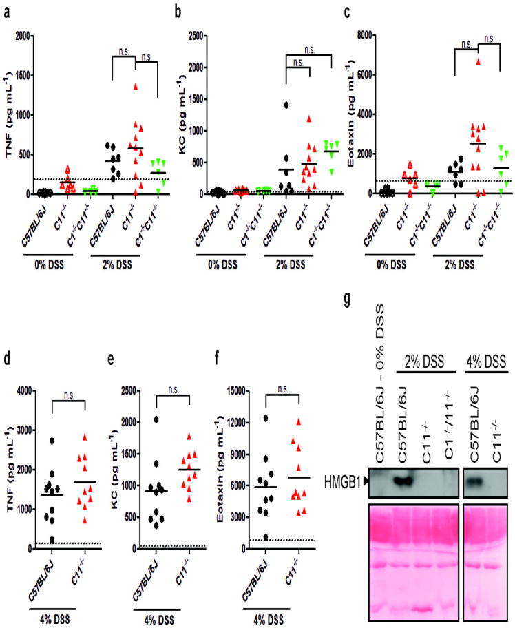
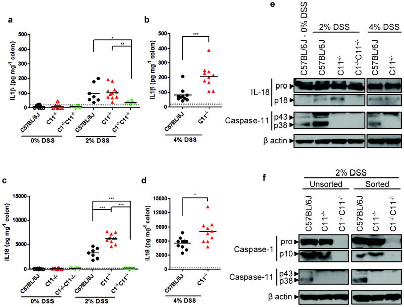
Ulcerative colitis and Crohn's disease are major inflammatory syndromes that affect millions of patients. Caspase-11 confers protection against Gram-negative enteropathogens, but its role during colitis is unknown. Here, we show that caspase-11 was constitutively expressed in the colon, and that caspase-11-deficient (caspase-11(-/-)) mice were hypersusceptible to dextran sodium sulfate (DSS)-induced colitis. Notably, pro-inflammatory Prevotella species were strongly reduced in the gut microbiota of caspase-11(-/-) mice. Co-housing with wild-type mice leveled Prevotella contents, but failed to protect caspase-11(-/-) mice from increased susceptibility to DSS-induced colitis. We therefore addressed the role of caspase-11 in immune signaling. DSS-induced tissue damage and inflammatory cell infiltration in the gut were markedly increased in caspase-11−/− mice, while release of the pyroptosis/necroptosis marker HMGB1 was abolished [Corrected]. Moreover, caspase-11(-/-) mice showed normal or increased production of mature interleukin (IL)-1β and IL-18, whereas IL-1β and IL-18 secretion was blunted in animals lacking both caspases 1 and 11. In conclusion, we showed that caspase-11 shapes the gut microbiota composition, and that caspase-11(-/-) mice are highly susceptible to DSS-induced colitis. Moreover, DSS-induced inflammasome activation relied on caspase-1, but not caspase-11. These results suggest a role for other caspase-11 effector mechanisms such as pyroptosis in protection against intestinal inflammation.


Conflict of interest statement

Conflict of interest statement

The authors declare no competing financial interests.

Figures

Figure 1
Figure 1. Caspase-11 deficiency sensitizes mice to DSS-induced morbidity and lethality
(a) Schematic of the design of the survival study and the studies for the collection of serum and colon samples. In general, mice were fed a 2% or 4% DSS solution in drinking water for 5 days, followed by normal drinking water until the end of the experiment. (b) Kaplan-Meier survival plot of C57BL/6J, caspase-11-/- (C11-/-) and caspase-1-/-/11-/- (C1-/-/11-/-) mice induced with 4% DSS (n=10/group). (c-d) The percentage of body weight (c) and the clinical score (d) of C57BL/6J, C11-/- and C1-/-/11-/- mice induced with 2% DSS. Data depict the mean ± SEM (n=10/group). Statistical significance between DSS-treated C57BL/6J and Casp11-/- mice is indicated. (e) Macroscopic pictures and (f) graphic presentation of the colon length of C57BL/6J, C11-/- and C1-/-/11-/- mice without (0% DSS) or 10 days after induction with 2% DSS. Data in panel f depict individual replicates with mean; C57BL/6J mice (0% DSS, n=8; 2%, n=7), Casp11-/- mice (0% DSS, n=6; 2%, n=11) and Casp1-/-/Casp11-/- mice (0% DSS, n=4; 2% DSS, n=6). Statistical significance between DSS-treated C57BL/6J, C11-/- and C1-/-/11-/- mice is indicated. *, P < 0.05; **, P < 0.01; ***, P < 0.001.
Figure 2
Figure 2. The microbiome of caspase-11-/- mice contains reduced Prevotellacea, but this is not associated with their hypersensitivity towards DSS-induced colitis
(a-b) Expression of the microbial phyla Prevotellacea (a) and Bacteroides (b) in the fecal samples of separately housed C57BL/6J and caspase-11-/- (C11-/-) mice before (day 0) and 10 days after the start of 2% DSS administration. Data depict the mean ± SD (n=6/group). (c-d) Expression of the microbial species Prevotellaceae (c) and Bacteroides (d) in the fecal samples of C57BL/6J and C11-/- mice that were first co-housed together for 4 weeks (day 0) and then induced with 2% DSS (day 10). Data depict the mean ± SD (n = 12/group). (e-h) The percentage of body weight (e), the clinical score (f), and macroscopic pictures and (h) graphic presentation of the colon length of C57BL/6J and C11-/- mice that were co-housed together for 4 weeks and afterwards induced with 2% DSS. Data depict the mean ± SEM (e-f; n=12/group) or individual replicates with mean (h; n=12/group). Statistical significance between DSS-treated C57BL/6J and C11-/- mice is indicated. *, P < 0.05; **, P < 0.01; ***, P < 0.001, n.s., not significant.
Figure 3
Figure 3. Caspase-11-/- mice show increased colonic tissue damage and leukocyte infiltration, despite normal apoptosis
(a-b) Representative microscopic pictures (a) and semi-quantitative scoring (b) of hematoxylin and eosin (H&E)-stained colon sections of DSS-treated C57BL/6J, caspase-11-/- (C11-/-) and Caspase-1-/-/11-/- (C1-/-/11-/-) mice without (0% DSS) or 10 days after induction with 2% DSS. (c-d) Representative microscopic pictures (c) and semi-quantitative scoring (d) of hematoxylin and eosin (H&E)-stained colon sections of DSS-treated C57BL/6J and C11-/- mice that were co-housed together for 4 weeks and afterwards induced with 2% DSS. Data depict the mean ± SD (b; n=4/group and d; n=5/group). Bars: 100 μm. (e-f) Representative microscopic pictures (e) and quantification of the number of TUNEL-positive cells per field (f) of DSS-treated C57BL/6J, C11-/- and C1-/-/11-/- mice 3 days after induction with 4% DSS. Bars: 50 μm. Data depict the mean ± SEM in 5 (0% DSS) and 10 (4% DSS) optical fields per mouse, respectively (n=4/group). Statistical significance between DSS-treated C57BL/6J, C11-/- and C1-/-/11-/- mice is indicated. n.s., not significant.
Figure 4
Figure 4. Increased colonic expression of CCL5 correlates with higher inflammatory cell infiltration in caspase-11-/- mice after DSS treatment
(a-c) Production of IL-6 (a), KC (b) and CCL5 (c) in the colon of C57BL/6J, caspase-11-/- (C11-/-) and caspase-1-/-/11-/- (C1-/-/11-/-) mice without (0% DSS) or 10 days after induction with 2% DSS. (d-f) Production of IL-6 (d), KC (e) and CCL5 (f) in the colon of C57BL/6J and C11-/- mice without (0% DSS) or 7 days after induction with 4% DSS. Data depict the individual replicates with mean; C57BL/6J mice (0% DSS, n=8; 2%, n=7 and 4%, n=10), C11-/- mice (0% DSS, n=6; 2%, n=11 and 4%, n=10) and C1-/-/11-/- mice (0% DSS, n=4 and 2%, n=6). Statistical significance between DSS-treated C57BL/6J, C11-/- and C1-/-/11-/- mice is indicated. *, P < 0.05; **, P < 0.01, ***, P < 0.001; n.s., not significant. (g-h) Photomicrographs of macrophage (F4/80) and neutrophil (Ly6G) staining on representative colon sections of DSS-treated C57BL/6J and C11-/- mice without (0% DSS; n=4/group) or 10 days after induction with 2% DSS (n=4/group). Bars: 100 μm.
Figure 5
Figure 5. Systemic cytokine and chemokine production is unaltered in DSS-treated caspase-11-/- mice
(a-c) Release of TNF (a), KC (b) and eotaxin (c) in the serum of C57BL/6J, caspase-11-/- (C11-/-) and caspase-1-/-/11-/- (C1-/-/11-/-) mice without (0% DSS) or 10 days after induction with 2% DSS. (d-f) Release of TNF (d), KC (e) and eotaxin (f) in the serum of C57BL/6J and C11-/- mice 7 days after induction with 4% DSS. Data are presented as individual replicates with mean; C57BL/6J mice (0% DSS, n=8; 2%, n=7 and 4%, n=10), C11-/- mice (0% DSS, n=6; 2%, n=11 and 4%, n=10) and C1-/-/11-/- mice (0% DSS, n=4 and 2%, n=6). Statistical significance between DSS-treated C57BL/6J, C11-/- and C1-/-/11-/- mice is indicated. n.s., not significant. (g) Representative serum samples of C57BL/6J, C11-/- and C1-/-/11-/- mice without (0% DSS), 10 days after induction with 2% DSS or 7 days after induction with 4% DSS were analyzed by Western blotting for HMGB1 release. Ponceau staining was used as loading control.
Figure 6
Figure 6. Increased secretion of IL-18 in colons of DSS-treated caspase-11-/- mice
(a, c) Production of IL-1β and IL-18 in the colon of C57BL/6J, caspase-11-/- (C11-/-) and caspase-1-/-/11-/- (C1-/-/11-/-) mice without (0% DSS) or 10 days after induction with 2% DSS. (b, d) Production of IL-1β and IL-18 in the colon of C57BL/6J and C11-/- mice 7 days after induction with 4% DSS. Data are presented as individual replicates with mean; C57BL/6J mice (0% DSS, n=8; 2%, n=7 and 4%, n=10), C11-/- mice (0% DSS, n=6; 2%, n=11 and 4%, n=10) and C1-/-/11-/- mice (0% DSS, n=4 and 2%, n=6). Statistical significance between DSS-treated C57BL/6J, C11-/- and C1-/-/11-/- mice is indicated. *, P < 0.05; **, P < 0.01; ***, P < 0.001. (e) Protein extracts of representative colon samples from (a) and (b) were analyzed by Western blotting for IL-18 maturation and caspase-11 induction. (f) Colonic epithelial cells from 2% DSS-fed C57BL/6J, C11-/- and C1-/-/11-/- mice (pool of n=5/group) were isolated and enriched by FACS-assisted sorting as described in the Methods section. Protein extracts of colonic epithelial cells before (unsorted) and after sorting were analyzed by Western blotting for caspase-1 processing and caspase-11 induction. β-actin was used as loading control in (e) and (f).

References

    1. Jostins L, et al. Host-microbe interactions have shaped the genetic architecture of inflammatory bowel disease. Nature. 2012;491:119–24. - PMC - PubMed
    1. Kitajima S, Takuma S, Morimoto M. Changes in colonic mucosal permeability in mouse colitis induced with dextran sulfate sodium. Exp Anim. 1999;48:137–43. - PubMed
    1. Dieleman LA, Ridwan BU, Tennyson GS, Beagley KW, Bucy RP, Elson CO. Dextran sulfate sodium-induced colitis occurs in severe combined immunodeficient mice. Gastroenterology. 1994;107:1643–52. - PubMed
    1. Lamkanfi M, Dixit VM. Inflammasomes and their roles in health and disease. Annu Rev Cell Dev Biol. 2012;28:137–61. - PubMed
    1. Villani AC, et al. Common variants in the NLRP3 region contribute to Crohn’s disease susceptibility. Nat Genet. 2009;41:71–6. - PMC - PubMed

Publication types

MeSH terms