

Persistent left superior vena cava in association with sinus venosus defect type of atrial septal defect and partial pulmonary venous return on 64-MDCT
- PMID: 24850552
- PMCID: PMC4039911
- DOI: 10.1136/bcr-2013-202999
Persistent left superior vena cava in association with sinus venosus defect type of atrial septal defect and partial pulmonary venous return on 64-MDCT
Abstract
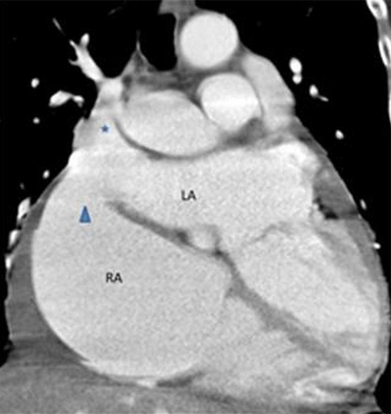
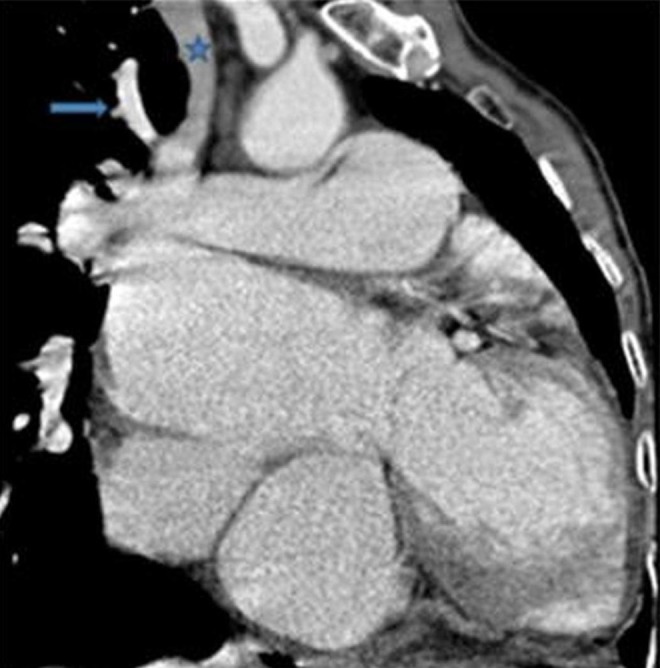
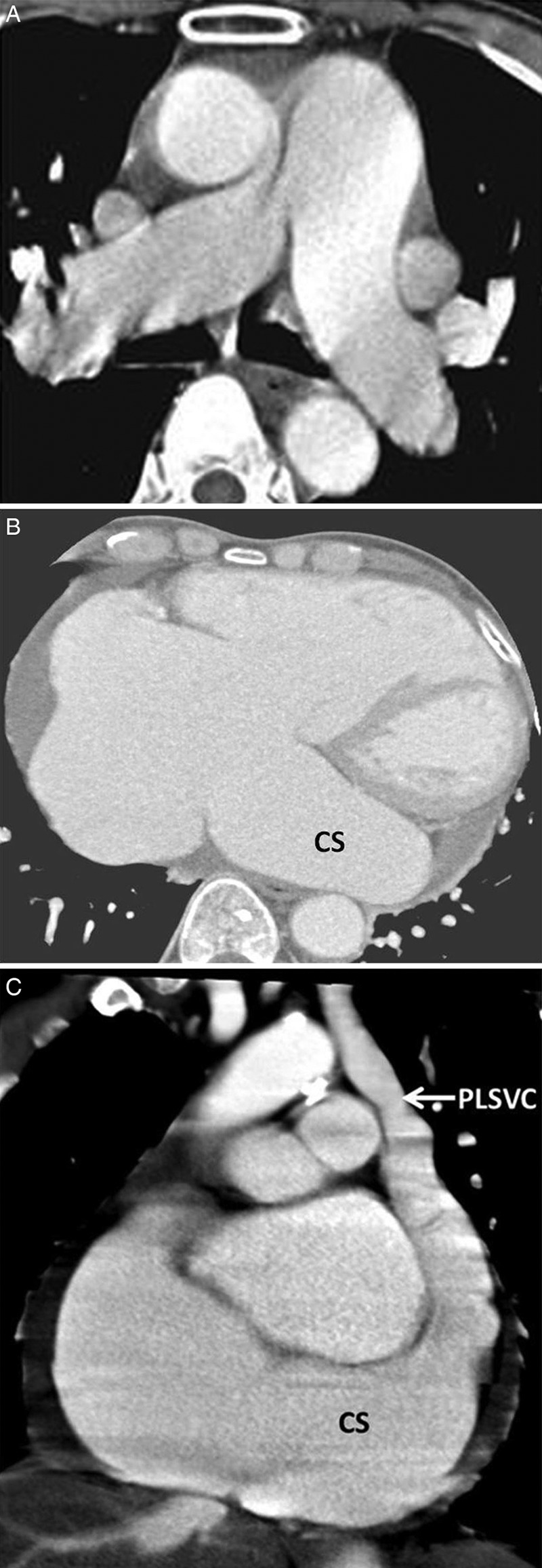
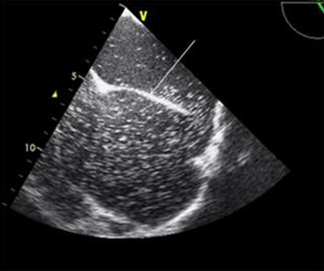
The most common venous abnormality of the thorax is persistent left superior vena cava (PLSVC), incidence being less than 0.5%. However, with congenital heart disease, it is about 6.1%. When the coronary sinus is dilated always search for PLSVC. The coronary sinus may communicate with the left atrium. This is known as an unroofed coronary sinus (UCS) and preoperatively documenting it is important. Of all the congenital cardiac anomalies, the sinus venosus defect (SVD) type of atrial septal defect (ASD) is most commonly associated with PLSVC and accounts for 4-11% of all ASDs. Multidetector CT can easily show all these abnormalities along with haemodynamics. On transoesophageal echocardiography it is difficult to characterise SVD and visualise a coronary sinus because of a limited window, contrast resolution and poor patient compliance. The complex of UCS and PLSVC is one such abnormality and its treatment requires careful assessment of other concomitant cardiac abnormalities to prevent post-treatment haemodynamic complications.
2014 BMJ Publishing Group Ltd.
Figures
Similar articles
-
[Anomalous right upper pulmonary venous drainage into the superior vena cava with coexistence sinus venosus atrial septal defect (ASD)].Przegl Lek. 2013;70(5):353-5. Przegl Lek. 2013. PMID: 23944110 Polish.
-
CT appearance of persistent left superior vena cava, anomalous right superior pulmonary venous return into the right-sided superior vena cava and a sinus venosus-type atrial septal defect.Br J Radiol. 2009 Nov;82(983):e235-9. doi: 10.1259/bjr/27663006. Br J Radiol. 2009. PMID: 19890118
-
Use of multi-modality imaging in a patient with a persistent left superior vena cava, partial anomalous pulmonary venous connection, and sinus venosus-type atrial septal defect.Eur Heart J Cardiovasc Imaging. 2012 Jun;13(6):499. doi: 10.1093/ejechocard/jer274. Epub 2011 Dec 6. Eur Heart J Cardiovasc Imaging. 2012. PMID: 22146782 No abstract available.
-
[A case report of coronary sinus septal defect associated with left superior vena cava, sinus venosus defect and partial anomalous pulmonary venous drainage].Nihon Geka Hokan. 1988 Sep 1;57(5):434-42. Nihon Geka Hokan. 1988. PMID: 3075110 Review. Japanese. No abstract available.
-
Partial anomalous pulmonary venous connection to the right side of the heart.J Thorac Cardiovasc Surg. 1989 Nov;98(5 Pt 2):861-8. J Thorac Cardiovasc Surg. 1989. PMID: 2682021 Review.
Cited by
-
Persistent Left-Sided Superior Vena Cava: A Case Report.Cureus. 2024 Aug 27;16(8):e67935. doi: 10.7759/cureus.67935. eCollection 2024 Aug. Cureus. 2024. PMID: 39328610 Free PMC article.
-
Septal Defects: Unveiling Sex-Based Disparities and Screening Challenges for Timely Intervention Through a Case Report and Systematic Literature Review.Cureus. 2024 Jul 30;16(7):e65752. doi: 10.7759/cureus.65752. eCollection 2024 Jul. Cureus. 2024. PMID: 39144879 Free PMC article.
-
The Presence of Patent Foramen Ovale in the Superior Type of Sinus Venosus Atrial Septal Defect.J Tehran Heart Cent. 2020 Jul;15(3):98-104. doi: 10.18502/jthc.v15i3.4218. J Tehran Heart Cent. 2020. PMID: 33552204 Free PMC article.
-
Sinus Venosus ASDs: Imaging and Percutaneous Closure.Curr Cardiol Rep. 2021 Aug 19;23(10):138. doi: 10.1007/s11886-021-01571-7. Curr Cardiol Rep. 2021. PMID: 34410510 Review.
References
-
- Kula S, Cevik A, Sanli C, et al. Persistent left superior vena cava: experience of a tertiary health-care center. Pediatr Int 2011;53:1066–9 - PubMed
-
- Quaegebeur J, Kirklin JW, Pacifico AD, et al. Surgical experience with unroofed coronary sinus. Ann Thorac Surg 1979;27:418–25 - PubMed
-
- Hahm JK, Park YW, Lee JK, et al. Magnetic resonance imaging of unroofed coronary sinus: three cases. Pediatr Cardiol 2000;21:382–7 - PubMed
-
- Brancaccio G, Miraldi F, Ventriglia F, et al. Multidetector-row helical computed tomography imaging of unroofed coronary sinus. Int J Cardiol 2003;91:251–3 - PubMed
Publication types
MeSH terms
LinkOut - more resources
Full Text Sources
Other Literature Sources