

Acute Marchiafava-Bignami disease: clinical and serial MRI correlation
- PMID: 24850553
- PMCID: PMC4039915
- DOI: 10.1136/bcr-2013-203442
Acute Marchiafava-Bignami disease: clinical and serial MRI correlation
Abstract
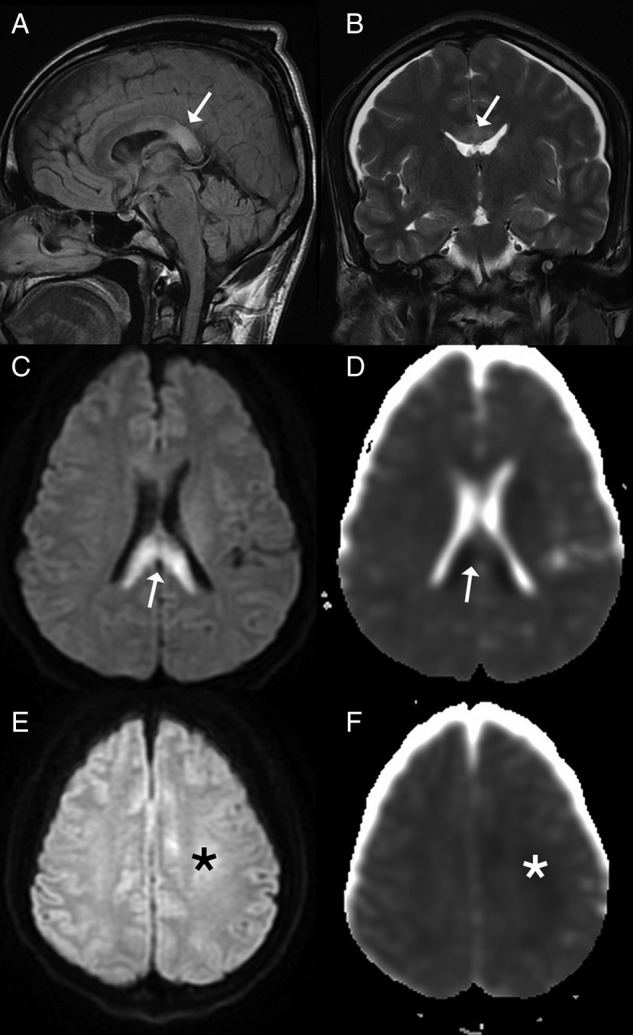
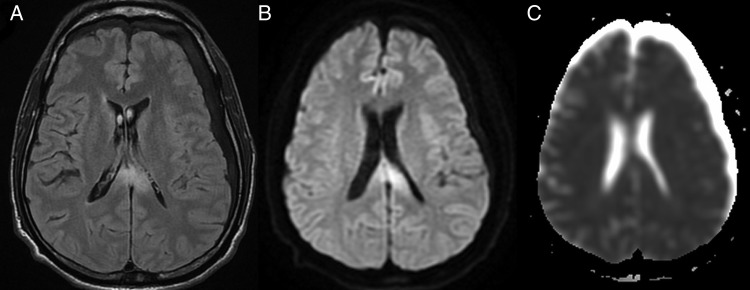
Marchiafava-Bignami disease (MBD) is a form of toxic demyelinating disease more often seen in chronic alcoholics. The disease process typically involves the corpus callosum and clinically often presents with altered sensorium, neurocognitive defects or seizures with acute cases often deteriorating to comatose state. The death rate is high. We report a rare case of MBD with complete clinical recovery. A 50-year-old male patient presented in an unconscious state and underwent MRI of the brain which showed significant lesions involving the corpus callosum. Following treatment with thiamine and supportive therapy, he improved clinically and a follow-up MRI revealed significant resolution of the earlier lesions. Diffusion-weighted MRI showed the changes more conspicuously as compared with conventional imaging. The clinical resolution corresponded well with the MRI pattern. The case highlights that diffusion-weighted MRI is an extremely useful tool in evaluation and prognostication of MBD.
2014 BMJ Publishing Group Ltd.
Figures
Similar articles
-
The value of diffusion-weighted imaging in the diagnosis of Marchiafava-Bignami disease: apropos of a case.J Neuroimaging. 2008 Apr;18(2):188-90. doi: 10.1111/j.1552-6569.2007.00202.x. Epub 2007 Dec 7. J Neuroimaging. 2008. PMID: 18318682
-
Marchiafava Bignami disease presenting as a cerebrovascular accident.Am J Emerg Med. 2024 Dec;86:190.e1-190.e3. doi: 10.1016/j.ajem.2024.09.058. Epub 2024 Sep 28. Am J Emerg Med. 2024. PMID: 39366786
-
Diagnosis and management of Marchiafava-Bignami disease: a review of CT/MRI confirmed cases.J Neurol Neurosurg Psychiatry. 2014 Feb;85(2):168-73. doi: 10.1136/jnnp-2013-305979. Epub 2013 Aug 26. J Neurol Neurosurg Psychiatry. 2014. PMID: 23978380 Free PMC article. Review.
-
Simultaneous acute Marchiafava-Bignami disease and central pontine myelinolysis: A case report of a challenging diagnosis.Medicine (Baltimore). 2018 Feb;97(8):e9878. doi: 10.1097/MD.0000000000009878. Medicine (Baltimore). 2018. PMID: 29465574 Free PMC article.
-
Acute Marchiafava-Bignami disease with extensive diffusion restriction and early recovery: case report and review of the literature.J Neuroimaging. 2014 Jul-Aug;24(4):421-4. doi: 10.1111/j.1552-6569.2012.00755.x. Epub 2012 Dec 17. J Neuroimaging. 2014. PMID: 23253188 Review.
Cited by
-
Clinical and radiological features of Marchiafava-Bignami disease.Medicine (Baltimore). 2018 Feb;97(5):e9626. doi: 10.1097/MD.0000000000009626. Medicine (Baltimore). 2018. PMID: 29384842 Free PMC article.
-
From Chronic Alcohol Consumption to Coma: Report of an Uncommon Cause.Cureus. 2023 Mar 20;15(3):e36411. doi: 10.7759/cureus.36411. eCollection 2023 Mar. Cureus. 2023. PMID: 37090382 Free PMC article.
-
Reversible MR Findings in Marchiafava-Bignami Disease.Case Rep Neurol Med. 2019 Feb 6;2019:1951030. doi: 10.1155/2019/1951030. eCollection 2019. Case Rep Neurol Med. 2019. PMID: 30881711 Free PMC article.
-
A rare neurological manifestation of a malnourished alcohol-dependent acute pancreatitis patient with Marchiafava-Bignami disease.Gastroenterol Rep (Oxf). 2020 Oct 1;9(2):179-181. doi: 10.1093/gastro/goaa062. eCollection 2021 Apr. Gastroenterol Rep (Oxf). 2020. PMID: 34026226 Free PMC article. No abstract available.
-
A Rare Case of Marchiafava-Bignami Disease With Reversible Splenial Lesion.Cureus. 2025 Apr 7;17(4):e81845. doi: 10.7759/cureus.81845. eCollection 2025 Apr. Cureus. 2025. PMID: 40337580 Free PMC article.
References
-
- Bourekas EC, Varakis K, Bruns D, et al. Lesions of the corpus callosum: MR imaging and differential considerations in adults and children. AJR Am J Roentgenol 2002;179:251–7 - PubMed
-
- Ellison D, Love S, Chimmeli L, et al. Neuropathology. A reference text of CNS pathology. 2nd edn Philadelphia: Mosby, 2004:489–90
-
- Ruiz-Martinez J, Martinez Perez-Balsa A, Ruibal M, et al. Marchiafava-Bignami disease with widespread extracallosal lesions and favourable course. Neuroradiology 1999;41:40–3 - PubMed
-
- Leong ASY. Marchiafava Bignami disease in a non-alcoholic Indian male. Pathology 1979;11:241–9 - PubMed
Publication types
MeSH terms
Substances
LinkOut - more resources
Full Text Sources
Other Literature Sources