. 2014 Jul 1;193(1):177-84.
doi: 10.4049/jimmunol.1400553. Epub 2014 May 21.

Rapid proliferation and differentiation impairs the development of memory CD8+ T cells in early life

Affiliations

Rapid proliferation and differentiation impairs the development of memory CD8+ T cells in early life

Norah L Smith et al. J Immunol. .

Abstract

Neonates often generate incomplete immunity against intracellular pathogens, although the mechanism of this defect is poorly understood. An important question is whether the impaired development of memory CD8+ T cells in neonates is due to an immature priming environment or lymphocyte-intrinsic defects. In this article, we show that neonatal and adult CD8+ T cells adopted different fates when responding to equal amounts of stimulation in the same host. Whereas adult CD8+ T cells differentiated into a heterogeneous pool of effector and memory cells, neonatal CD8+ T cells preferentially gave rise to short-lived effector cells and exhibited a distinct gene expression profile. Surprisingly, impaired neonatal memory formation was not due to a lack of responsiveness, but instead because neonatal CD8+ T cells expanded more rapidly than adult cells and quickly became terminally differentiated. Collectively, these findings demonstrate that neonatal CD8+ T cells exhibit an imbalance in effector and memory CD8+ T cell differentiation, which impairs the formation of memory CD8+ T cells in early life.


Figures

Figure 1
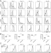
Figure 1. Neonatal CD8+ T Cells proliferate more than their adult counterparts
(A) Untouched CD8+ T cells from adult (shaded) or neonate (line) C57Bl/6 mice were labeled with CFSE and stimulated with plate bound anti-CD3/anti-CD28. (B) CFSE dilution in gB peptide (10−9 M)-stimulated gBT-I CD8+ T cells from adult or neonatal mice. Data are representative of at least 3 independent experiments.
Figure 2
Figure 2. Neonatal CD8+ T cells fail to transition into the memory pool
(A) Schematic of experimental design: 1×10⁴ gBT-I CD8+ T cells from congenic adult (Thy1.1) and neonatal (Thy1.2) donors were co-transferred intravenously to congenic, wild type recipients (CD45.1). These recipients were then infected with 5×10³ CFU WT Lm-gB and serially bled to monitor donor specific CD8+ T cell responses. (B) Relative numbers of donor adult (solid) or neonatal (dashed) gBT-I CD8+ T cells following infection and subsequent challenge (5×10⁴ CFU WT Lm gB). (C) The ratio of the donor populations with respect to time. Dashed line indicates starting ratio of donor cells. (D) At 70 days post infection, peripheral organs were harvested and the ratio of adult to neonatal CD8+ donor cells was assessed. Significance was determined by one-way ANOVA followed by Dunnett's multiple comparisons test. Data representative of 2–4 experiments, n=4−8 mice/group.
Figure 3
Figure 3. Neonatal CD8+ T cells exhibit a more differentiated phenotype
(A) Splenocytes were harvested at indicated days post infection and donor CD8+ T cells from congenically marked adult (shaded) and neonatal (line) were assessed for surface expression of CD127, KLRG1, CD62L and CD27. Histograms show representative data from each timepoint. (B) Statistical analysis of phenotypic markers over all recipient mice on day 7 post infection. (C) IFNγ production was assayed by peptide re-stimulation of splenocytes on indicated days post infection. Histograms show representative data from each timpoint. Significance was determined by paired T test. Data representative of 4 experiments, n=4−8 mice/group.
Figure 4
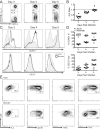
Figure 4. Neonatal CD8+ T cells rapidly become terminally differentiated
5×10⁴ congenically marked gBT-I cells from each age group (adult and neonate) were transferred into congenically marked recipients and spleens and lymph nodes were harvested at the indicated timepoints. (A) Neonatal donor gBT-I (Thy1.1−) cells are more numerous than adult donor gBT-I (Thy1.1+) early in infection; representative plots of donor cells are shown. (B) Ratio of adult to neonatal donors in the spleens of all recipients at early time points; dashed line indicates starting ratio of donor cells. (C) Representative histograms show that neonatal donor cells (black line) rapidly acquire the phenotype of short-lived effector cells (KLRG1hiCD127low). (D) Percentage of donor neonatal and adult CD8+ T cells that are either KLRG1hi or CD127low. (E) Acquisition of effector functions by neonatal and adult donor CD8+ T cells in the lymph nodes at 4 days post-infection. Data representative of 2–3 experiments, n=4−8 mice/group.
Figure 5
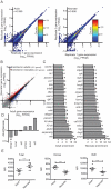
Figure 5. Gene expression differences between adult and neonatal effector cells
Gene expression differences between adult and neonatal effector cells. (A) Gene expression values (FPKM) for each gene from RNA-seq data for two biological replicates of adults and of neonates were plotted. Adult sample pairs had Pearson correlation coefficient of 0.999 (p-value <2.2 × 10⁻¹⁶), and neonate sample pairs had a Pearson correlation coefficient of 0.998 (p-value <2.2 × 10⁻¹⁶). (B) Gene expression values for each gene were plotted as FPKM values from adult (x-axis) and neonatal (y-axis) samples. Genes in which the FPKM value was at least 1 in either sample were considered expressed (11,446 in total). The remaining 13,296 genes that did not reach that cutoff are shown in gray. Genes that are expressed at significantly (q-value <0.05) higher or lower levels in adults as compared to neonates are highlighted in blue (321 genes) and red (261 genes), respectively. (C) Fold differences in expression for the most differentially expressed genes in adult and neonate effector cells. (D) Differential expression levels between adults and neonates are shown for transcription factors associated with effector cell and memory cell fate. In panels C and D, genes marked with an asterisk are significantly differentially expressed (q-value <0.05). (E) Co-transferred cells were recovered on day 7 post-infection (5×10³ WT LmgB) and stained for intracellular expression of transcription factors. Significance was assessed by paired t test, n=8.
Figure 6
Figure 6. Failure to form CD8+ memory is a general feature of neonatal CD8+ mediated immune responses
1×10⁴ gBT-I CD8+ T cells from both adult and neonatal donors were co-transferred intravenously to congenic, wild type recipients. These recipients were then infected intraperitoneally with 2×10⁵ PFU VACV-gB. Recipient mice were serially bled to monitor donor specific CD8+ T cell responses. (A) Percentage of gBT-1 CD8+ T cell in donor adult (solid) or neonatal (dashed) cells following infection and subsequent intravenous challenge with WT Lm gB (5×10⁴ CFU). (B) The ratio of the donor populations with respect to time indicates that adult donors become the majority population. (C) Phenotypic distribution of adult and neonatal donors at peak of primary response (day 6). Significance was determined by paired T test. Data are representative of 2 experiments, n=8–12.

References

    1. Siegrist CA. Neonatal and early life vaccinology. Vaccine. 2001;19:3331–3346. - PubMed
    1. Marchant A, Goldman M. T cell-mediated immune responses in human newborns: ready to learn? Clin Exp Immunol. 2005;141:10–18. - PMC - PubMed
    1. Adkins B, Leclerc C, Marshall-Clarke S. Neonatal adaptive immunity comes of age. Nat Rev Immunol. 2004;4:553–564. - PubMed
    1. Chang J, Braciale TJ. Respiratory syncytial virus infection suppresses lung CD8+ T-cell effector activity and peripheral CD8+ T-cell memory in the respiratory tract. Nat Med. 2002;8:54–60. - PubMed
    1. Zinkernagel RM, Doherty PC. Immunological surveillance against altered self components by sensitised T lymphocytes in lymphocytic choriomeningitis. Nature. 1974;251:547–548. - PubMed

Publication types