Comparative Study
. 2014 Aug;88(15):8514-27.
doi: 10.1128/JVI.00752-14. Epub 2014 May 21.

Human immunodeficiency virus reverse transcriptase displays dramatically higher fidelity under physiological magnesium conditions in vitro

Affiliations
Comparative Study

Human immunodeficiency virus reverse transcriptase displays dramatically higher fidelity under physiological magnesium conditions in vitro

Vasudevan Achuthan et al. J Virol. 2014 Aug.

Erratum in

  • J Virol. 2015 Feb;89(3):1944

Abstract

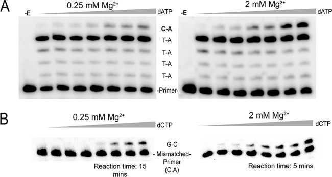
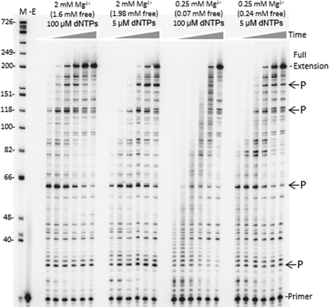
The fidelity of human immunodeficiency virus (HIV) reverse transcriptase (RT) has been a subject of intensive investigation. The mutation frequencies for the purified enzyme in vitro vary widely but are typically in the 10(-4) range (per nucleotide addition), making the enzyme severalfold less accurate than most polymerases, including other RTs. This has often been cited as a factor in HIV's accelerated generation of genetic diversity. However, cellular experiments suggest that HIV does not have significantly lower fidelity than other retroviruses and shows a mutation frequency in the 10(-5) range. In this report, we reconcile, at least in part, these discrepancies by showing that HIV RT fidelity in vitro is in the same range as cellular results from experiments conducted with physiological (for lymphocytes) concentrations of free Mg(2+) (~0.25 mM) and is comparable to Moloney murine leukemia virus (MuLV) RT fidelity. The physiological conditions produced mutation rates that were 5 to 10 times lower than those obtained under typically employed in vitro conditions optimized for RT activity (5 to 10 mM Mg(2+)). These results were consistent in both commonly used lacZα complementation and steady-state fidelity assays. Interestingly, although HIV RT showed severalfold-lower fidelity under high-Mg(2+) (6 mM) conditions, MuLV RT fidelity was insensitive to Mg(2+). Overall, the results indicate that the fidelity of HIV replication in cells is compatible with findings of experiments carried out in vitro with purified HIV RT, providing more physiological conditions are used.

Importance: Human immunodeficiency virus rapidly evolves through the generation and subsequent selection of mutants that can circumvent the immune response and escape drug therapy. This process is fueled, in part, by the presumably highly error-prone HIV polymerase reverse transcriptase (RT). Paradoxically, results of studies examining HIV replication in cells indicate an error frequency that is ~10 times lower than the rate for RT in the test tube, which invokes the possibility of factors that make RT more accurate in cells. This study brings the cellular and test tube results in closer agreement by showing that HIV RT is not more error prone than other RTs and, when assayed under physiological magnesium conditions, has a much lower error rate than in typical assays conducted using conditions optimized for enzyme activity.


Figures

FIG 1
FIG 1
PCR-based lacZα complementation system used to determine the fidelity of HIV RT. (A) An overview of the procedure used to assess polymerase fidelity is presented. RNAs are indicated by broken lines and DNAs by solid lines. Primers have arrowheads at the 3′ end. The ∼760-nucleotide template RNA used as the initial template for HIV RT RNA-directed DNA synthesis is shown at the top with the 3′ and 5′ ends indicated. The positions of PvuII and EcoRI restriction sites are indicated for reference to the vector. The filled box at the bottom of is the 115-base region of the lacZα gene that was scored in the assay. Details for specific steps are provided in Materials and Methods. (B) Plasmid pBSM13ΔPvuII1146 is shown. Relevant sites on the plasmid are indicated; numbering is based on that for the parent plasmid (pBSM13+ [Stratagene]). (C) The nucleotide and amino acid sequences for the 115-base region of the lacZα gene that was scored in the assay are shown. Both strands of the DNA plasmid are shown since HIV RT synthesis was performed in both directions (see panel A). A line is drawn above the 92 nt that are in the detectable area for substitution mutations, while frameshifts can be detected over the entire 115-nucleotide region. Based on a previous cataloging of mutations in this gene (19), the assay can detect 116 different substitutions (33.6% of the 345 possible substitutions in the 115-nucleotide sequence) and 100% of the frameshift mutations.
FIG 2
FIG 2
DNA sequence analysis from the PCR-based lacZα complementation fidelity assay. The 115-base region analyzed for mutations is shown (see Fig. 1). The coding strand for lacZα is shown in the 5′-3′ direction (bottom strand in Fig. 1C). Numbering is as shown in Fig. 1C. Deletions are shown as regular triangles, insertions are shown as downward triangles with the inserted base shown adjacent to the downward triangle, unless it was same as the base in a nucleotide run, and base substitutions are shown directly above or below the sequence. Substitutions shown correspond to the recovered sequence for the coding strand; however, these mutations could have occurred during synthesis of the noncoding strand as well (i.e., a C-to-A change shown here could have resulted from a C-to-A change during synthesis of the coding strand or a G-to-T change during synthesis of the noncoding strand) (see Fig. 1 and Results). Mutations recovered from HIV RT at 6 mM Mg2+ and 100 μM dNTPs and mutations from background controls are shown above the sequence as open triangles and normal text or filled triangles and bold italicized text, respectively. Mutations from HIV RT at 0.25 mM Mg2+ and 5 μM dNTPs are shown below the sequence. Individual sequence clones which had multiple mutations (more than one mutation event) are marked with subscripts adjacent to the mutations. Several clones with deletions (either single or multiple deletions) at positions 181 to 183, just outside the scored region, were also recovered (data not shown). This was the dominant mutation type recovered in background controls (19 out of 24 total sequences) and probably resulted from improper ligation events or damaged plasmid vectors (see Results). Two out of 22 HIV RT-derived sequences at 6 mM Mg2+ and 62 out of 162 HIV RT-derived sequences at 0.25 mM Mg2+ also had these deletions.
FIG 3
FIG 3
Constructs used in mismatched primer extension and running-start misincorporation assays. The sequence of the DNA constructs used in each assay type is shown. The underlined nucleotides show the only differences between the two templates. Only one primer was used in the running-start assays, and it terminated at the 3′ C nucleotide before the dashes. The four dashes indicate the 4 A nucleotides that must be incorporated before RT incorporates the target nucleotide (denoted by X or Y).
FIG 4
FIG 4
Representative experiments for running-start and mismatch extension assays. (A) Running-start misincorporation of C·A base pair at 0.25 and 2 mM Mg2+. Reactions were performed on the primer-template shown in Fig. 3 for 3 min with a final free Mg2+ concentration of 0.25 or 2 mM (adjusted according to the total concentration of dNTPs in each reaction using the KD values of Mg2+ and ATP). A fixed concentration of dATP (55 μM) was used in all running-start reactions for elongation of the primer to the target site. The concentration of the target nucleotide (dATP for C·A insertion) in each lane was, from left to right, 0, 400, 630, 1,380, 2,610, and 3,660 μM. For other base pair misinsertions noted in Table 4, the target nucleotide was changed according to the desired misinsertion. −E, no enzyme was added. (B) Extension of a mismatched primer-template with a C·A 3′ terminus, using 0.25 and 2 mM Mg2+. Reactions were performed on the primer-template shown in Fig. 3 for the indicated time with a final free Mg2+ concentration of 0.25 or 2 mM (adjusted as described above). The concentration of the next correct nucleotide (dCTP) in each lane was, from left to right, 0, 50, 100, 200, 400, 630, 1,200 and 1,870 μM. All mismatched primer-templates were extended for 5 min at 2 mM Mg2+, whereas in 0.25 mM Mg2+, primer-template constructs with 3′ termini of C·C and G·A were extended for 20 min and all other mismatched primer-templates were extended for 15 min.
FIG 5
FIG 5
Time course of HIV RT synthesis on the ∼760-nucleotide RNA template used in the PCR-based α complementation assay. Shown is an autoradiogram with extension of a 20-nucleotide 5′-, 32P-end-labeled DNA primer on the RNA template used for round 1 synthesis by HIV RT (see Fig. 1). Full extension of the primer resulted in a 199-nucleotide product. A DNA ladder with nucleotide size positions is shown on the left. Concentrations of total and free Mg2+ and dNTPs are indicated above each lane. Positions of some prominent DNA synthesis pause sites (P) are indicated on the right. Reactions were performed for (1eft to right) 15 s, 30 s, 1 min, 2 min, 4 min, or 8 min under each condition. A no-enzyme control (−E) is also shown. Refer to Materials and Methods for details.

References

    1. Arts EJ, Hazuda DJ. 2012. HIV-1 antiretroviral drug therapy, p 321–343 In Bushman FD, Nabel GJ, Swanstrom R. (ed), HIV from biology to prevention and treatment. Cold Spring Harbor Laboratory Press, Cold Spring Harbor, NY - PMC - PubMed
    1. Telesnitsky A, Goff SP. 1993. Reverse transcriptase. Cold Spring Harbor Laboratory Press, Cold Spring Harbor, NY - PubMed
    1. Cowan JA, Ohyama T, Howard K, Rausch JW, Cowan SM, Le Grice SF. 2000. Metal-ion stoichiometry of the HIV-1 RT ribonuclease H domain: evidence for two mutually exclusive sites leads to new mechanistic insights on metal-mediated hydrolysis in nucleic acid biochemistry. J. Biol. Inorg. Chem. 5:67–74. 10.1007/s007750050009 - DOI - PubMed
    1. Johnson KA. 2010. The kinetic and chemical mechanism of high-fidelity DNA polymerases. Biochim. Biophys. Acta 1804:1041–1048. 10.1016/j.bbapap.2010.01.006 - DOI - PMC - PubMed
    1. Joyce CM, Steitz TA. 1994. Function and structure relationships in DNA polymerases. Annu. Rev. Biochem. 63:777–822. 10.1146/annurev.bi.63.070194.004021 - DOI - PubMed

Publication types

MeSH terms

LinkOut - more resources