. 2014 Aug;88(16):8783-94.
doi: 10.1128/JVI.00531-14. Epub 2014 May 21.

Discovery and evolution of bunyavirids in arctic phantom midges and ancient bunyavirid-like sequences in insect genomes

Affiliations

Discovery and evolution of bunyavirids in arctic phantom midges and ancient bunyavirid-like sequences in insect genomes

Matthew J Ballinger et al. J Virol. 2014 Aug.

Abstract

Bunyaviridae is a large family of RNA viruses chiefly comprised of vertebrate and plant pathogens. We discovered novel bunyavirids that are approximately equally divergent from each of the five known genera. We characterized novel genome sequences for two bunyavirids, namely, Kigluaik phantom virus (KIGV), from tundra-native phantom midges (Chaoborus), and Nome phantom virus (NOMV), from tundra-invading phantom midges, and demonstrated that these bunyavirid-like sequences belong to an infectious virus by passaging KIGV in mosquito cell culture, although the infection does not seem to be well sustained beyond a few passages. Virus and host gene sequences from individuals collected on opposite ends of North America, a region spanning 4,000 km, support a long-term, vertically transmitted infection of KIGV in Chaoborus trivittatus. KIGV-like sequences ranging from single genes to full genomes are present in transcriptomes and genomes of insects belonging to six taxonomic orders, suggesting an ancient association of this clade with insect hosts. In Drosophila, endogenous virus genes have been coopted, forming an orthologous tandem gene family that has been maintained by selection during the radiation of the host genus. Our findings indicate that bunyavirid-host interactions in nonbloodsucking arthropods have been much more extensive than previously thought.

Importance: Very little is known about the viral diversity in polar freshwater ponds, and perhaps less is known about the effects that climate-induced habitat changes in these regions will have on virus-host interactions in the coming years. Our results show that at the tundra-boreal boundary, a hidden viral landscape is being altered as infected boreal phantom midges colonize tundra ponds. Likewise, relatively little is known of the deeper evolutionary history of bunyavirids that has led to the stark lifestyle contrasts between some genera. The discovery of this novel bunyavirid group suggests that ancient and highly divergent bunyavirid lineages remain undetected in nature and may offer fresh insight into host reservoirs, potential sources of emerging disease, and major lifestyle shifts in the evolutionary history of viruses in the family Bunyaviridae.


Figures

FIG 1
FIG 1
Evolutionary relationships between bunyavirid polymerase sequences. A phylogram of RdRp amino acid sequences belonging to representatives of the five established genera of Bunyaviridae, currently unclassified bunyavirids, and the novel bunyavirids described in this study supports the evolutionary position of the novel viruses as members of the family Bunyaviridae. Branches are labeled with Bayesian posterior probabilities/approximate likelihood ratio test scores. Taxonomic groups are labeled with their accepted or proposed names.
FIG 2
FIG 2
Sequence, genome structure, and PCR evidence of a novel, exogenous bunyavirid. (A) A graphical representation of the genomic structure for each of the three genome segments of KIGV is shown in positive-sense directionality. On the M segment, locations of predicted signal peptides and cleavage sites within the glycoprotein precursor protein are indicated by pink blocks and scissors, respectively. The predicted Gn-Gc cleavage site (pink asterisk) is shown to the right, as a sequence alignment between KIGV, Nome phantom virus (NOMV), and four phleboviruses (Jacunda virus [JANV] [included because this is the closest BLASTp match to KIGV Gn-Gc], sandfly fever Naples virus [SFNV], Rift Valley fever virus [RVFV], and Uukuniemi virus [UUKV]). Above the M segment is the result of transmembrane domain (TMD) prediction. Gray blocks represent predicted TMDs, dark blue blocks represent external domains, and light blue blocks represent internal domains. Y's represent predicted glycosylation sites. (B) Sequences and predicted secondary structures of the complementary terminal NCRs of the three KIGV segments and of the M and S segments of NOMV. Sequences underlined in green are those that NOMV shares with KIGV. (C) Agarose gel electrophoresis following RT-PCR experiments targeting the putative viral segments of KIGV demonstrates that products were amplified from RNA templates and not from DNA templates. Primer targets and treatment conditions are displayed above the appropriate lanes. (D) Agarose gel electrophoresis following RT-PCR experiments targeting the putative viral segments of KIGV, enriched by CsCl ribonucleoprotein particle isolation, shows that each putative viral segment was enriched relative to the host-encoded transcript.
FIG 3
FIG 3
KIGV infection experiments in mosquito cell culture. (A) Agarose gel electrophoresis of RT-PCR products amplified with primers targeting the KIGV polymerase in an Aedes albopictus c6/36 cell culture inoculated with KIGV-infected Chaoborus trivittatus tissue homogenate shows increasing viral RNA detection with days postinoculation. (B) RT-PCR results of a 2-week passage of KIGV in Aedes albopictus cells. Numbers above lanes indicate the days on which samples were tested.
FIG 4
FIG 4
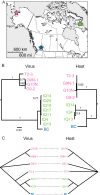
Evidence for long-term, vertically transmitted KIGV infection in Chaoborus trivittatus. (A) Map of the northern United States and Canada, with colored stars indicating locations from which KIGV-positive Chaoborus trivittatus flies were collected. Pink, Seward Peninsula of Alaska; green, Iqaluit, Baffin Island, Canada; blue, Salmon Arm, British Columbia, Canada. (B) Virus and host nucleotide sequence-based maximum likelihood phylogenies showing topological congruence between the viral polymerase and the host mitochondrial COI gene. Branches are labeled with approximate likelihood ratio test scores. Tips are colored to match the host collection site to the geographic locations shown in panel A. (C) Parsimony tanglegram built from the virus and host sequences used for panel B, shown to provide resolution to within-region relationships.
FIG 5
FIG 5
An ancient, orthologous gene family in Drosophila is derived from a phasmavirus nucleoprotein gene. (A) A maximum likelihood phylogram shows the evolutionary relationships between the two clades of phasmavirus nucleoprotein (N)-like paleoviruses and exogenous phasmavirus N amino acid sequences. Branches are labeled with filled and hollow circles to indicate approximate likelihood ratio test scores of >0.95 and >0.8, respectively. Tips are labeled with species names. (B) Microsynteny map of the genomic features flanking the N-like paleoviruses of each species. Green arrows indicate the positions of clade 1 paleoviruses, and blue arrows indicate the positions of clade 2 paleoviruses. Arrow direction indicates gene directionality. The Greek letter ψ indicates a pseudogenized paleovirus. Pseudogenized paleoviruses shorter than 100 amino acids were excluded from the phylogenetic analysis. Regions of the genome maps that remain unshaded are those where the assembly is incomplete. Genes of host origin used to establish positional homology were as follows: DPEP, dipeptidase; CCNA, cyclin A; IAP-1, inhibitor of apoptosis 1; GnT-IV, N-acetylglucosaminyltransferase IV.
FIG 6
FIG 6
Phylogenetic relationships between phasmavirus genes and phasmavirus-like paleoviruses in insects. Maximum likelihood phylograms show predicted relationships between extant phasmavirus genes and paleoviruses identified by significant tBLASTn sequence matches in insect genomes and transcriptomes. Individual trees are labeled with gene names or abbreviations. Branches are labeled with aLRT support values of ≥0.70. Branch tips are labeled with host species names, and GenBank accession numbers are provided in Table S2 in the supplemental material. Taxa for which virus-like sequences corresponding to each of the 3 genome segments were identified in transcriptomes are marked with pink asterisks. The nucleoprotein-like paleoviruses of Drosophila are represented by a subset of taxa in the interest of conserving space. See Fig. 5 for a complete phylogenetic analysis of this paleovirus family.

References

    1. Jorgenson MT, Shur YL, Pullman ER. 2006. Abrupt increase in permafrost degradation in Arctic Alaska. Geophys. Res. Lett. 33:L02503. 10.1029/2005GL024960 - DOI
    1. Prowse TD, Wrona FJ, Reist JD, Gibson JJ, Hobbie JE, Levesque LM, Vincent WF. 2006. Climate change effects on hydroecology of arctic freshwater ecosystems. Ambio 35:347–358. 10.1579/0044-7447(2006)35[347:CCEOHO]2.0.CO;2 - DOI - PubMed
    1. Ruhland KM, Paterson AM, Keller W, Michelutti N, Smol JP. 2013. Global warming triggers the loss of a key Arctic refugium. Proc. Biol. Sci. 280:20131887. 10.1098/rspb.2013.1887 - DOI - PMC - PubMed
    1. Elliott RM. 2009. Bunyaviruses and climate change. Clin. Microbiol. Infect. 15:510–517. 10.1111/j.1469-0691.2009.02849.x - DOI - PubMed
    1. Maltezou HC, Papa A. 2010. Crimean-Congo hemorrhagic fever: risk for emergence of new endemic foci in Europe? Travel Med. Infect. Dis. 8:139–143. 10.1016/j.tmaid.2010.04.008 - DOI - PubMed

Publication types

LinkOut - more resources