. 2014 Aug;88(15):8667-77.
doi: 10.1128/JVI.01092-14. Epub 2014 May 21.

Structural insight into BH3 domain binding of vaccinia virus antiapoptotic F1L

Affiliations

Structural insight into BH3 domain binding of vaccinia virus antiapoptotic F1L

Stephanie Campbell et al. J Virol. 2014 Aug.

Abstract

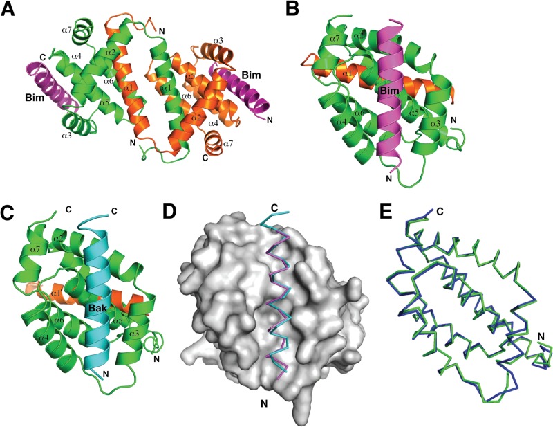
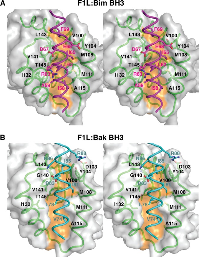
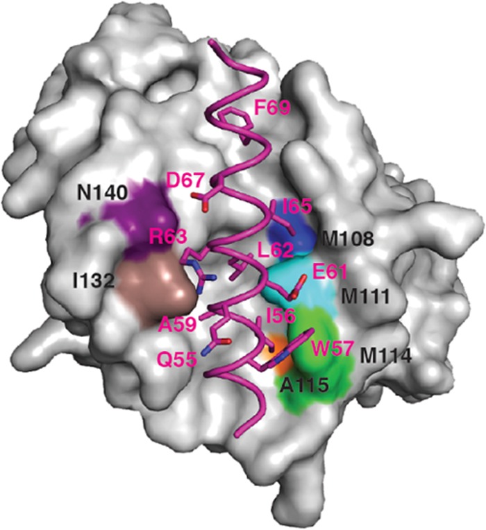
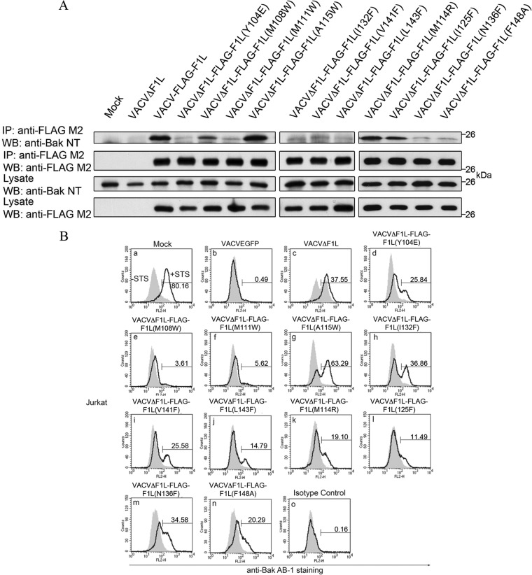
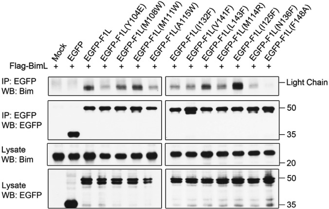
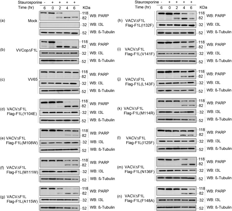
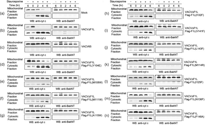
Apoptosis is a tightly regulated process that plays a crucial role in the removal of virus-infected cells, a process controlled by both pro- and antiapoptotic members of the Bcl-2 family. The proapoptotic proteins Bak and Bax are regulated by antiapoptotic Bcl-2 proteins and are also activated by a subset of proteins known as BH3-only proteins that perform dual functions by directly activating Bak and Bax or by sequestering and neutralizing antiapoptotic family members. Numerous viruses express proteins that prevent premature host cell apoptosis. Vaccinia virus encodes F1L, an antiapoptotic protein essential for survival of infected cells that bears no discernible sequence homology to mammalian cell death inhibitors. Despite the limited sequence similarities, F1L has been shown to adopt a novel dimeric Bcl-2-like fold that enables hetero-oligomeric binding to both Bak and the proapoptotic BH3-only protein Bim that ultimately prevents Bak and Bax homo-oligomerization. However, no structural data on the mode of engagement of F1L and its Bcl-2 counterparts are available. Here we solved the crystal structures of F1L in complex with two ligands, Bim and Bak. Our structures indicate that F1L can engage two BH3 ligands simultaneously via the canonical Bcl-2 ligand binding grooves. Furthermore, by structure-guided mutagenesis, we generated point mutations within the binding pocket of F1L in order to elucidate the residues responsible for both Bim and Bak binding and prevention of apoptosis. We propose that the sequestration of Bim by F1L is primarily responsible for preventing apoptosis during vaccinia virus infection.

Importance: Numerous viruses have adapted strategies to counteract apoptosis by encoding proteins responsible for sequestering proapoptotic components. Vaccinia virus, the prototypical member of the family Orthopoxviridae, encodes a protein known as F1L that functions to prevent apoptosis by interacting with Bak and the BH3-only protein Bim. Despite recent structural advances, little is known regarding the mechanics of binding between F1L and the proapoptotic Bcl-2 family members. Utilizing three-dimensional structures of F1L bound to host proapoptotic proteins, we generated variants of F1L that neutralize Bim and/or Bak. We demonstrate that during vaccinia virus infection, engagement of Bim and Bak by F1L is crucial for subversion of host cell apoptosis.


Figures

FIG 1
FIG 1
Crystal structures of F1L in complex with BH3 peptides of both Bak and Bim. (A) Cartoon representation of F1L bound to the Bim BH3 domain. The F1L dimer is shown as a cartoon (one monomer is green, and the other is orange) with helices α1 to α7 labeled. Two BimL BH3 molecules are shown in magenta. The view is down the 2-fold symmetry axis between the domain-swapped α1 helices. (B) Cartoon representation of F1L bound to the BimL BH3 domain. This view is into the hydrophobic binding grove formed by helices α2 to α5. F1L helices α2 to α7 from monomer 1 (green) are labeled, as is helix α1′ from monomer 2 (orange). BimL BH3 is shown in magenta. (C) Cartoon representation of F1L bound to the Bak BH3 complex. F1L (green and orange) is in complex with Bak BH3 (cyan). The view is the same as in panel B. (D) BH3 domain binding to F1L. The BimL (magenta) and Bak (cyan) BH3 domains are shown as traces bound to the hydrophobic binding groove on F1L, which is shown as a molecular surface (gray). (E) Superimposition of the F1L main chains from the two complexes formed with the BimL (green) and Bak (blue) BH3 domains.
FIG 2
FIG 2
Stereo diagrams of F1L in complex with the BH3 peptides of both Bak and Bim. (A) Stereo diagram of the binding interface formed by the F1L (green):Bim (magenta) complex. The F1L surface is gray, except for the orange shading indicating the bottom of the peptide binding groove. F1L residues are labeled in black, and BimL BH3 residues are labeled in magenta. (B) Stereo diagram of the binding interface formed by the F1L (green):Bak (cyan) complex. The view and labeling are the same as in panel A, except for the labeling of Bak residues in cyan.
FIG 3
FIG 3
Structure of F1L in complex with the Bim BH3 peptide highlighting the F1L binding pocket hydrophobic residues. Shown is the molecular surface of F1L in complex with the BH3 peptide of BimL (in magenta). The FIL binding pocket residues that facilitate hydrophobic contact with specific amino acids of BimL are highlighted in colors. Included are F1L(N140), which is purple; F1L(I132), which is brown; F1L(A119W), which is orange; F1L(M114), which is green; F1L(M111), which is aqua; and F1L(M108), which is blue.
FIG 4
FIG 4
F1L binding pocket residues are responsible for Bak interaction and prevention of Bak activation. (A) HeLa cells were mock infected or infected with VACVΔF1L, VACV-FLAG-F1L, or recombinant virus expressing the F1L binding pocket mutations. Infected-cell lysates were then immunoprecipitated (IP) with a monoclonal antibody recognizing FLAG, and Bak was detected by blotting with an anti-Bak monoclonal antibody (40, 41). (B) Jurkat cells were infected with WT VACV expressing EGFP, VACVΔF1L, or a panel of recombinant VACVs carrying F1L point mutations (VACVΔF1L-F1L) for 4 h at an MOI of 10 before treatment with 250 nM STS for 1.5 h to induce apoptosis. Bak N-terminal exposure was monitored by staining cells with the conformation-specific anti-Bak AB-1 antibody (40, 41) or an anti-NK1.1 antibody (42) as an isotype control. Shaded histograms, untreated cells; open histograms, STS-treated cells. WB, Western blotting.
FIG 5
FIG 5
F1L binding pocket residues facilitate interactions with the BH3-only protein BimL. HEK 293T cells were cotransfected with EGFP, EGFP-F1L, or EGFP-tagged F1L containing the binding cleft mutation with FLAG-BimL. Cells were lysed and immunoprecipitated (IP) with goat anti-GFP antibody. Samples were separated by SDS-PAGE, and BimL was detected with anti-Bim antibody (Enzo Life Sciences). WB, Western blotting.
FIG 6
FIG 6
Prevention of cytochrome c release by infection with VACV encoding F1L binding pocket mutants. Jurkat cells (1 × 10⁶) were mock infected (a) or infected with VACVΔF1L (b), VACV65 (c), or a panel of recombinant VACVs carrying F1L point mutations (d to n) at an MOI of 10 for 6, 8, 10, or 12 h. At 6 h postinfection, cells were treated with 2 μM STS. Mitochondrial and cytoplasmic fractions were separated in lysis buffer containing digitonin. Mitochondrial fractions were subsequently resuspended in 0.1% Triton X-100 lysis buffer. Twenty percent of the mitochondrial fractions and 50% of the cytoplasmic fractions were Western blotted (WB) with anti-cytochrome c antibody to determine the presence of cytochrome c in the separated fractions or with anti-BakNT antibody to ensure the separation of the two fractions.
FIG 7
FIG 7
Inhibition of PARP cleavage by infection with VACV encoding F1L binding pocket mutations. Jurkat cells (1 × 10⁶) were mock infected (a) or infected with VACV65 (b), VACVΔF1L (c), or a panel of recombinant VACVs carrying the F1L binding pocket point mutations (d to n) at an MOI of 10. After 6 h of infection, cells were treated with 2 μM STS for 2, 4, or 6 h. Whole-cell lysates were then collected in SDS lysis buffer containing 8 M urea. Samples were subjected to SDS-PAGE and Western blotted (WB) for PARP to determine apoptosis, β-tubulin as a loading control, and I3L as a sign of infection (44).

References

    1. Cuconati A, White E. 2002. Viral homologs of BCL-2: role of apoptosis in the regulation of virus infection. Genes Dev. 16:2465–2478. 10.1101/gad.1012702 - DOI - PubMed
    1. Galluzzi L, Brenner C, Morselli E, Touat Z, Kroemer G. 2008. Viral control of mitochondrial apoptosis. PLoS Pathog. 4:e1000018. 10.1371/journal.ppat.1000018 - DOI - PMC - PubMed
    1. Taylor JM, Barry M. 2006. Near death experiences: poxvirus regulation of apoptotic death. Virology 344:139–150. 10.1016/j.virol.2005.09.032 - DOI - PubMed
    1. Youle RJ, Strasser A. 2008. The BCL-2 protein family: opposing activities that mediate cell death. Nat. Rev. Mol. Cell Biol. 9:47–59. 10.1038/nrm2308 - DOI - PubMed
    1. Chipuk JE, Moldoveanu T, Llambi F, Parsons MJ, Green DR. 2010. The BCL-2 family reunion. Mol. Cell 37:299–310. 10.1016/j.molcel.2010.01.025 - DOI - PMC - PubMed

Publication types

MeSH terms

LinkOut - more resources